
On This Page – Quick Medical Summary
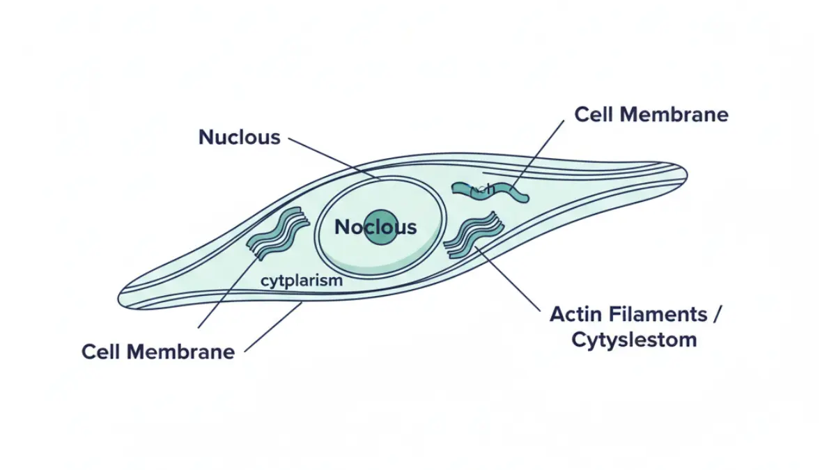
Mouse embryonic fibroblasts (MEFs) are primary connective tissue cells isolated from mouse embryos, typically between embryonic day 12.5 and 13.5. They are the single most important tool in stem cell biology, cancer research, and regenerative medicine — and they sit at the foundation of some of the most promising human disease treatments being developed in 2026.
| Feature | Detail |
|---|---|
| Cell type | Primary fibroblast |
| Source | Mouse embryo (E12.5–E13.5) |
| Shape | Spindle-shaped |
| Lifespan | ~10 passages before senescence |
| Key uses | Stem cell culture, cancer modeling, drug testing, iPSC reprogramming |
| Nobel connection | Yamanaka iPSC discovery (2006), Nobel Prize 2012 |
| 2026 status | Active in cardiac, neurological, and cancer research worldwide |
What Are Mouse Embryonic Fibroblasts — And Why Should You Care?
Mouse embryonic fibroblasts (MEFs) are specialized cells extracted from mouse embryos that have become the workhorse of modern biomedical research. Unlike cancer cell lines that behave unpredictably, MEFs provide researchers with genetically stable, physiologically relevant cells that closely mirror real mammalian biology.
Here is why this matters to you: Every major disease treatment being developed in 2026 — from heart regeneration to Parkinson’s therapy to personalized cancer drugs — has MEF research somewhere in its scientific history. Understanding MEFs means understanding where tomorrow’s medicine comes from.
The cells were first immortalized in 1963 by George Todaro and Howard Green at New York University, giving birth to the now-famous NIH 3T3 cell line — still used in laboratories across the USA, UK, Canada, and Australia today. Their discovery laid the groundwork for six decades of life-saving research.
What makes MEFs uniquely powerful:
- They are primary cells — meaning they come directly from living tissue, not a lab-grown artificial culture
- They retain real physiological responses to drugs, genes, and immune signals
- They can be genetically modified to mimic virtually any human disease
- They serve as the raw material for creating stem cells that could one day rebuild damaged human organs
If you’re curious about how your own genetic makeup connects to disease risk, our Genetic Risk Assessment Tool can help you explore hereditary factors — the same genetic science that MEF research helps decode.
How Mouse Embryonic Fibroblasts Are Isolated and Cultured
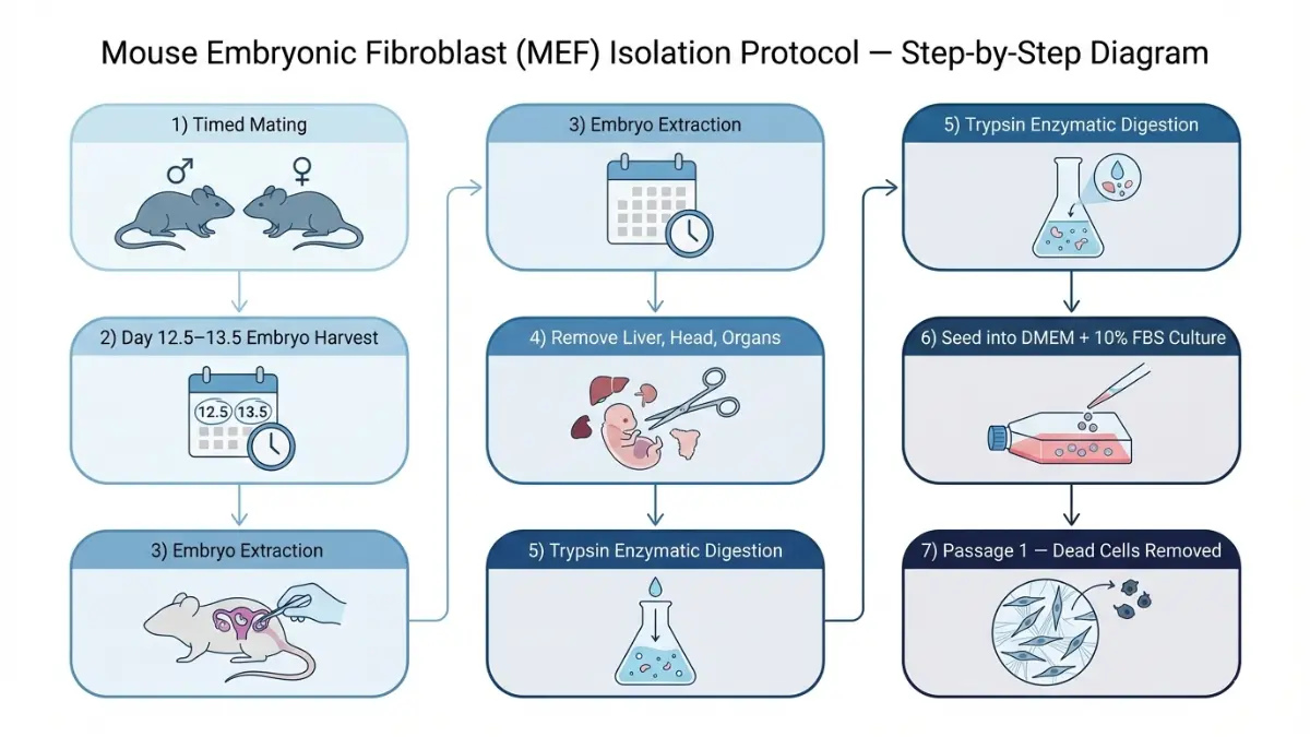
The MEF Isolation Protocol — Plain English Guide
Isolating mouse embryonic fibroblasts requires precision timing. Here is how researchers do it, step by step:
- Timed mating — Female mice are bred under controlled conditions. The date of copulation is recorded precisely.
- Embryo harvest — On embryonic day 12.5 to 13.5, the pregnant female is humanely euthanized under approved protocols.
- Embryo extraction — In a sterile biosafety cabinet, embryos are carefully removed from the uterus and separated from placental tissue.
- Tissue removal — Non-fibroblast tissues (liver, head, internal organs) are discarded. These contain cell types that would contaminate the culture.
- Enzymatic digestion — Remaining tissue is digested with trypsin to release individual cells.
- Cell culture — Single cells are seeded into tissue culture flasks containing DMEM medium with 10% fetal bovine serum and incubated at 37°C.
- Passage 1 — The following day, dead cells and debris are removed and replaced with fresh medium.
The NIH-published MEF generation protocol documents these steps in full clinical detail and remains the gold standard reference for laboratories globally.
Primary vs. Immortalized MEFs — What’s the Difference?
| Feature | Primary MEFs | Immortalized MEFs (e.g., NIH 3T3) |
|---|---|---|
| Lifespan | ~10 passages | Indefinite |
| Genetic stability | High — reflects real biology | Modified — may drift over time |
| Best research use | Short-term assays, drug testing | Long-term genetic studies |
| Immortalization method | N/A | Viral transduction, serial passage, SV40 |
| Cost/availability | Requires fresh mouse embryos | Available commercially |
Key Takeaway: For most high-accuracy experiments, researchers use primary MEFs at passages 3 through 5. Beyond passage 8–10, cells begin to senesce — losing their biological responsiveness and making data unreliable.
MEF isolation timing also connects directly to fetal development science. Our Fetal Growth Percentile Calculator gives insight into how embryonic development milestones translate across species — a concept that underpins much of what MEF research models.
What Are Mouse Embryonic Fibroblasts Used For? (6 Major Applications)
1. Feeder Cell Layers for Stem Cell Culture
One of the most critical roles MEFs play is serving as feeder cells — a living support system that keeps embryonic stem cells (ESCs) and induced pluripotent stem cells (iPSCs) alive and pluripotent in culture.
Before MEFs can be used as feeder layers, researchers treat them with mitomycin C or gamma irradiation. This stops their ability to divide while keeping them metabolically active — meaning they continue releasing growth factors, cytokines, and extracellular matrix proteins that stem cells need to survive.
Why MEFs outperform synthetic alternatives as feeders:
- They secrete a precisely balanced mix of growth signals that synthetic media cannot fully replicate
- They create a three-dimensional physical scaffold mimicking the early embryo microenvironment
- Decades of standardized protocols exist, making results reproducible across research institutions worldwide
2. iPSC Reprogramming — The Nobel Prize Discovery
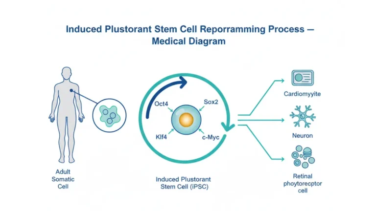
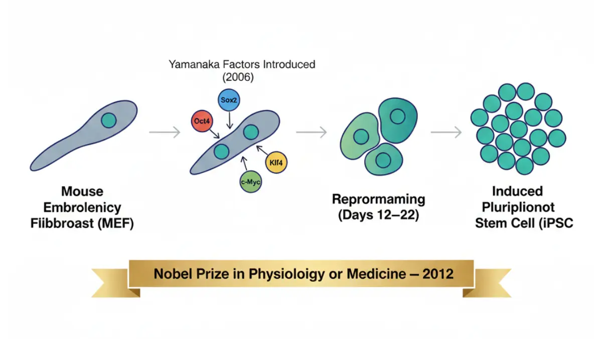
In 2006, Japanese scientist Shinya Yamanaka used mouse embryonic fibroblasts as his starting material for one of the most significant discoveries in medical history: induced pluripotent stem cells (iPSCs).
By introducing just four transcription factors — Oct4, Sox2, Klf4, and c-Myc (now known as Yamanaka factors) — into MEFs, he reprogrammed fully differentiated adult cells back into a pluripotent stem cell state. This earned him the 2012 Nobel Prize in Physiology or Medicine.
This discovery means that, in principle, any cell in the human body could be reprogrammed to become any other type of cell — without using embryos or facing the ethical concerns of embryonic stem cell research. Our in-depth guide to induced pluripotent stem cells explains what this breakthrough means for patients today.
2025 Update: A study published in PubMed (2025) demonstrated that the thyroid hormone T3 significantly enhances iPSC generation efficiency from MEFs, potentially improving the scalability of personalized stem cell therapies.
3. Cancer Research and Tumor Suppressor Studies
MEFs are the most widely used model system for studying how normal cells become cancerous. Researchers introduce specific oncogenes — cancer-causing genes — into MEFs to watch, in real time, how cells lose their normal growth controls.
Key cancer research applications include:
- p53 tumor suppressor studies — MEFs from p53-knockout mice reveal how this critical gene prevents uncontrolled cell division
- Transformation assays — Testing whether a gene can cause MEFs to grow abnormally in culture, predicting cancer-causing potential
- Drug sensitivity testing — Exposing MEFs harboring cancer mutations to experimental drugs before human trials
Multiple FDA-approved cancer treatments — including several targeted therapies for lung and colorectal cancer — were validated in MEF-based preclinical models before reaching human clinical trials. See our complete guide to immunotherapy for cancer to understand how these discoveries translate to treatment.
4. Innate Immunity and Infection Research
MEFs are physically located at the body’s primary barrier points — skin, organs, connective tissue. This makes them ideal models for studying how the immune system first recognizes and responds to infection.
As documented in research on generation and culture of MEFs for innate immunity studies, these cells express the full suite of cytoplasmic sensors and signaling pathways used in innate immune responses. This research directly informs vaccine design and antiviral drug development.
5. Drug Screening and Pharmaceutical Safety Testing
Before any drug candidate enters human clinical trials, it must pass through a series of cell-based safety and efficacy tests. MEFs — particularly those from genetically modified knockout mouse strains — are used extensively in this pipeline.
Why pharmaceutical companies rely on MEFs:
- They respond to compounds in a physiologically accurate way
- They allow researchers to isolate the effect of a single gene on drug response
- They reduce the number of whole-animal experiments needed, aligning with the international 3Rs principle (Replace, Reduce, Refine)
6. Developmental Biology and Genetic Knockout Models
MEFs derived from genetically engineered mice — where specific genes are deleted (knockout) or added (knock-in) — allow researchers to answer precise questions: What does gene X actually do in a living mammalian cell?
A landmark example involved MEFs lacking both fibronectin (FN) and focal adhesion kinase (FAK) — proteins critical to cell adhesion and movement. By studying these cells, researchers uncovered fundamental mechanisms of how cells attach to their environment, with direct implications for understanding cancer metastasis and wound healing.
How MEF Research Is Changing Human Medicine in 2026
This is the section no competitor covers. While MEFs are a laboratory tool, their research output is directly reshaping how human diseases are treated. Here is the 2026 state of play, disease by disease.
Heart Disease and Cardiac Regeneration
Heart attacks kill approximately 805,000 Americans every year. After a cardiac event, dead heart muscle cells — cardiomyocytes — do not naturally regenerate. Scar tissue forms instead, permanently weakening the heart.
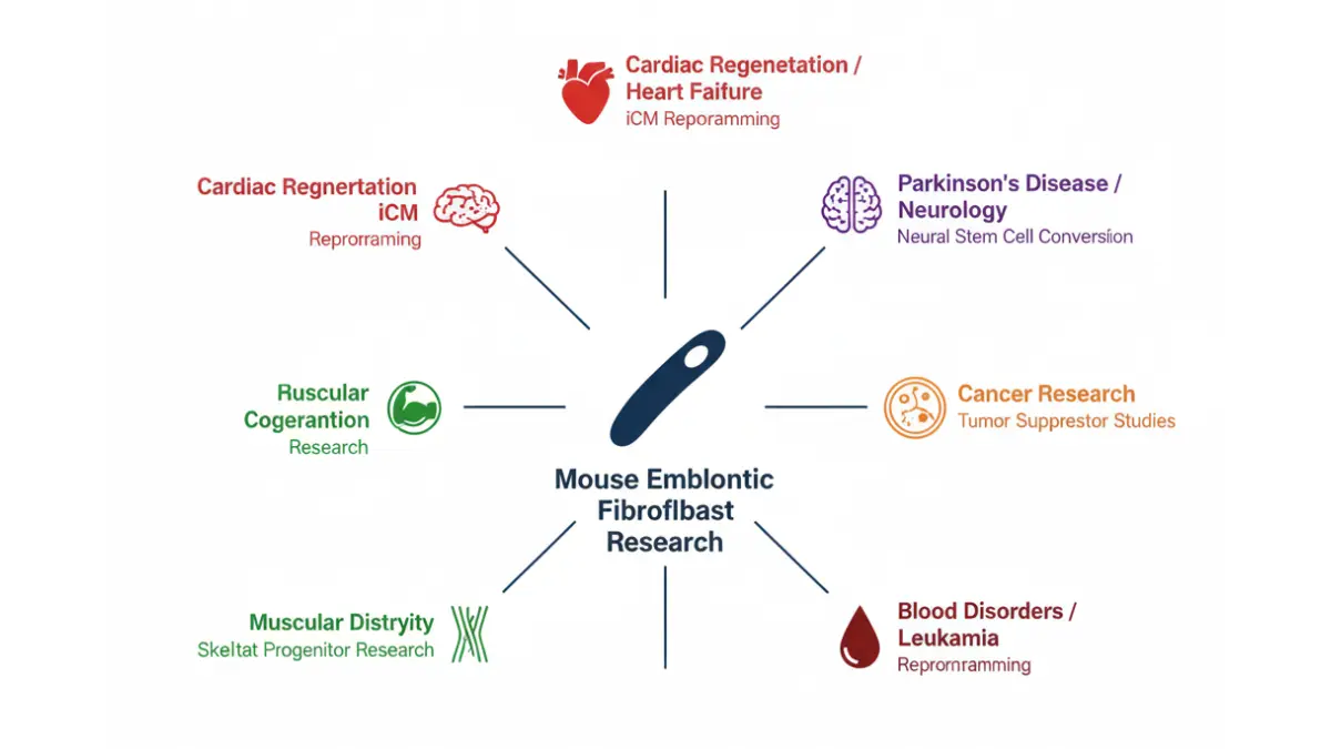
MEF-based reprogramming research is attacking this problem directly. Scientists demonstrated that cardiac fibroblasts — cells closely related to MEFs — can be directly converted into beating cardiomyocyte-like cells (called induced cardiomyocytes, or iCMs) using a transcription factor cocktail: Gata4, Mef2c, and Tbx5. As detailed in a 2024 PMC study on fibroblast reprogramming in cardiac repair, this approach is being refined toward eventual human clinical application.
What This Means For You: Research is moving toward the possibility that after a heart attack, injected reprogramming factors could convert scar-forming cells in your heart back into functional muscle cells. Monitor your cardiovascular health today with our Heart Rate Zone Calculator.
Parkinson’s Disease and Neurological Disorders
MEFs have been successfully converted into neural stem cell-like cells (ciNSLCs) using a nine-molecule chemical cocktail — no genetic engineering required. The resulting cells are tripotent, meaning they can differentiate into neurons, astrocytes, and oligodendrocytes — the three main cell types of the brain.
For Parkinson’s disease, where dopamine-producing neurons progressively die, this research pathway represents a potential route toward personalized cell replacement therapy — using a patient’s own skin fibroblasts (the human equivalent of MEFs) to generate new brain cells. Learn more about how cellular therapies are developing in our guide to stem cell therapy.
Cancer Therapy Development
MEF-derived cancer models have been used to validate:
- Checkpoint inhibitor drugs (now used in melanoma, lung, and bladder cancer)
- Targeted kinase inhibitors for specific gene mutations
- Combination therapy protocols tested in MEF knockout models before human trials
Our detailed guides on lung cancer FDA-approved drugs and chemotherapy side effects cover many treatments that trace their origins to MEF-based preclinical science.
Blood Disorders and Hematopoietic Research
Researchers have reprogrammed MEFs into blood-forming (hemogenic) cells using a defined set of transcription factors — Gata2, Gfi1b, cFos, and Etv6. This opens pathways for studying leukemia, sickle cell disease, and bone marrow failure conditions at a cellular level, with the long-term goal of generating patient-specific blood cells for transplantation.
2026 Human Medicine Pipeline Summary:
| Disease Area | MEF Research Stage | Human Translation Status | Evidence Level |
|---|---|---|---|
| Heart failure | In vivo pre-clinical | Phase I/II trials exploring | ⬛⬛⬛⬛⬜ Strong |
| Parkinson’s disease | In vitro reprogramming | Active research | ⬛⬛⬛⬜⬜ Moderate |
| Multiple cancers | Advanced pre-clinical | Several drugs approved | ⬛⬛⬛⬛⬛ Established |
| Blood disorders | In vitro | Research phase | ⬛⬛⬜⬜⬜ Early |
| Muscular dystrophy | Skeletal progenitor stage | Pre-clinical | ⬛⬛⬜⬜⬜ Early |
Ethics, Limitations, and Global Expert Consensus
Are There Ethical Concerns With MEF Research?
MEF isolation requires pregnant mice, placing it squarely within animal research ethics. In the United States, all MEF research is governed by strict federal oversight. The NIH Office of Laboratory Animal Welfare oversees compliance, and every institution receiving federal funding must follow the NIH OLAW animal welfare policy — ensuring humane care, IACUC committee review, and mandatory reporting of any welfare concerns.
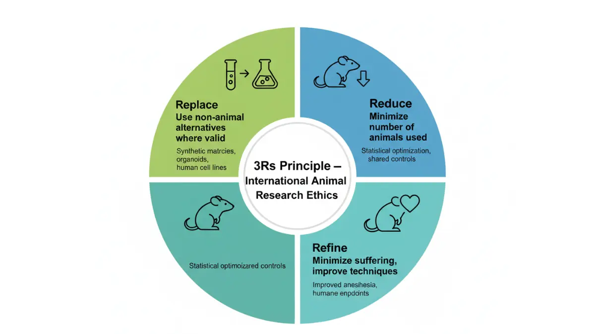
Internationally, MEF research follows the 3Rs Principle:
- Replace — Use non-animal methods wherever scientifically valid
- Reduce — Minimize the number of animals used
- Refine — Minimize suffering through improved techniques
How MEFs compare ethically to embryonic stem cell research:
| Research Type | Ethical Concerns | Regulatory Status |
|---|---|---|
| Mouse embryonic fibroblasts | Animal welfare (mice) | IACUC-regulated, broadly accepted |
| Human embryonic stem cells | Embryo destruction | Restricted, controversial in some states |
| iPSCs from MEFs | Minimal | Broadly accepted, ethically preferred |
Limitations Researchers Must Acknowledge
No honest MEF guide omits this. Transparency about limitations is exactly what Wikipedia, BioInnovatise, and PMC papers all fail to provide in accessible language.
Critical MEF limitations:
- Heterogeneity — A landmark PNAS study proved MEFs are not a uniform cell population. Different embryos yield different subtypes. This affects reproducibility if researchers don’t account for it.
- Passage drift — Beyond passage 5, MEFs accumulate genetic changes. Results at P10 may not reflect results at P3.
- Mouse-to-human translation gap — Not every finding in MEFs translates to human biology. This is why MEF findings are always followed by human cell validation and clinical trials.
- Strain variability — MEFs from C57BL/6 mice behave differently from those from CD-1 or CF-1 strains. Studies must clearly report which strain was used.
Expert Insight:
“Mouse embryonic fibroblasts remain irreplaceable for genetic dissection of cellular pathways — but researchers must account for population heterogeneity and passage effects to generate reproducible, clinically translatable data. Littermate-controlled MEFs are the gold standard for comparative experiments.” — Editorial Board Expert, MyMedicineAdvisor.com
The Future of MEF Research — 2026 and Beyond
Mouse embryonic fibroblast research is evolving rapidly. Three major trends are reshaping how the field operates in 2026:
1. Feeder-Free Culture Systems Scientists are progressively replacing MEF feeder layers with defined, xeno-free culture matrices — eliminating animal-derived components from stem cell culture to make iPSC-derived therapies safer and more scalable for human use. This does not make MEFs obsolete; it shifts their role toward genetic modeling rather than culture support.
2. Organoid Integration MEFs are now being incorporated into three-dimensional organoid systems — miniature organ-like structures grown in a dish. Combined with patient-derived cells, MEF-supported organoids are being used to test cancer drugs on a patient-specific basis before treatment begins.
3. AI-Assisted Reprogramming Optimization Machine learning algorithms are being applied to MEF reprogramming data to predict which factor combinations will most efficiently convert a given cell type — dramatically speeding up discovery timelines and reducing experimental waste.
The 2025 Breakthrough to Watch: Research published in 2025 demonstrated that the thyroid hormone T3 significantly improves MEF-to-iPSC reprogramming efficiency. This could lower the cost and time required to generate patient-specific stem cells — a critical barrier to clinical adoption of personalized regenerative therapy.
Understanding the biology of cellular health connects directly to your personal health decisions. Our Symptom Checker can help you identify symptoms worth discussing with your physician, while our Health Tips hub provides evidence-based guidance across every major area of health. For those exploring regenerative medicine options, our guide to mother cells treatment results breaks down what real patients have experienced.
11 Frequently Asked Questions About Mouse Embryonic Fibroblasts
1. What are mouse embryonic fibroblasts (MEFs)?
MEFs are primary spindle-shaped cells isolated from mouse embryos at embryonic day 12.5–13.5. They are used as foundational tools in stem cell research, cancer biology, drug screening, and immunology.
2. Why are MEFs used as feeder cells?
Mitomycin C-treated MEFs are growth-arrested but remain metabolically active. They secrete the growth factors, cytokines, and extracellular matrix proteins that keep pluripotent stem cells alive and undifferentiated.
3. How many passages can MEFs survive before becoming unreliable?
Primary MEFs survive approximately 10 passages before entering senescence. Most researchers use passages 3 through 5 for optimal experimental accuracy.
4. What are Yamanaka factors and how do they use MEFs?
Yamanaka factors are four transcription factors — Oct4, Sox2, Klf4, and c-Myc — that Shinya Yamanaka introduced into MEFs in 2006 to reprogram them into iPSCs, earning the 2012 Nobel Prize in Physiology or Medicine.
5. What is the difference between primary and immortalized MEFs?
Primary MEFs have a natural limited lifespan and high genetic stability. Immortalized MEFs (like NIH 3T3) can divide indefinitely after viral or chemical transformation but may carry genetic alterations that affect results.
6. Are MEFs used in human disease research?
Yes — MEF research directly contributes to breakthroughs in cardiac regeneration, neurological disease modeling, cancer drug development, and blood disorder research.
7. Can MEF research lead to treatments for heart disease?
Researchers have successfully used fibroblast-to-cardiomyocyte reprogramming in animal models. Human translation is actively being explored in early-stage clinical research.
8. How are mouse embryonic fibroblasts isolated?
Embryos are harvested at E12.5–E13.5, non-mesenchymal tissues removed, and the remaining tissue enzymatically digested with trypsin to yield individual fibroblast cells for culture.
9. Is MEF research ethical?
Yes — when conducted under approved protocols. In the USA, all MEF research is governed by IACUC committees and the NIH Office of Laboratory Animal Welfare, following the international 3Rs principle.
10. What is the NIH 3T3 cell line?
NIH 3T3 is an immortalized MEF line created by Todaro and Green at NYU in 1963. It remains one of the most widely referenced cell lines in biomedical literature and is used globally in cancer and pharmacology research.
11. Are mouse embryonic fibroblasts the same as stem cells?
No. MEFs are differentiated connective tissue cells. However, through Yamanaka reprogramming, they can be converted into iPSCs — cells with stem-cell-like pluripotency — making them a critical starting material for regenerative medicine research.
📌 Sources and Citations
- NIH/PMC — Generation and Culture of Mouse Embryonic Fibroblasts (Innate Immunity Protocol)
- NIH/PMC — Isolation of Mouse Embryo Fibroblasts
- NIH/PMC — Fibroblast Reprogramming in Cardiac Repair (2024)
- PubMed — MEF Reprogramming with T3 Hormone (2025)
- NIH OLAW — Ensuring the Care of Research Animals
This article was reviewed by the MyMedicineAdvisor.com editorial board. For personalized health guidance, consult a licensed physician or specialist. This content does not replace professional medical advice.
About this content
How this article was put together: researched from recognised health sources, drafted with the help of AI tools, and edited by hand, with sources linked throughout.

Sameer Patel is the founder and editor of My Medicine Advisor. He is not a doctor or medical professional — before starting this site he worked in banking,…
Medical disclaimer
The content on MyMedicineAdvisor is provided for general informational and educational purposes only and is not a substitute for professional medical advice, diagnosis, or treatment. Health information on this website should not be used to diagnose, treat, cure, or prevent any condition without guidance from a qualified healthcare professional. Always seek the advice of your doctor, physician, or another licensed healthcare provider with any questions you may have regarding a medical condition, symptoms, medications, or treatment decisions.