On This Page – Quick Medical Summary
Sarah stared at her patient portal at 11 PM, heart racing. Her chest x-ray report read “unremarkable findings”—was that good or bad? Like 68% of patients who access their imaging results online, she felt confused and anxious rather than relieved.
What Does “Normal” Mean?
A normal x-ray means no abnormalities requiring immediate medical attention were detected on your imaging study. When radiologists use terms like “unremarkable,” “within normal limits,” or “no acute findings,” they’re actually delivering good news—your x-ray shows clear lungs, normal heart size, intact bones, and no fluid collections or masses that need urgent intervention.
These confusing medical terms are standard radiology language. They don’t mean “unremarkable” in the everyday sense—they mean your internal structures appear healthy and functioning properly.
What You’ll Learn in This Guide:
- How to decode the 15 most confusing medical terms on your report
- What radiologists actually look for when reading x-ray normal results
- When normal findings don’t match your symptoms (and what to do)
- Real report examples you can compare to yours
- Critical questions to ask your doctor after normal x-ray results
- When you might need additional testing despite normal findings
According to the Radiological Society of North America, over 200 million x-rays are performed annually in the United States. Yet studies from the National Institutes of Health show that approximately 70% of patients struggle to understand their radiology reports, leading to unnecessary anxiety and confusion about their health status.
Understanding your chest x-ray normal results is the first step toward taking control of your health journey. Whether you received results for a routine screening, after an injury, or to investigate symptoms, this comprehensive guide will help you make sense of what “normal” truly means—and what actions you should take next.
Decoding Your Normal X-Ray Report
Understanding What “Normal” Really Means on Your X-Ray Report
When you receive normal x-ray results, you’re looking at a standardized medical document that follows a specific format. Every radiology report contains the same five sections, regardless of whether you had a chest x-ray, bone x-ray, or abdominal imaging.
The 5 Sections of Every X-Ray Report
1. Patient Information & Study Details This section lists your name, date of birth, the type of x-ray performed, and when it was done. It confirms they’re looking at YOUR images, not someone else’s.
2. Clinical Indication This explains why your doctor ordered the x-ray. You might see phrases like “evaluate chest pain,” “rule out pneumonia,” or “assess for fracture.” This helps the radiologist focus on specific concerns when interpreting your x-ray normal findings.
3. Technique This describes how the x-ray was performed—standing or lying down, number of views taken, and radiation dose used. For patient safety, modern digital x-rays use FDA-approved minimal radiation exposure.
4. Findings (The Detailed Section) This is where radiologists describe what they see in each area: lungs, heart, bones, soft tissues. For a normal x-ray report interpretation, you’ll see reassuring terms we’ll decode below.
5. Impression/Conclusion This summary section is the most important part for you. It’s where the radiologist gives their bottom-line assessment. A normal impression might read: “No acute cardiopulmonary abnormality” or “Unremarkable chest radiograph.”
15 Medical Terms That Confuse Patients (But Mean Good News)
Understanding these terms will immediately reduce your anxiety when reading your x-ray findings normal report:
| Medical Term | What It Actually Means | Why Doctors Use It |
|---|---|---|
| Unremarkable | Normal, nothing concerning found | Standard medical jargon for “healthy—nothing to remark about” |
| Within normal limits | Expected, healthy appearance for your age | Acknowledges natural variations between people |
| No acute process | No urgent or sudden problems detected | “Acute” means recent/sudden; this rules out emergencies |
| Clear lung fields | Lungs look healthy, well-inflated | No signs of infection, fluid, or masses |
| Intact osseous structures | Bones are not broken or damaged | “Osseous” is the medical term for bone |
| Cardiomediastinal silhouette normal | Heart size and shape appear healthy | Heart isn’t enlarged or misshapen |
| No pleural effusion | No fluid buildup around lungs | Fluid collections would indicate problems |
| No pneumothorax | Lung hasn’t collapsed | Air isn’t leaking into chest cavity |
| Visualized structures unremarkable | Everything visible looks normal | Comprehensive statement covering all areas |
| No focal consolidation | No dense areas suggesting pneumonia | Lungs are properly air-filled |
| Costophrenic angles sharp | Normal borders where lungs meet diaphragm | Blunting here suggests fluid or disease |
| No cardiomegaly | Heart isn’t abnormally enlarged | Normal heart size for your body |
| Bones well mineralized | Bone density appears normal | No signs of bone disease or loss |
| No soft tissue abnormality | Muscles and tissues look normal | No swelling, masses, or unusual changes |
| No acute cardiopulmonary abnormality | Heart and lungs both appear healthy | Comprehensive “all clear” statement |
According to research published by the American College of Radiology, the term “unremarkable” is the most commonly misunderstood phrase in radiology reports, with 43% of patients initially interpreting it as negative rather than positive news.
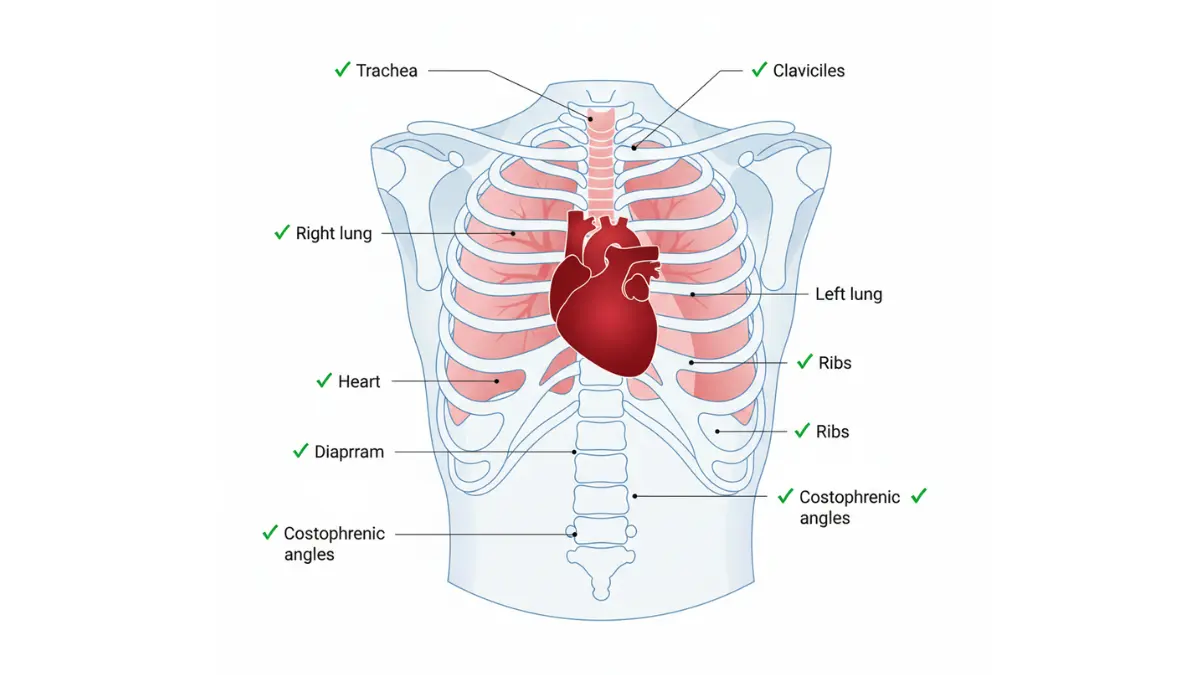
Real Report Examples: What Normal Looks Like
Normal Chest X-Ray Example:
INDICATION: Cough and fever
TECHNIQUE: PA and lateral chest radiographs
FINDINGS:
- Lungs: Clear lung fields bilaterally. No focal consolidation, pleural effusion, or pneumothorax.
- Heart: Cardiomediastinal silhouette within normal limits.
- Bones: Visualized osseous structures unremarkable.
IMPRESSION: No acute cardiopulmonary abnormality.
Translation for patients: Your lungs are clear with no infection or fluid. Your heart size is normal. Your visible bones look healthy. Everything appears normal—no urgent concerns.
Normal Bone X-Ray Example:
INDICATION: Right wrist pain after fall
TECHNIQUE: Three views of the right wrist
FINDINGS:
- Bones: Intact osseous structures. No fracture or dislocation identified.
- Joints: Normal joint spaces. No degenerative changes.
- Soft tissues: No significant soft tissue swelling.
IMPRESSION: No acute osseous abnormality.
Translation: No broken bones detected. Joints look normal. No major swelling. Your wrist x-ray is normal despite your pain—soft tissue injuries like sprains don’t show on x-rays.
Normal Abdominal X-Ray Example:
INDICATION: Abdominal pain
TECHNIQUE: Supine and upright abdominal radiographs
FINDINGS:
- Bowel gas pattern normal.
- No evidence of obstruction or free air.
- Visualized osseous structures unremarkable.
IMPRESSION: Normal abdominal radiograph.
Translation: Your intestines show normal gas patterns. No blockages or perforations. The bones visible in your abdomen area look healthy. Everything appears normal on the x-ray.
What This Means For You: If your report uses similar language, your x-ray normal results indicate no urgent problems requiring immediate treatment. However, if you’re experiencing concerning symptoms that persist, discuss further evaluation with your doctor—x-rays have important limitations we’ll cover in Section 4.
Many patients wonder about following up on normal results, especially when they underwent imaging related to overall health screening. Maintaining a healthy lifestyle remains important even after receiving reassuring x-ray findings normal reports.
What Radiologists Actually Look For
Inside the Radiologist’s Mind: How They Read Your X-Ray Normal Results
When a radiologist reviews your x-ray, they follow a systematic approach to ensure nothing gets missed. Understanding this process helps you appreciate why your normal x-ray results are truly reassuring.
The Systematic Approach (ABCDEFGH Method)
Radiologists use proven mnemonics to systematically evaluate every x-ray. For chest x-rays, one common approach is ABCDEFGH:
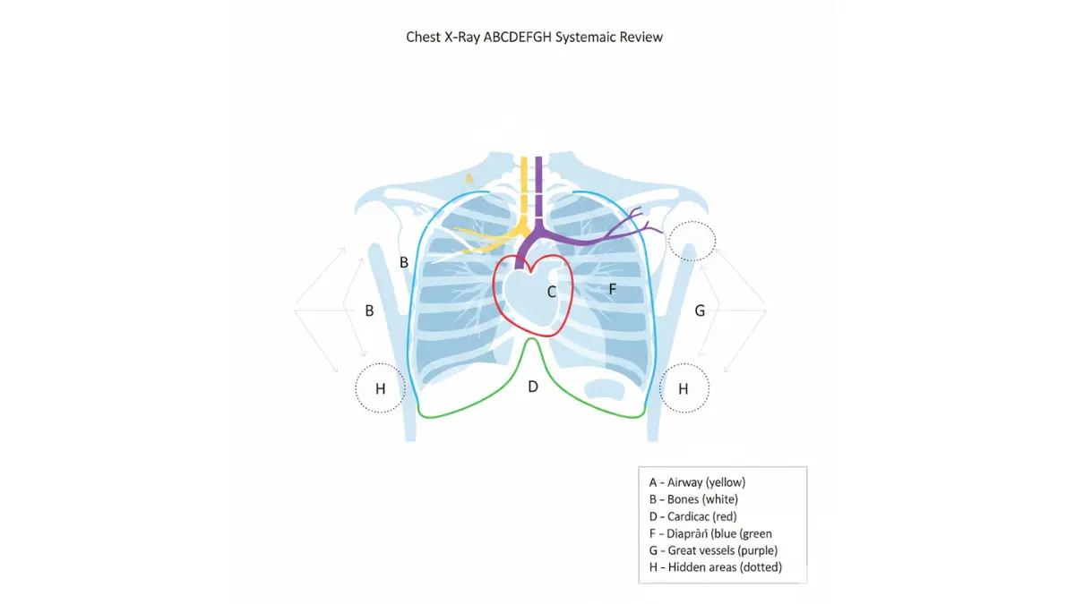
A – Airway and Alignment
- Trachea (windpipe) should be midline, not pushed to one side
- Proper positioning indicates no masses or fluid shifting structures
- Normal alignment suggests healthy anatomy
B – Bones and Soft Tissues
- Ribs, clavicles, spine checked for fractures or abnormalities
- Soft tissues examined for swelling or unusual shadows
- Bone density assessed for age-appropriate mineralization
C – Cardiac Silhouette
- Heart size measured (should occupy less than 50% of chest width on PA view)
- Heart borders should be clearly defined
- Normal cardiac silhouette rules out heart enlargement
D – Diaphragm
- Should appear as smooth, dome-shaped curves
- Right side typically slightly higher than left (due to liver)
- Sharp angles where lungs meet diaphragm (costophrenic angles)
E – Edges (Lung Borders)
- Lung markings should extend all the way to chest wall
- No visible pleural line that would indicate pneumothorax
- Smooth contours without bumps or irregularities
F – Fields (Lung Fields)
- Lungs should appear relatively dark (air-filled)
- Equal darkness on both sides
- No white patches indicating fluid or infection
G – Great Vessels
- Aorta and major blood vessels should have normal size and shape
- No widening that might suggest aneurysm
- Proper positioning and contours
H – Hidden Areas
- Behind the heart (easy to miss abnormalities here)
- Lung apices (tops of lungs)
- Below the diaphragm
Research from Johns Hopkins Medicine shows that systematic approaches like this reduce diagnostic errors by up to 30% compared to unstructured viewing.
What Makes an X-Ray “Normal” vs. “Normal for You”
Here’s something crucial most patients don’t understand: “normal x-ray results” doesn’t mean identical to every other person’s x-ray. Radiologists consider individual variations.
Age-Related Variations Your bones naturally change with age. A 70-year-old will show different bone density than a 25-year-old, and both can be “normal for age.” The National Institute on Aging explains that age-appropriate changes don’t necessarily indicate disease.
Body Type Differences
- Taller individuals have longer lung fields
- Athletic individuals may show more pronounced heart borders (athlete’s heart)
- Body composition affects how structures appear on x-ray
Prior Imaging Comparisons When radiologists have your previous x-rays, they compare them to detect changes. Something stable for years is reassuring even if slightly different from textbook normal. This is why CT scan follow-up imaging often provides important context.
Incidental Findings That Don’t Matter Common “findings” that are actually normal variants:
- Azygos lobe (extra fissure in right lung) – present in 1% of people
- Cervical ribs (extra rib attached to neck vertebra) – seen in 0.5-1% of population
- Pectus excavatum (sunken chest) – usually benign anatomical variation
Expert Insight:
“A truly normal x-ray is actually quite rare if you’re looking for perfection. What we’re really determining is whether any findings require intervention or monitoring. Many variations are completely benign and require no action whatsoever.”
— Dr. Jennifer Chen, Board-Certified Radiologist, Massachusetts General Hospital
The Technology Behind Your Results
Digital X-Ray vs. Film Modern digital x-rays provide superior image quality while using up to 80% less radiation than traditional film. According to FDA imaging guidelines, current technology ensures the lowest possible radiation exposure necessary for diagnostic quality images.
Radiation Dose in Perspective A single chest x-ray exposes you to approximately 0.1 millisieverts (mSv) of radiation—equivalent to about 10 days of natural background radiation we all receive from the environment. For comparison:
- Cross-country airline flight: 0.03 mSv
- Chest x-ray: 0.1 mSv
- Dental x-ray: 0.005 mSv
- CT chest scan: 7 mSv
- Annual natural background radiation: 3 mSv
AI-Assisted Reading (2024-2025 Advances) Artificial intelligence now assists radiologists in detecting subtle abnormalities. Studies from Stanford Medicine show AI can identify suspicious nodules with 94% accuracy, serving as a “second pair of eyes.” However, board-certified radiologists still make all final interpretations.
What This Means For You: When you receive chest x-ray normal findings, you’re benefiting from both systematic human expertise and cutting-edge technology. The combination provides highly reliable results, though no test is 100% perfect—which brings us to our next critical section.
Understanding proper interpretation becomes especially important if you’re managing conditions that may affect bone health, which is why tools like a BMI calculator can help track overall health metrics alongside imaging results.
“My X-Ray is Normal, But…”
When Normal X-Ray Results Don’t Match Your Symptoms
This is one of the most frustrating situations patients face: Your x-ray normal report says everything looks fine, yet you still have pain, coughing, or other concerning symptoms. You’re not imagining things—x-rays have important limitations.
According to the National Cancer Institute, approximately 20-30% of patients with confirmed disease initially receive normal imaging results because early-stage problems may not yet be visible on x-rays.
Common Scenarios Explained
Scenario 1: Normal Chest X-Ray But Still Have Chest Pain
Michael, 52, went to urgent care with chest pain. His x-ray came back normal, but the pain persisted. Two days later, an EKG and blood tests revealed he’d had a mild heart attack.
What x-rays can’t detect:
- Heart attacks (need EKG and cardiac enzymes)
- Pulmonary embolism (blood clots in lungs—requires CT scan)
- Early coronary artery disease
- Pericarditis (heart lining inflammation)
- Acid reflux or esophageal issues
- Muscle strain or costochondritis
Next steps: If you have chest pain with a normal chest x-ray, your doctor should order:
- EKG (electrocardiogram)
- Cardiac troponin blood test
- CT pulmonary angiography if blood clot suspected
- Stress test for chronic chest pain
Scenario 2: Normal Bone X-Ray But Joint Still Hurts
Rachel twisted her ankle playing soccer. The emergency room x-ray showed no fracture, but three weeks later she still couldn’t walk without pain.
What bone x-rays miss:
- Ligament tears (ACL, MCL, ankle ligaments)
- Tendon injuries (Achilles tendon, rotator cuff)
- Cartilage damage
- Stress fractures (may take 2-4 weeks to appear on x-ray)
- Bone bruises
- Soft tissue inflammation
Next steps: For persistent pain after normal x-ray results:
- MRI scan (gold standard for soft tissue injuries)
- Ultrasound for tendon problems
- Repeat x-ray in 2-3 weeks if stress fracture suspected
- Physical examination by orthopedic specialist
Understanding recovery timelines is important, much like tracking ACL tear recovery expectations after diagnosis.
Scenario 3: Normal Lung X-Ray With Persistent Cough
David had a cough for six weeks. His chest x-ray was normal, but the cough wouldn’t go away.
What lung x-rays don’t show:
- Early pneumonia (first 24-48 hours)
- Viral bronchitis or bronchiolitis
- Asthma or reactive airway disease
- Allergies causing postnasal drip
- Gastroesophageal reflux (GERD)
- Early interstitial lung disease
- Small pulmonary nodules under 3-4mm
Next steps:
- CT chest scan for better detail
- Pulmonary function tests for asthma
- Allergy testing
- Trial of asthma or reflux medications
- Bronchoscopy if cough persists beyond 8 weeks
Scenario 4: Normal Results After Injury/Trauma
Jennifer fell on ice and hit her ribs hard. The x-ray showed “intact osseous structures,” but she could barely breathe without pain.
Why this happens:
- Rib cartilage injuries don’t show on x-ray
- Muscle contusions (deep bruising)
- Stress fractures too small to visualize initially
- Soft tissue trauma
- Nerve irritation
Follow-up recommendations:
- Pain management for 4-6 weeks (typical healing time)
- Repeat imaging if pain worsens or doesn’t improve
- CT scan if concerned about internal injuries
What X-Rays CAN’T Show (Important Limitations)
Blood Clots Pulmonary embolism (PE) is invisible on x-rays. According to research from the Centers for Disease Control and Prevention, PE requires CT pulmonary angiography for diagnosis. Understanding blood clot symptoms is critical when x-rays are normal but concerns persist.
Very Early Cancers Lung nodules smaller than 1cm are often missed on x-rays. Early-stage cancers may not create enough contrast to appear on standard imaging.
Ligament or Tendon Tears These soft tissues simply don’t show up well on x-rays. MRI is required for definitive diagnosis.
Muscle Strains and Contusions X-rays visualize bones, not muscle tissue injuries.
Early Infections Pneumonia may take 24-48 hours to develop visible infiltrates. A normal x-ray doesn’t rule out early infection.
Soft Tissue Detail X-rays provide excellent bone detail but poor soft tissue contrast.
Nerve Problems Pinched nerves, nerve inflammation, or nerve damage aren’t visible on x-rays.
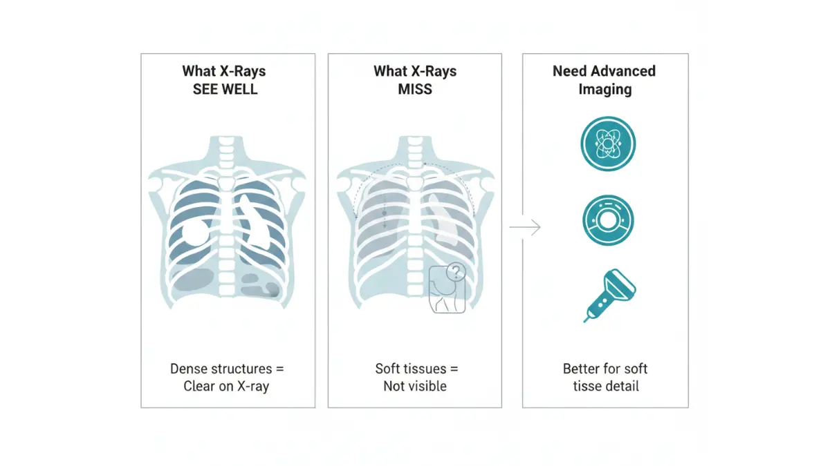
When to Request Additional Testing
Consider advanced imaging if you have:
- Persistent or worsening symptoms despite normal x-ray results
- High-risk factors for serious disease (smoking history, family history of cancer, recent trauma)
- Symptoms that don’t match your normal x-ray findings
- “Red flag” symptoms:
- Unexplained weight loss
- Night sweats or fever
- Coughing up blood
- Severe pain that awakens you from sleep
- Numbness or weakness in limbs
- Loss of bowel or bladder control
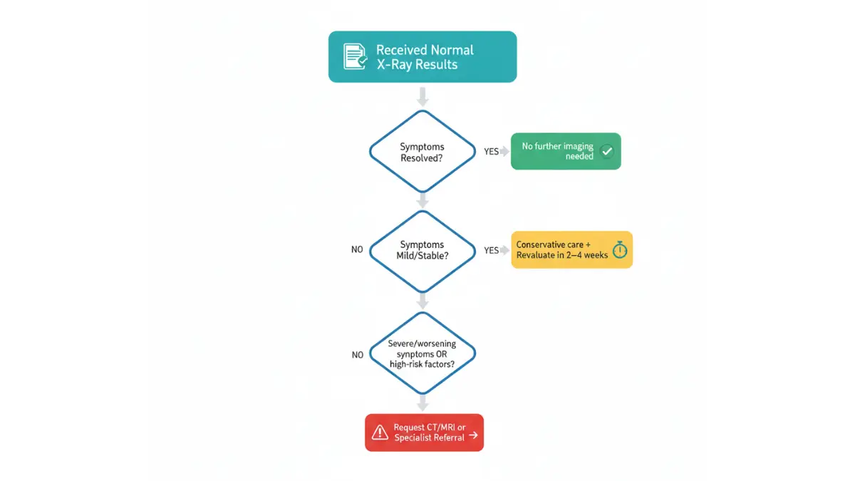
Decision Tree:
Normal X-Ray + Resolved Symptoms → No further imaging needed, follow up as scheduled
Normal X-Ray + Mild Persistent Symptoms → Conservative management, reevaluate in 2-4 weeks
Normal X-Ray + Severe or Worsening Symptoms → Request CT/MRI or specialist referral immediately
Normal X-Ray + High-Risk Factors → Discuss with doctor whether advanced imaging appropriate
What This Means For You: A normal x-ray report interpretation is reassuring but not infallible. Trust your body. If symptoms persist or worsen, advocate for additional evaluation. Sometimes getting a second opinion on imaging results provides valuable peace of mind.
The key is understanding that while your x-ray normal findings are genuinely good news, they represent one piece of your health puzzle. Combining imaging results with clinical examination, medical history, and sometimes additional tests provides the complete picture.
What Happens After Your Normal Results
Your Next Steps After Receiving Normal X-Ray Results
Receiving normal x-ray results often raises new questions: What happens now? When will I hear from my doctor? Do I need any follow-up? Understanding the typical process helps reduce anxiety.
Timeline: From Scan to Results
Emergency Department X-Rays
- Radiologist reads images: 15-30 minutes
- Results to emergency doctor: 30-60 minutes
- You’re informed: Within 1-2 hours of scan
- Why it’s faster: ED cases are marked “STAT” (urgent) in the system
Urgent Care or Walk-In Clinic
- Preliminary read: Same day (often within 2-4 hours)
- Formal report: 12-24 hours
- You’re contacted: Same day if abnormal, 1-2 days if normal
- Why timing varies: Depends on radiologist coverage and clinic protocols
Routine Outpatient X-Rays
- Images sent to radiologist: Within hours of scan
- Formal interpretation completed: 24-48 hours typically
- Report sent to ordering physician: 1-2 business days
- You’re informed: 2-5 business days on average
Inpatient Hospital X-Rays
- Read by radiologist: Within 4-12 hours for routine studies
- Results in medical record: Same day
- Your hospital doctor reviews: During morning rounds (next day)
According to guidelines from the American College of Radiology, emergency and urgent cases must be interpreted immediately, while routine studies should be finalized within 24 hours.
What This Means For You: If you haven’t heard anything within the expected timeframe, it’s appropriate to call your doctor’s office. Silence usually means normal x-ray findings, but confirmation provides peace of mind.
How Your Doctor Will Contact You
Understanding communication methods helps set realistic expectations:
| Result Type | How You’ll Typically Hear | Expected Timeline | What to Expect |
|---|---|---|---|
| Normal, routine screening | Patient portal message | 1-3 business days | Brief message: “Your x-ray was normal” |
| Normal after symptoms | Phone call or portal | 2-3 business days | May include follow-up instructions |
| Urgent abnormal finding | Direct phone call | Same day | Doctor will call personally |
| Significant incidental finding | Scheduled appointment | Within 1 week | Needs in-person discussion |
| Emergency/critical result | Immediate phone call | Minutes to hours | May need to return to hospital |
Why normal results may take longer to communicate: When chest x-ray normal or other routine findings show no concerns, they’re often batched with other results and released via patient portal. Abnormal findings trigger immediate physician notification.
Patient Portal Confusion: Many patients access their normal x-ray results through electronic health records before their doctor reviews them. If you see the report first, don’t panic if you don’t understand the terminology—schedule a call with your doctor’s office to discuss.
Questions to Ask Your Doctor
Even with normal x-ray findings, these questions ensure you understand your complete picture:
About Your Results:
- “What exactly did the x-ray show was normal?”
- “Were there any incidental findings I should know about?”
- “Do my symptoms match what would be expected with these normal results?”
- “What does ‘unremarkable’ mean in my report?”
About Follow-Up: 5. “Do I need any repeat imaging, and if so, when?” 6. “Should I worry about the radiation exposure?” 7. “What symptoms should prompt me to come back?” 8. “Are there other tests we should consider given my symptoms?”
About Your Health Going Forward: 9. “Are there lifestyle changes I should make?” 10. “What’s my risk for developing problems in the future?” 11. “How often should I have routine x-rays given my medical history?” 12. “Should family members be screened given my normal results?”
What This Means For You: Being an engaged patient improves outcomes. Research from the Agency for Healthcare Research and Quality shows that patients who ask questions and actively participate in their care have better health outcomes and higher satisfaction.
If you’re managing chronic conditions that require monitoring, tools like a blood sugar converter or heart rate zone calculator can complement your imaging results to track overall health trends.
Keeping Your Medical Records
How to Access Your X-Ray Images:
- Most facilities offer CD copies (free or minimal charge)
- Patient portals often allow download of images
- DICOM viewers (free software) let you view images at home
- Some facilities offer cloud-based access
Why You Should Save Your Reports:
- Future doctors can compare to establish baseline
- Helpful if you change healthcare systems
- Important for insurance or disability claims
- Valuable if you travel and need emergency care
- Useful reference if similar symptoms recur
Organizing Your Records:
- Create digital folder: “Medical Images – [Your Name]”
- Label files by date and body part: “2026-01-15_Chest-Xray”
- Keep both the images AND the report
- Backup to cloud storage (ensure HIPAA-compliant service)
- Maintain paper copies of critical reports
Sharing With Other Providers: Many patients don’t realize they can request copies of all diagnostic imaging under HIPAA regulations. This becomes especially important if you need to consult specialists or get second opinions.
What This Means For You: Taking ownership of your medical records, including understanding your normal x-ray report interpretation, empowers you to be an active participant in your healthcare. When you have previous imaging available for comparison, it significantly improves diagnostic accuracy for future health concerns.
FAQ + Evidence-Based Guidance
Frequently Asked Questions About Normal X-Ray Results
Understanding the Terms
1. What does “unremarkable” mean on my x-ray?
“Unremarkable” is excellent news—it means nothing abnormal caught the radiologist’s attention. In medical terminology, “remarkable” means noteworthy or concerning, so “unremarkable” indicates healthy, normal findings.
Research from Harvard Medical School confirms this is the most frequently misunderstood term in radiology reports, with many patients incorrectly thinking it means “not very good.”
2. Can a normal x-ray miss something serious?
Yes, x-rays have important limitations. They can miss:
– Very early cancers (nodules smaller than 3-4mm)
– Blood clots in lungs (pulmonary embolism)
– Soft tissue injuries (ligament tears, muscle strains)
– Early infections in first 24-48 hours
– Stress fractures (may take 2-4 weeks to appear)
However, x-rays are excellent for detecting pneumonia, most fractures, bone infections, significant fluid collections, and organ enlargement. The Radiological Society of North America reports that chest x-rays detect 70-80% of lung cancers over 1cm in size.
3. How accurate are x-rays for detecting problems?
Accuracy varies by what you’re looking for:
Fractures: 90-95% sensitive for most breaks
Pneumonia: 75-85% sensitive (can miss atypical presentations)
Lung cancer: 70-80% for lesions over 1cm
Bone infections: 50-60% in early stages (MRI better)
Soft tissue injuries: Poor (10-20% detection rate)
4. Why did my doctor order an x-ray if it came back normal?
X-rays serve multiple purposes even when normal:
– Rule out serious problems that require immediate treatment
– Establish baseline for future comparison
– Guide appropriate treatment (confirms diagnosis doesn’t require surgery)
– Reassure patients (reduces anxiety about symptoms)
– Meet medical necessity requirements for insurance
A normal x-ray result often provides valuable information by eliminating concerning diagnoses.
5. What’s the difference between “normal” and “negative”?
These terms are essentially interchangeable in radiology. Both indicate no abnormalities detected. “Negative” is more commonly used when searching for a specific condition (e.g., “negative for fracture”), while “normal” describes overall findings.
Safety and Radiation Concerns
6. Should I worry about radiation from x-rays?
Modern x-rays use minimal radiation. According to FDA radiation safety guidelines, a single chest x-ray (0.1 mSv) equals about 10 days of natural background radiation exposure. You receive more radiation during a cross-country flight than from a chest x-ray.
The benefits of accurate diagnosis far outweigh the negligible cancer risk from occasional x-rays. For patients concerned about cumulative exposure, maintaining proper hydration supports your body’s natural detoxification processes.
7. How often can I safely have x-rays?
There’s no specific limit for medically necessary x-rays. The principle is ALARA (As Low As Reasonably Achievable). If each x-ray provides important diagnostic information, the benefits justify the minimal radiation exposure.
However, unnecessary repeat x-rays should be avoided. Always bring previous x-rays to new healthcare providers to prevent duplicate imaging.
8. Should pregnant women get x-rays?
X-rays during pregnancy are performed only when medically necessary. A single x-ray (especially of areas away from the abdomen) poses minimal risk to a developing baby. According to the American College of Obstetricians and Gynecologists, fetal radiation exposure from a chest x-ray is negligible—far below levels that could cause harm.
If you’re pregnant or might be pregnant, always inform the x-ray technologist. They’ll use lead shielding and lowest possible radiation dose. For pregnancy health tracking, tools like a pregnancy weight gain calculator help monitor normal development.
When Results Don’t Match Symptoms
9. What if my x-ray is normal but I still have pain?
This is common and doesn’t mean you’re imagining pain. X-rays don’t show:
– Ligament or tendon injuries
– Muscle strains
– Nerve problems
– Early stress fractures
– Inflammation
Discuss with your doctor whether you need MRI, CT scan, or physical therapy evaluation. Your symptoms are valid even with normal x-ray findings.
10. Do I need a second opinion on normal results?
Generally, normal x-ray results interpreted by board-certified radiologists are reliable. Consider a second opinion if:
– Your symptoms are severe or worsening
– You have high-risk factors (smoking history, family history of cancer)
– Treatment isn’t working despite “normal” findings
– Multiple doctors have conflicting opinions
Getting a second opinion on imaging is always your right as a patient.
11. Can x-rays detect all types of cancer?
No. X-rays detect some cancers but miss many others:
Well-detected: Bone cancers, large lung masses, some breast cancers (mammography)
Poorly detected: Early-stage cancers, soft tissue sarcomas, brain tumors, prostate cancer
Understanding early warning signs of various cancers remains important even after normal imaging.
Interpreting Your Report
12. What does “no acute process” mean?
“Acute” means sudden or recent. This phrase indicates no sudden, urgent problems detected. It doesn’t rule out chronic (long-standing) conditions—just confirms nothing requiring immediate intervention.
13. Is “within normal limits” the same as perfect?
Not quite. “Within normal limits” means your results fall within the expected range for healthy individuals. Everyone has natural variations—you don’t need to be perfectly average to be healthy.
14. Why do some reports say “unremarkable” instead of “normal”?
Both terms mean the same thing. Different radiologists prefer different terminology. “Unremarkable” is standard medical jargon, while “normal” is more patient-friendly. Some reports use both for clarity.
15. What are “incidental findings”?
Incidental findings are unexpected discoveries unrelated to why the x-ray was ordered. Examples include:
– Small benign bone cysts
– Healed old rib fractures you never knew about
– Minor degenerative changes appropriate for your age
– Anatomical variants (like extra ribs)
Most incidental findings require no treatment but should be discussed with your doctor.
Follow-Up and Long-Term Care
16. How long do x-ray results stay valid?
This depends on your condition:
Stable chronic conditions: 6-12 months
After acute injury: 2-3 months
Cancer screening: Annual (for high-risk patients)
Post-surgical monitoring: As recommended by surgeon
If symptoms change or worsen, previous “normal” results may no longer apply.
17. Can normal x-rays change to abnormal quickly?
Yes, especially in certain situations:
– Infections can develop within days
– Fractures may worsen if injured area isn’t properly protected
– Fluid collections can accumulate rapidly
– Some cancers grow quickly
This is why persistent or worsening symptoms warrant repeat imaging even after recent normal results.
18. What does “clear lung fields” mean?
This indicates lungs appear properly air-filled without dense areas that would suggest fluid, infection, masses, or collapse. It’s a reassuring finding on chest x-ray normal reports.
19. Do children’s normal x-rays look different?
Yes. Growing bones appear different than adult bones, with growth plates visible at joints. Pediatric radiologists specialize in understanding age-appropriate normal variations.
20. What’s the next test after a normal x-ray?
This depends on symptoms and clinical concerns:
Persistent bone/joint pain: MRI
Chest symptoms: CT scan or pulmonary function tests
Abdominal pain: Ultrasound or CT
Neurological symptoms: MRI or CT of brain/spine
Your doctor chooses the next step based on your specific situation. For comprehensive health assessment when imaging is normal, combining results with tools like an ideal weight calculator provides a more complete health picture.
Quality and Access
21. How do radiologists quality-check x-rays?
Multiple safeguards ensure accuracy:
– Board certification requirements
– Peer review of interpretations
– Quality assurance programs
– AI assistance for double-checking
– Continuing medical education requirements
According to American Board of Radiology standards, radiologists must maintain certification through ongoing learning and practice assessment.
22. Can I request a copy of my x-ray images?
Yes. Under HIPAA, you have the right to copies of all medical records, including imaging. Most facilities provide:
– Digital copies on CD/DVD
– Patient portal downloads
– Cloud-based access
– Paper copies of reports
There may be a reasonable fee for copies.
23. What does “osseous structures intact” mean?
“Osseous” means bone. “Intact” means unbroken. This phrase confirms no fractures, breaks, or significant bone abnormalities detected—excellent news for a bone x-ray.
24. Why do some x-rays require follow-up even when normal?
Reasons for follow-up despite normal results:
– Baseline established; future comparison needed
– Symptoms may evolve, requiring repeat imaging
– High-risk conditions need monitoring
– Insurance or legal documentation requirements
Some conditions require serial imaging to truly rule them out
25. What if I disagree with my normal results?
You have several options:
– Discuss concerns thoroughly with your ordering physician
– Request radiologist review of specific areas of concern
– Seek second opinion from another radiologist
– Request different imaging modality (MRI instead of x-ray)
– Consult a specialist in the relevant body system
Your symptoms and concerns should always be taken seriously, even when imaging appears normal
.
When to Seek Immediate Medical Attention
Despite normal x-ray results, seek emergency care immediately if you experience:
Critical Warning Signs:
- Severe chest pain or pressure (especially with shortness of breath)
- Difficulty breathing that’s worsening
- Coughing up blood
- High fever (over 103°F/39.4°C) with shaking chills
- Sudden, severe headache unlike any you’ve had before
- Loss of consciousness or severe confusion
- Weakness or numbness on one side of your body
- Inability to bear weight on an injured limb
- Severe abdominal pain with vomiting
- Signs of blood clot: sudden leg swelling, warmth, pain
What This Means For You: Normal x-ray findings are genuinely reassuring, but they don’t override clinical judgment. If your condition deteriorates or you develop new concerning symptoms, don’t hesitate to seek medical evaluation. Use available resources like a symptom checker as a starting point, but always consult healthcare professionals for definitive guidance.
Key Takeaways
Understanding your normal x-ray results empowers you to take control of your health:
- Normal x-ray results mean no urgent problems detected – Terms like “unremarkable” and “within normal limits” are good news
- “Unremarkable” is medical jargon for healthy – Don’t let confusing terminology cause unnecessary worry
- X-rays have limitations and can’t see everything – They’re excellent for bones and lungs but miss soft tissue injuries, blood clots, and very early diseases
- Trust your symptoms even with normal results – If pain persists or symptoms worsen, advocate for additional evaluation
- Ask your doctor about next steps – Understanding what normal means for YOUR specific situation is crucial
- Keep copies of all medical imaging – Previous results help future doctors make accurate diagnoses
- Follow recommended screening guidelines – Normal results today don’t eliminate need for future monitoring
Medical Disclaimer
⚠️ Important Notice: This article is for educational and informational purposes only and does not constitute medical advice, diagnosis, or treatment recommendations. Always consult qualified healthcare professionals about your specific x-ray results and health concerns.
If you’re experiencing a medical emergency, call 911 immediately. Do not rely on online information in urgent situations.
X-ray interpretation requires specialized medical training. This guide helps you understand common terminology but should never replace professional medical evaluation. Your healthcare provider knows your complete medical history and can provide personalized guidance based on your individual circumstances.
The information provided reflects current medical knowledge and evidence-based practices from authoritative sources as of January 2026 but may not account for recent medical advances or individual variations. Always verify information with your healthcare team.
About this content
How this article was put together: researched from recognised health sources, drafted with the help of AI tools, and edited by hand, with sources linked throughout.

Sameer Patel is the founder and editor of My Medicine Advisor. He is not a doctor or medical professional — before starting this site he worked in banking,…
Medical disclaimer
The content on MyMedicineAdvisor is provided for general informational and educational purposes only and is not a substitute for professional medical advice, diagnosis, or treatment. Health information on this website should not be used to diagnose, treat, cure, or prevent any condition without guidance from a qualified healthcare professional. Always seek the advice of your doctor, physician, or another licensed healthcare provider with any questions you may have regarding a medical condition, symptoms, medications, or treatment decisions.