
On This Page – Quick Medical Summary
When Nicholas, a 61-year-old teacher from Ohio, noticed a faint blurry patch during a routine vision check, he assumed he needed stronger glasses. His optometrist saw something else entirely — a dark mass growing silently behind his retina. He had uveal melanoma: the most common primary eye cancer in adults, and one of the most misunderstood.
Uveal melanoma is a malignant tumor that arises from pigment cells (melanocytes) within the uveal tract — the middle layer of the eye. It accounts for approximately 5% of all melanoma cases and affects roughly 2,500 Americans each year. Most patients feel no pain. Many have no symptoms at all.
The stakes are high: up to 50% of patients develop liver metastasis, often years after their eye is successfully treated. But 2022 brought a pivotal change — the first-ever FDA-approved systemic therapy for this disease is now reshaping outcomes.
⚠️ Disclaimer: This article is for educational purposes only and does not constitute medical advice. Always consult a qualified healthcare professional for diagnosis and treatment decisions.
What Is Uveal Melanoma? The Eye Cancer That Hides in Plain Sight
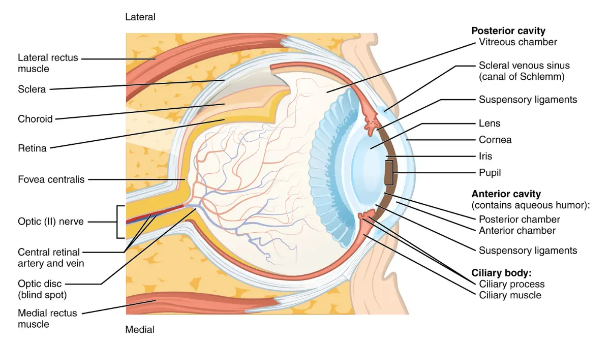
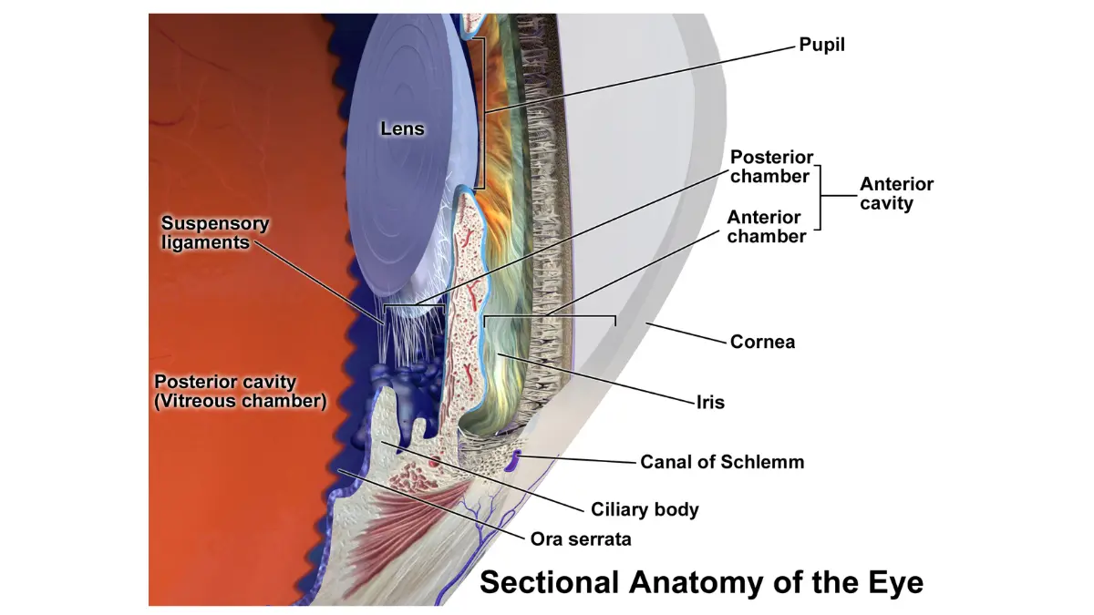
Uveal melanoma — also called ocular melanoma, intraocular melanoma, or choroidal melanoma — develops from melanocytes found within the uveal tract. This structure sits between the white outer layer of the eye (sclera) and the light-sensitive retina, and it has three anatomical regions where melanoma can originate.
Where Does Uveal Melanoma Start?
| Location | % of Uveal Melanomas | Key Clinical Note |
|---|---|---|
| Choroid | ~90% | Hidden behind retina; most often asymptomatic at first |
| Ciliary body | ~6% | Often larger at diagnosis; associated with worse prognosis |
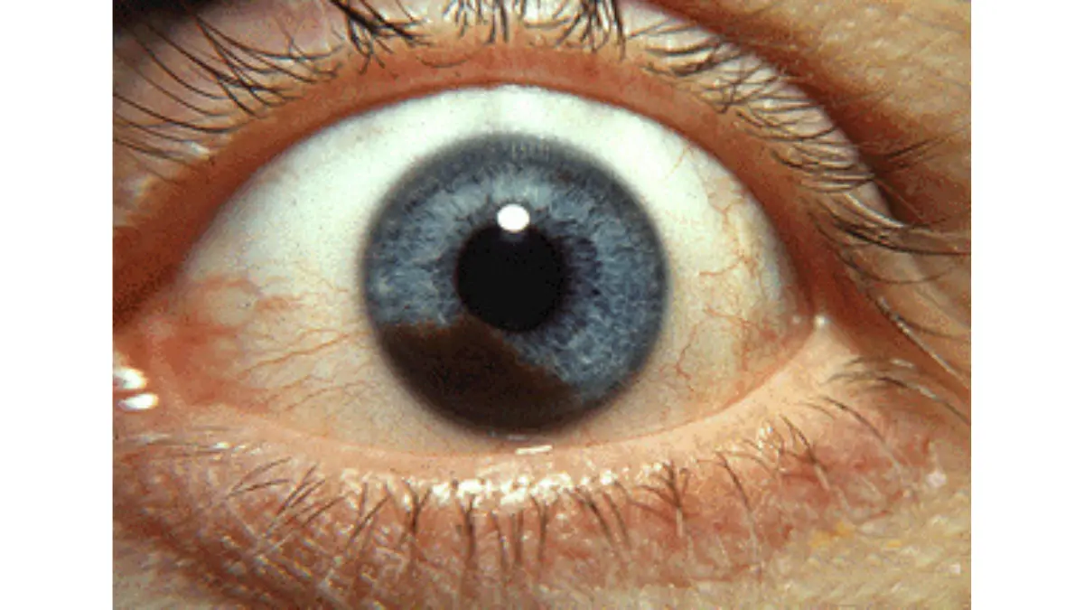
| Iris | ~4% | Only visible type; appears as a dark spot on the colored part of the eye |
The choroid is the most common site because it is richly vascularized and contains the highest density of melanocytes. As the American Society of Retina Specialists confirms, the incidence in the US is approximately 6 cases per million people per year, making this a rare but serious diagnosis.
How Is Uveal Melanoma Different From Skin Melanoma?
Many patients assume uveal melanoma behaves like skin melanoma. It does not — and that misunderstanding has real treatment consequences.
- Different genetic drivers: Uveal melanoma is driven by mutations in GNAQ, GNA11, PLCB4, or CYSLTR2 — not the BRAF mutations found in most cutaneous melanomas
- Different immune profile: Uveal melanoma has one of the lowest tumor mutational burdens of any cancer (~0.5 mutations per megabase), which is why checkpoint inhibitors largely fail here
- Different spread pattern: Metastasis goes primarily to the liver (in ~90% of metastatic cases), not to lymph nodes
For a detailed comparison, see our guide on melanoma vs. skin cancer differences.
What This Means For You: Treatments proven for skin melanoma often do not work for uveal melanoma. This is why referral to a specialist ocular oncologist — not just a general oncologist — is critical.
Who Gets Uveal Melanoma? — 2026 Risk Factor Profile
Uveal melanoma is primarily a disease of fair-skinned, light-eyed adults over 60. According to StatPearls (NCBI), the following significantly increase risk:
- Light-colored eyes (blue, gray, or green)
- Fair skin with limited ability to tan
- Ocular melanocytosis (excess pigmentation of the uvea)
- BAP1 gene mutation — a hereditary risk factor
- Male sex (5.8 per million in men vs. 4.4 per million in women)
- Age 60+ (mean diagnosis age has risen from 55 to 62 in recent decades)
Unlike skin melanoma, UV sun exposure plays a less clearly established role. If you have a family history of uveal melanoma or BAP1 mutations, our Genetic Risk Assessment Tool can help you understand inherited cancer risk factors to discuss with your physician.
Uveal Melanoma Symptoms — The Silent Warning Signs You Must Not Ignore
Here is what makes uveal melanoma so dangerous: the vast majority of patients have no symptoms at all. Studies suggest that up to 40% of diagnoses are made incidentally during routine dilated eye exams — before a single symptom appears.
When Symptoms Do Appear — 7 Warning Signs
If uveal melanoma does cause symptoms, they typically include:
- Blurred or distorted vision in one eye
- Floaters (new or sudden increase in dark specks)
- Photopsia (flashing lights or visual disturbances)
- Visual field loss — a shadow, curtain, or dark area
- Change in pupil shape (more common in ciliary body tumors)
- Visible dark spot on the iris (iris melanoma only — the only externally visible type)
- Eye redness or pressure (rare; typically late-stage or ciliary body involvement)
Bold Takeaway: The absence of pain does NOT mean your eye is healthy. Uveal melanoma rarely causes pain until it is advanced. If you are experiencing any unusual visual changes, use our Symptom Checker to identify what to discuss with your doctor.
Learn more about general melanoma warning signs and symptoms across all types.
Choroidal Nevus vs. Uveal Melanoma — The “Eye Freckle” Question
One of the most common concerns patients bring to search engines: “I was told I have a freckle on my eye — could it be melanoma?” Choroidal nevi (benign eye freckles) occur in approximately 20% of the population, but some carry elevated melanoma risk.
| Feature | Benign Choroidal Nevus | Uveal Melanoma |
|---|---|---|
| Thickness | Less than 2 mm | Greater than 2 mm |
| Subretinal fluid | Absent | Often present |
| Orange pigment (lipofuscin) | Absent | Often present |
| Margin near optic disc | Not typical | Common in melanoma |
| Growth on follow-up | Stable | Progressive |
| Symptoms | None | May cause visual changes |
What This Means For You: Ocular oncologists use the TFSOM mnemonic (Thickness, Fluid, Symptoms, Orange pigment, Margin) to flag high-risk lesions. Each additional factor raises the probability of melanoma. If your eye doctor identifies a suspicious lesion, ask for a referral to an ocular oncologist.
To better understand your eye exam findings, explore our free Eye Exam Tool.
How Uveal Melanoma Is Diagnosed — From Eye Exam to Genetic Profiling
Unlike most cancers, uveal melanoma is usually diagnosed without a biopsy. Approximately 95% of diagnoses are made non-invasively through clinical examination and imaging. The National Cancer Institute outlines the following standard diagnostic pathway:
The Diagnostic Pathway — Step by Step
Step 1 — Dilated fundus exam: A dilated eye exam with indirect ophthalmoscopy allows visualization of the choroid, the most common tumor site.
Step 2 — B-scan ocular ultrasound: The most critical diagnostic tool. Ultrasound measures tumor thickness, identifies acoustic hollowness (a sign of malignancy), and detects subretinal fluid.
Step 3 — Fundus photography + fluorescein angiography: Documents the tumor’s vascular supply and identifies orange pigment characteristic of uveal melanoma.
Step 4 — OCT and OCT-A: Optical coherence tomography angiography provides high-resolution cross-sectional imaging, especially useful for detecting subretinal fluid.
Step 5 — MRI: Used when extrascleral extension is suspected or when CT cannot provide sufficient detail. The Wills Eye Hospital Oncology Service considers MRI essential for large tumors near the optic nerve.
Step 6 — Fine-needle aspiration biopsy (FNAB): Only needed in approximately 5% of cases. However, FNAB is increasingly performed not for diagnosis but for genetic profiling — even after a clinical diagnosis is confirmed.
Genetic Profiling — The 2025–2026 Game-Changer
Gene Expression Profiling (GEP) is now a standard-of-care consideration for all patients, according to the Melanoma Research Alliance. It stratifies tumors into three risk classes:
| GEP Class | Metastatic Risk (5-Year) | Recommended Surveillance |
|---|---|---|
| Class 1A | ~2% | Annual imaging sufficient |
| Class 1B | ~21% | 6-monthly liver imaging |
| Class 2 | ~72% | 6-monthly MRI/ultrasound + enrollment in trials |
In addition, BAP1 mutation, chromosome 3 monosomy, and SF3B1 mutations are key prognostic markers. Patients with BAP1 loss and chromosome 3 deletion carry the highest metastatic risk. Asking your oncologist about FNAB for genetic profiling is one of the most important steps any uveal melanoma patient can take in 2026.
Tumor Size Categories That Drive Treatment
| Category | Thickness | Width | Treatment Implication |
|---|---|---|---|
| Small (atypical nevus) | <2.5 mm | <10 mm | Watchful waiting or TTT |
| Medium | 2.5–10 mm | Any | Plaque brachytherapy preferred |
| Large | >10 mm | >16 mm | Proton beam or enucleation |
Uveal Melanoma Treatment Options — Eye-Preserving Radiation to the 2026 FDA Breakthrough
The goal of treatment in 2026 is threefold: destroy the tumor, preserve vision where possible, and prevent metastatic spread. The MD Anderson Cancer Center confirms that over 90% of patients with medium-sized tumors are now candidates for eye-preserving therapy.
Can You Keep Your Eye? Eye-Preserving Treatments
Enucleation (eye removal) is now a last resort — not the default.
1. Plaque Brachytherapy (Gold Standard) A custom radioactive plaque (most commonly iodine-125 or ruthenium-106) is surgically attached to the outside of the eye over the tumor for 4–7 days, then removed. The Wills Eye Hospital reports 98% local tumor control with combination plaque radiotherapy and transpupillary thermotherapy (TTT). This is the most widely used eye-preserving approach for small, medium, and select large tumors.
2. Proton Beam Therapy Used for large or posteriorly located tumors. Delivers highly focused radiation with minimal damage to surrounding structures. Available at specialized centers including Massachusetts General Hospital and UCSF.
3. Transpupillary Thermotherapy (TTT) Infrared laser treatment effective for small tumors away from the optic nerve. Often combined with plaque brachytherapy for enhanced control.
4. Stereotactic Radiosurgery (SRS) Non-invasive; uses focused radiation beams (CyberKnife, Gamma Knife). Used in select cases where plaque cannot be placed. For a deeper understanding of how radiation works in cancer treatment, see our radiation therapy guide.
The 2022 FDA Breakthrough: Tebentafusp (Kimmtrak)
This is what every patient with metastatic uveal melanoma needs to know — and what none of the top-ranking competitor sites adequately covers.
Tebentafusp (brand name Kimmtrak) is the first and only FDA-approved systemic therapy for metastatic uveal melanoma. Approved in January 2022, it represents the single biggest advance in this disease in decades.
How it works: Tebentafusp is a T-cell receptor bispecific fusion protein (gp100 × CD3). It bridges the immune system’s T cells directly to gp100-positive melanoma cells, activating tumor killing. Critically, it targets an intracellular antigen — something most immunotherapies cannot do.
The landmark Phase 3 trial published in the New England Journal of Medicine (2023) showed:
- Median overall survival: 21.6 months (tebentafusp) vs. 16.9 months (control)
- 3-year survival rate: 27% (tebentafusp) vs. 18% (control)
- Hazard ratio for death: 0.68 — a 32% reduction in mortality risk
Who is eligible? Patients must be HLA-A*02:01 positive — a genetic marker present in approximately 40–50% of Caucasian patients. Testing for this marker is now standard before initiating treatment for metastatic disease.
| Tebentafusp Side Effect | Frequency | Management |
|---|---|---|
| Rash | 83% | Mostly Grade 1–2; topical treatment |
| Fever (pyrexia) | 76% | Premedication reduces severity |
| Pruritus (itching) | 70% | Antihistamines; improves after Dose 3 |
| Hypotension | 38% | Monitored in clinical settings post-infusion |
What This Means For You: If you or a loved one has metastatic uveal melanoma, immediately ask your oncologist: “Am I HLA-A*02:01 positive, and am I eligible for tebentafusp?” This is now the standard of care for eligible patients.
For a broader picture of how immunotherapy works against melanoma, our immunotherapy for melanoma guide covers the mechanisms and current options in detail.
Treating Liver Metastases
The liver is the primary target in metastatic uveal melanoma. Current options include:
- Hepatic arterial chemoembolization (HACE) — delivers chemotherapy directly to liver tumors
- Isolated hepatic perfusion (IHP) — high-dose chemotherapy isolated to the liver
- Combination immunotherapy — ipilimumab + nivolumab (limited efficacy but used in HLA-A*02:01 negative patients)
- Clinical trials — actively recruiting at major centers; search current uveal melanoma clinical trials on ClinicalTrials.gov
You can also explore our dedicated guide on melanoma clinical trials to understand what participation involves.
Uveal Melanoma Prognosis & Survival Rates — What 2025–2026 Data Shows
Prognosis for uveal melanoma depends heavily on whether the disease is localized or metastatic. Understanding both scenarios — with current data — gives patients realistic expectations and better questions to ask their care team.
Localized vs. Metastatic — Two Very Different Outcomes
Localized uveal melanoma treated with modern eye-preserving methods carries a favorable prognosis, with 5-year survival rates exceeding 80% for most patients. Local recurrence affects fewer than 5% of patients treated with plaque brachytherapy.
Metastatic uveal melanoma has historically been grim, with median overall survival of approximately 12 months. Tebentafusp has extended this to 21.6 months in eligible patients, with 27% alive at 3 years — a meaningful but still modest improvement that underscores the need for continued research.
According to the Melanoma Focus patient resource, the risk of developing metastases can be predicted using genetic profiling, allowing high-risk patients to be enrolled in surveillance programs and clinical trials earlier.
2026 Survival Rate Summary
| Disease Stage | 5-Year Survival | Best Current Treatment |
|---|---|---|
| Small localized | 90–95% | TTT or watchful waiting |
| Medium localized | 80–88% | Plaque brachytherapy |
| Large localized | 70–80% | Proton beam / enucleation |
| Metastatic (HLA+) | ~27% at 3 yrs | Tebentafusp (Kimmtrak) |
| Metastatic (HLA−) | ~18% at 3 yrs | Pembrolizumab / ipilimumab / dacarbazine |
Factors That Most Affect Your Prognosis
- Tumor size and ciliary body involvement (ciliary body melanoma has worse prognosis due to delayed diagnosis)
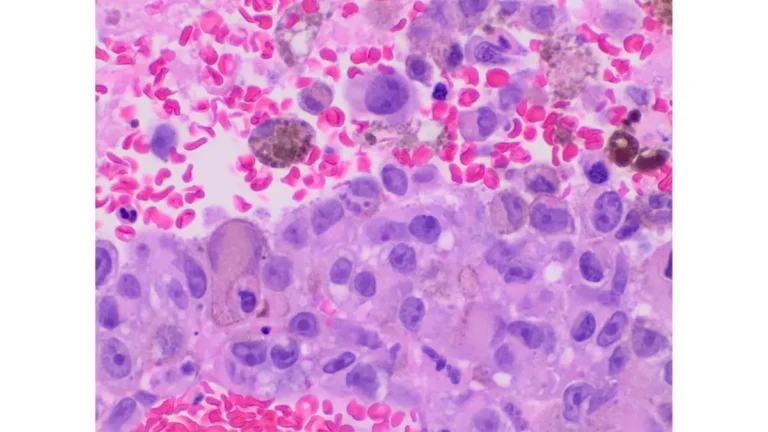
- Histologic cell type: Epithelioid cells = worst prognosis; spindle A cells = best
- GEP genetic class: Class 2 = 72% metastatic risk at 5 years
- Chromosome 3 monosomy + BAP1 loss = highest-risk molecular combination
- Liver involvement at diagnosis (most common metastatic site; liver-only disease has better outcomes than multiorgan spread)
For broader context on how cancer staging affects outcomes, explore our melanoma survival rate by stage guide.
What This Means For You: Early detection during a routine dilated eye exam remains the single most powerful factor in improving your outcome. Annual dilated eye exams — especially for those over 50 with light-colored eyes — are not optional. They are potentially life-saving.
Our free Eye Exam Tool can help you understand what your eye test results mean and what to ask your optometrist.
Life After Uveal Melanoma — Monitoring, Mental Health & Key Questions for Your Doctor
Completing treatment for uveal melanoma is not the end of the journey. It is the beginning of a structured, long-term surveillance plan designed to catch metastatic spread as early as possible.

Post-Treatment Surveillance Schedule
Because uveal melanoma can spread years — or even a decade — after the primary tumor is treated, lifelong monitoring is essential. The PMC clinical review on uveal melanoma management recommends the following schedule:
| Risk Level | Ocular Follow-Up | Systemic Imaging |
|---|---|---|
| Low risk (Class 1A GEP) | Every 6 months (Yr 1–2); annual thereafter | Annual chest X-ray; liver function tests |
| Intermediate (Class 1B) | Every 3–6 months | Liver ultrasound or MRI every 6 months |
| High risk (Class 2 GEP) | Every 3 months (Yr 1–3) | MRI liver every 6 months; consider clinical trial enrollment |
What imaging matters most? Liver MRI is more sensitive than ultrasound for detecting early liver metastases and is increasingly preferred for high-risk patients. Chest X-rays help screen for the less common but possible pulmonary spread.
Emotional Health After an Eye Cancer Diagnosis
A uveal melanoma diagnosis affects far more than vision. Research consistently shows that 40–60% of ocular melanoma survivors experience clinically significant anxiety, depression, or fear of recurrence. The threat of losing an eye — or facing a potentially fatal metastatic diagnosis — carries real psychological weight.
Key support resources include:
- Ocular Melanoma Foundation (ocularmelanoma.org) — peer support, specialist directories, and patient advocacy
- Melanoma Research Foundation at curemelanoma.org — education and clinical trial access
For patients concerned about hereditary risk for themselves or their family members, our detailed guide on whether melanoma is hereditary explains the genetics behind BAP1 mutations and what genetic counseling involves.
10 Questions to Ask Your Ocular Oncologist
These questions are designed to help you take an active, informed role in your care:
- What is my GEP class — Class 1A, 1B, or Class 2?
- Am I HLA-A*02:01 positive — and am I eligible for tebentafusp?
- Can my tumor be treated while preserving my eye?
- Which radiation modality is most appropriate for my tumor location and size?
- Should I have an FNAB for genetic profiling, even if my diagnosis is already confirmed?
- How often should I have liver MRI or ultrasound?
- Are there active clinical trials I qualify for right now?
- Should I be referred to a specialized ocular oncology center?
- What are the signs of metastatic recurrence I should watch for?
- What does my full 5-year surveillance schedule look like?
For a broader overview of where uveal melanoma fits within the landscape of melanoma types, visit our comprehensive melanoma symptoms, stages, and treatment guide.
Frequently Asked Questions About Uveal Melanoma
1. Is uveal melanoma the same as skin melanoma?
No. Uveal melanoma has entirely different genetic drivers, does not respond to BRAF or PD-1 inhibitors the way cutaneous melanoma does, and spreads primarily to the liver rather than lymph nodes.
2. Can uveal melanoma be cured?
Localized uveal melanoma is often treated successfully with eye-preserving radiation, achieving 98% local tumor control. Metastatic uveal melanoma is not yet curable, but tebentafusp has meaningfully improved survival in eligible patients.
3. What does uveal melanoma look like?
Most forms are invisible to the naked eye. Iris melanoma may appear as a darkening or irregular pigmentation of the colored part of the eye. Choroidal melanoma is detectable only through a dilated fundus exam.
4. How fast does uveal melanoma grow?
Growth rate is highly variable. Small lesions may remain stable for years; others enlarge rapidly. GEP genetic class (Class 1 vs Class 2) is currently the best predictor of biological behavior.
5. Can uveal melanoma spread to the brain?
Brain metastasis is uncommon but possible. The liver is the dominant metastatic site in approximately 90% of cases. Lung and skin are the next most frequent sites.
6. Is uveal melanoma hereditary?
In most cases it is not inherited. However, BAP1 gene mutations do run in families and significantly elevate lifetime risk. Genetic counseling is recommended for first-degree relatives of BAP1 mutation carriers.
7. What is the survival rate for uveal melanoma?
For localized disease, 5-year survival exceeds 80%. For metastatic disease, median survival with tebentafusp is now 21.6 months (NEJM Phase 3, 2023), with 27% alive at 3 years among eligible patients.
8. What causes uveal melanoma?
The exact cause remains unclear. Initiating mutations in GNAQ or GNA11 appear spontaneously in most cases. Risk factors include light eye color, fair skin, ocular melanocytosis, and BAP1 mutations.
9. How is uveal melanoma diagnosed without a biopsy?
Diagnosis in ~95% of cases is made through dilated fundus examination combined with B-scan ultrasound, fundus photography, and fluorescein angiography. These non-invasive tools are sufficient for a definitive diagnosis in the vast majority of patients.
10. What is tebentafusp (Kimmtrak) and who qualifies?
Tebentafusp (Kimmtrak) is the first FDA-approved systemic therapy for metastatic uveal melanoma, approved in January 2022. It is a T-cell receptor bispecific protein targeting gp100. Patients must be HLA-A*02:01 positive — approximately 40–50% of Caucasian patients carry this allele.
11. Can uveal melanoma be prevented?
There is no confirmed prevention strategy. The strongest protective action is a yearly dilated eye exam, particularly for individuals over 50 with light-colored eyes, fair skin, or a family history of BAP1 mutations. Early detection before symptoms develop remains the most effective intervention available.
This article was reviewed by the mymedicineadvisor.com medical expert panel in April 2026. Content is updated regularly to reflect current clinical guidelines and research findings. For personalized medical advice, please consult a licensed healthcare professional or a board-certified ocular oncologist.
About this content
How this article was put together: researched from recognised health sources, drafted with the help of AI tools, and edited by hand, with sources linked throughout.

Sameer Patel is the founder and editor of My Medicine Advisor. He is not a doctor or medical professional — before starting this site he worked in banking,…
Medical disclaimer
The content on MyMedicineAdvisor is provided for general informational and educational purposes only and is not a substitute for professional medical advice, diagnosis, or treatment. Health information on this website should not be used to diagnose, treat, cure, or prevent any condition without guidance from a qualified healthcare professional. Always seek the advice of your doctor, physician, or another licensed healthcare provider with any questions you may have regarding a medical condition, symptoms, medications, or treatment decisions.