On This Page – Quick Medical Summary
In 2023, Marcus, a 41-year-old construction worker from Texas, noticed a dark streak forming under his thumbnail. He assumed it was a bruise from a job site accident. Eight months later, his dermatologist delivered a crushing diagnosis: Stage III acral lentiginous melanoma. “This is one of the signs we fear most,” his doctor told him, “because it hides in plain sight.”
Marcus is not alone. Melanoma kills someone every 54 minutes in the United States. Yet the majority of those deaths are preventable with one powerful weapon: knowing exactly what to look for.
This guide reveals all 9 melanoma warning signs — including the 4 dangerous ones your doctor fears most and competitors like WebMD and Healthline routinely miss.
Why Melanoma Warning Signs Can’t Wait in 2026
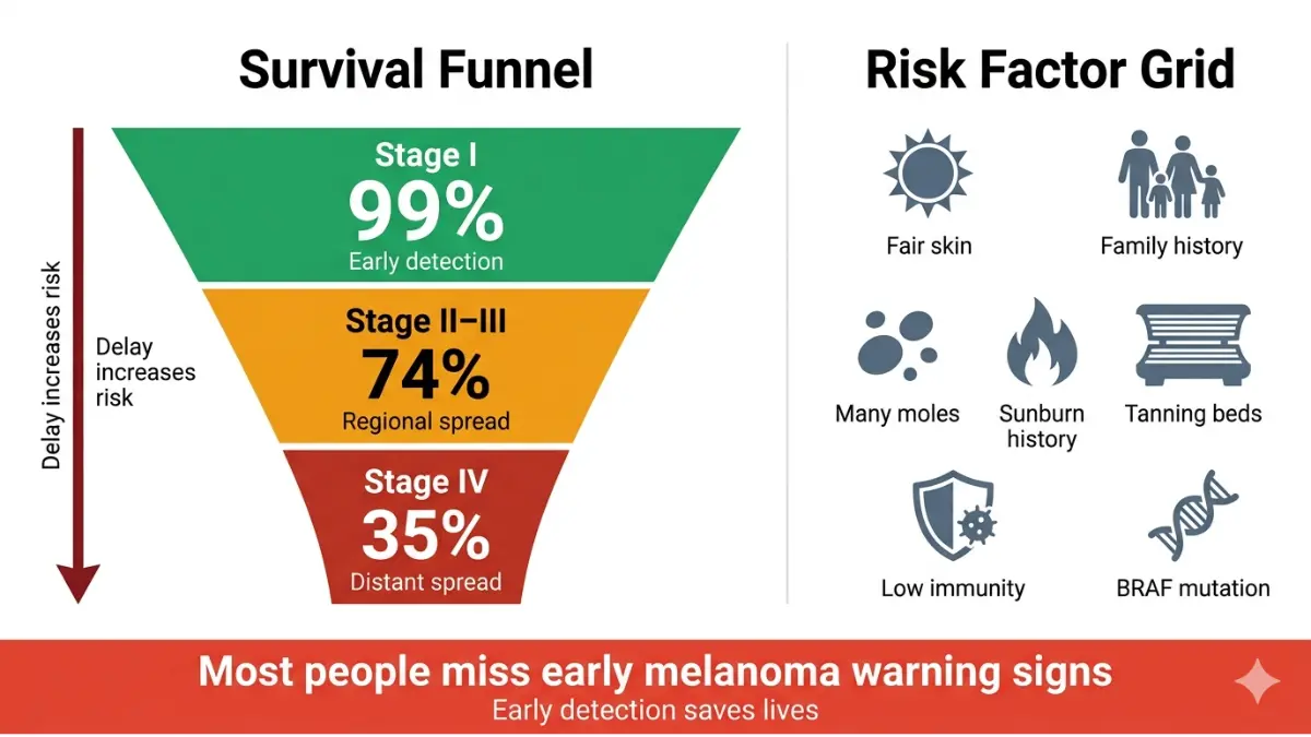
The short answer: Melanoma caught at Stage I carries a 99% five-year survival rate. Caught after it spreads to distant organs, that number collapses to just 35%.
That gap — 99% versus 35% — is what makes early recognition of melanoma warning signs one of the most critical medical skills you can develop.
According to melanoma statistics tracked by the CDC, melanoma rates have doubled over the past three decades. In 2026, approximately 104,960 Americans will receive a new melanoma diagnosis. It accounts for only 1% of all skin cancers — but causes the overwhelming majority of skin cancer-related deaths.
| Stage at Detection | 5-Year Survival Rate |
|---|---|
| Stage I (Localised) | 99% |
| Stage II (Regional lymph nodes) | 74% |
| Stage III–IV (Distant spread) | 35% |
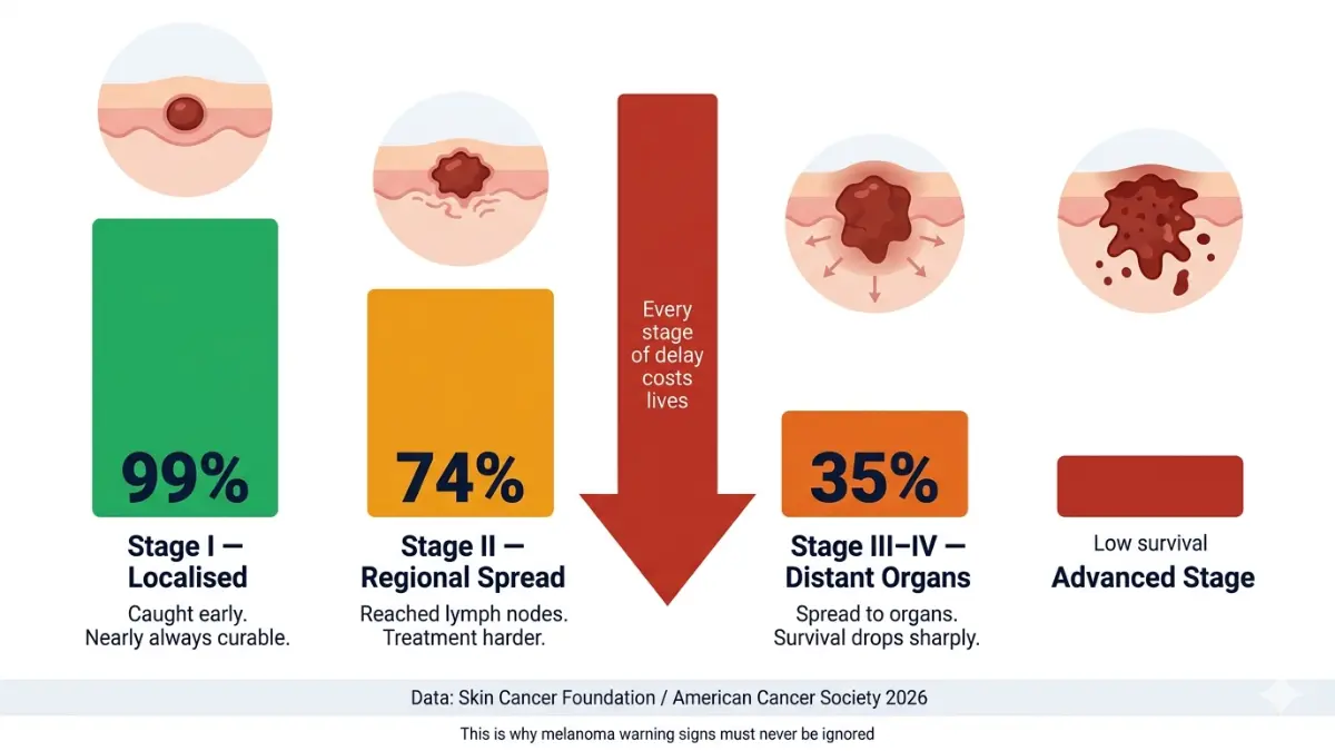
Source: Skin Cancer Foundation / American Cancer Society
What this means for you: Every week of delay matters. If you notice any change on your skin, use our free Symptom Checker as a first step while you arrange a dermatologist appointment.
Early melanoma detection is almost always visible to the naked eye. This puts power directly in your hands — if you know what you’re looking for.
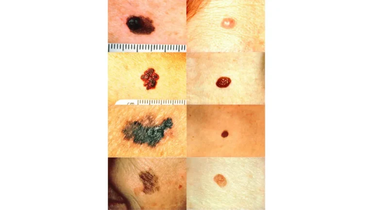
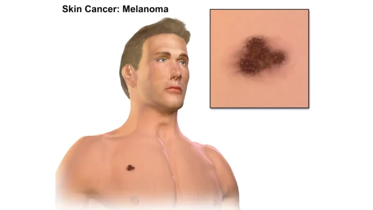
The ABCDE Rule — 5 Classic Melanoma Warning Signs You Must Know
The ABCDE rule is the foundation of melanoma warning signs, recommended by the National Cancer Institute and dermatologists worldwide. Master these five signals first.
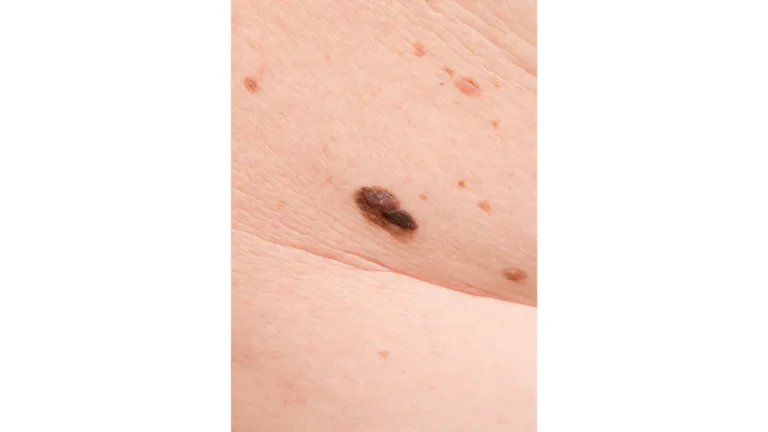
Sign 1: A — Asymmetry
A healthy mole is symmetrical. If you draw an imaginary line down the centre of a mole and the two halves don’t match, that asymmetry is a clinical red flag.
Normal: Round, even shape on both sides. Warning Sign: One half appears larger, irregular, or raised differently than the other.
Sign 2: B — Border Irregularity
Normal moles have smooth, clearly defined edges. Melanoma borders are different — they appear ragged, notched, scalloped, or blurred, often looking like the pigment is “escaping” into surrounding skin.
This spreading edge pattern is one of the earliest detectable signs of invasive growth. If a mole’s border looks like a coastline on a map rather than a smooth circle, see a dermatologist immediately.
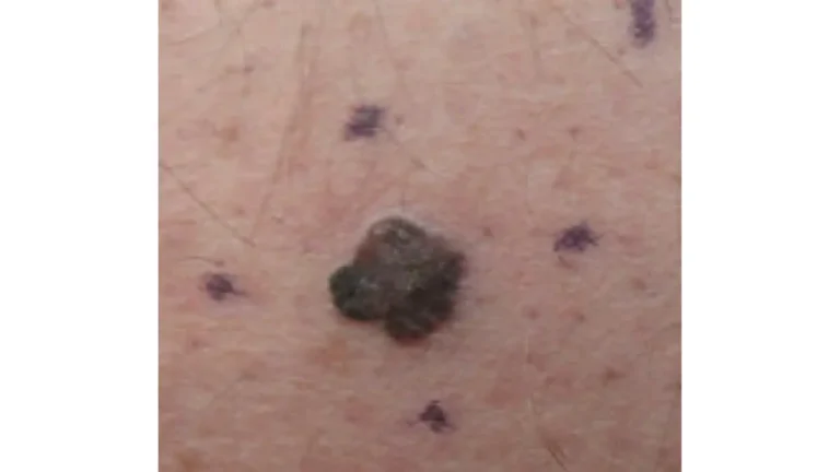
Sign 3: C — Color Variation
Each mole should be one uniform shade of brown or tan. A melanoma warning sign is the presence of multiple colors within a single lesion.
Shades to watch for within a single mole:
- Multiple shades of brown, tan, or black
- Patches of pink, red, or white
- Areas of blue or dark grey (indicates deeper invasion)
Skin-tone note: In people with darker complexions, colour variation can be subtler. Look for areas of grey, blue-black, or reddish patches rather than dramatic colour contrast. Competitors almost never address this. For a deeper understanding, read our guide on melanoma vs. other skin cancers.
Sign 4: D — Diameter AND Darkness
The traditional guideline is larger than 6mm (the size of a pencil eraser). However, the updated “D” now also includes dark lesions of any size — because small nodular melanomas under 6mm can be lethal if missed.
Key 2026 update: Don’t wait for a lesion to grow to pencil-eraser size. Any lesion that appears significantly darker than surrounding moles warrants evaluation, regardless of size.
Sign 5: E — Evolving
According to the CDC’s skin cancer symptoms page, evolving is the most critical ABCDE sign. Any change in size, shape, color, or elevation — or any new symptom such as bleeding, itching, or crusting — is an urgent warning.

Think of it this way: Normal moles are stable for years. A mole that is actively changing is biologically doing something. That “something” may be cancer.
| Feature | Normal Mole | Melanoma Warning Sign |
|---|---|---|
| Shape | Round/oval, symmetrical | Asymmetrical, irregular |
| Border | Smooth, defined | Ragged, notched, blurred |
| Color | Single shade of brown | Multiple colors, spreading |
| Size | Stable, typically <6mm | Growing, or >6mm |
| Change | Unchanged for years | Evolving over weeks/months |
The 4 Hidden Melanoma Warning Signs Doctors Fear Most
This is where our article separates from every competitor. These four signs are the ones most commonly missed — and the ones dermatologists genuinely fear, because patients arrive too late.
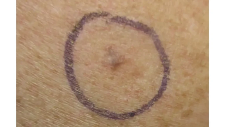
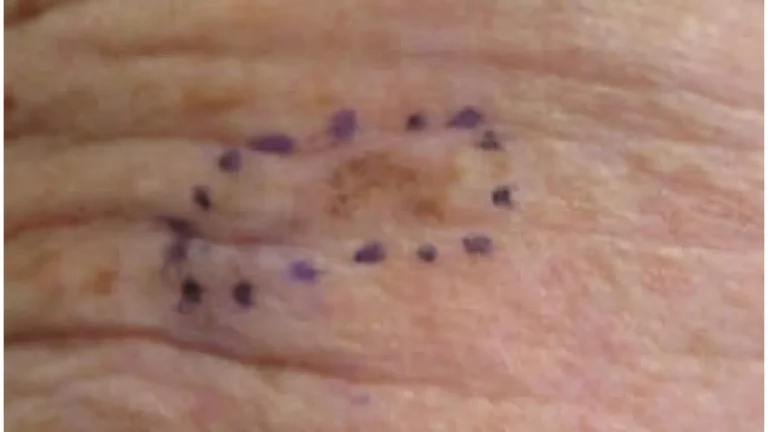
Sign 6: Amelanotic Melanoma — The “Invisible” Warning Sign
Most people picture melanoma as a dark, pigmented mole. But up to 5% of melanomas contain no dark pigment at all.
Called amelanotic melanoma, these lesions appear pink, light red, skin-colored, or even clear. They are frequently misdiagnosed as:
- A common rash
- An irritated pimple
- Eczema or psoriasis
The danger: If you’re only looking for dark moles, you will miss this entirely. A pink spot that doesn’t heal, continues to grow, or fails to respond to standard skincare treatments should be evaluated urgently.
For a full breakdown of what melanoma looks like in all its forms, see the NCI’s melanoma photo reference library.
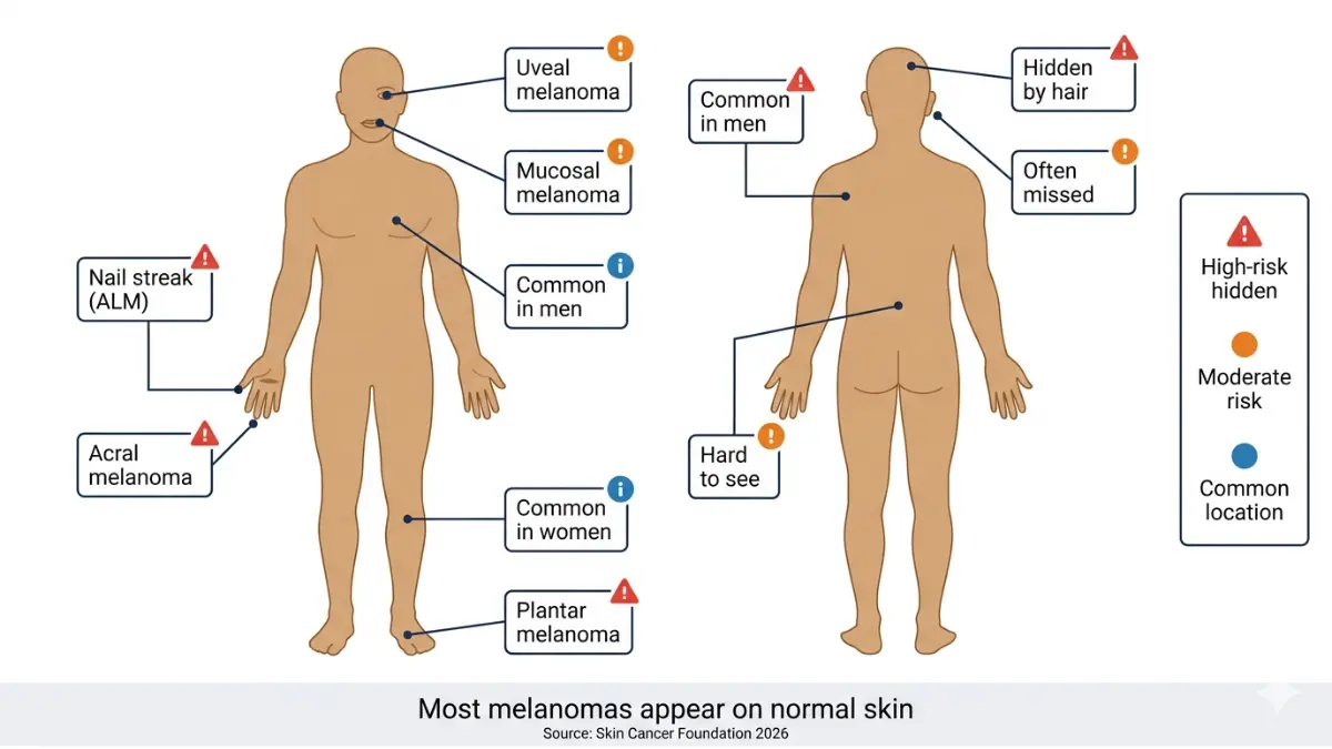
Sign 7: Nail Streak Melanoma (Acral Lentiginous Melanoma)
This is the warning sign that sent Marcus — from our opening story — to Stage III before he was even diagnosed.
Acral lentiginous melanoma (ALM) appears as a dark brown or black longitudinal streak under a fingernail or toenail. Unlike most melanomas, it is not caused by sun exposure and is significantly more common in people with darker skin tones.
Watch for:
- A dark vertical band running the full length of a nail
- Darkening of the skin around the nail base (Hutchinson’s sign — a critical warning)
- A nail that begins to crack, separate, or distort
Critical fact: Many patients assume nail streaks are bruises. If the streak doesn’t grow out with the nail within 8–12 weeks, see a dermatologist immediately.
ALM can also appear on the palms, soles of the feet, and fingers. To understand your genetic vulnerability to this type of melanoma, explore our Genetic Risk Assessment Tool.
Sign 8: Non-Healing Sore or Bleeding Spot
A lesion that bleeds, crusts, appears to heal, then returns within 4–6 weeks is a clinical red flag.
Unlike a standard healing wound, a melanoma-related sore has no clean resolution. The cycle of partial healing followed by re-bleeding or re-crusting is the body’s failed attempt to manage uncontrolled cancerous cell growth beneath the surface.
Do not apply over-the-counter creams and wait. If a sore fails to heal completely within three to four weeks, that lesion requires a biopsy. According to Moffitt Cancer Center, non-healing sores are among the most reliably clinical indicators of an underlying melanoma, especially in later-stage presentations.
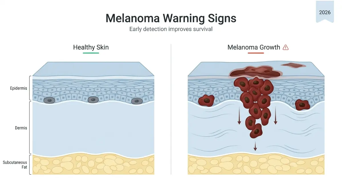
Sign 9: Rapid Vertical Growth — Nodular Melanoma
Nodular melanoma is the most aggressive melanoma subtype. Unlike superficial spreading melanoma, which grows horizontally along the skin over months or years, nodular melanoma grows vertically into deeper skin layers rapidly — sometimes within 2–4 weeks.
Early signs include:
- A firm, raised bump (often dome-shaped) that was not present before
- Black, blue-black, or occasionally red/pink coloring
- Rapid increase in height over days or weeks
- A mole that suddenly feels “different” or “solid”
Research published in PMC (National Institutes of Health) confirmed that patients with nodular melanoma reported bleeding and rapid shape changes as the earliest detectable patient-reported warning signs — with changes occurring over as little as two weeks before diagnosis.
This is why it is doctors’ most feared subtype. By the time a patient notices it, it may already be deep. For context on how this affects long-term outcomes, read our guide on stage 4 melanoma survival and therapy options.
How to Do a 10-Minute Melanoma Self-Exam at Home
The American Cancer Society recommends monthly head-to-toe self-exams. Here is exactly how to do one.
What you need: A well-lit room, a full-length mirror, a hand mirror, and good lighting.
Step-by-Step Self-Exam:
- Face, ears, scalp: Use a comb to part hair and examine the scalp in sections. Ask a partner to help check the back of the scalp.
- Neck, chest, and abdomen: Examine front and sides in the full-length mirror.
- Arms: Check both sides — top, underside, armpits, and between fingers.
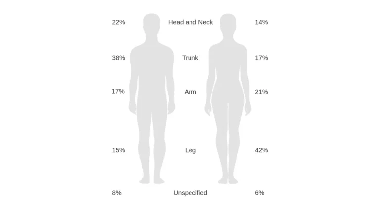
- Back and buttocks: Use a hand mirror to reflect into the full-length mirror. Pay special attention to the upper back — the #1 site for melanoma in men.
- Legs: Check front and back of each leg, including behind the knee.
- Feet and nails: Check soles, between toes, and examine each nail for dark streaks.
- Genital area: Mucosal melanoma can develop here; examine regularly.
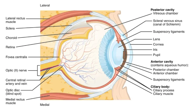
- Eyes: Changes in the iris or dark spots in the eye may indicate uveal melanoma. Use our online Eye Exam tool as an initial screening aid.
The Ugly Duckling Technique: Compare moles to each other. Normal moles on your body tend to look similar. A mole that looks dramatically different from all the others — larger, darker, differently shaped — is an “ugly duckling” and warrants a professional evaluation.
Track with photographs: Take close-up photos of all suspicious moles and date them. Compare month-to-month. Any visible change is a signal to act.
When to See a Doctor IMMEDIATELY
| Warning Signal | Urgency Level | Recommended Action |
|---|---|---|
| Any ABCDE warning sign | High | Book dermatologist within 1 week |
| Non-healing sore (3+ weeks) | High | See doctor within 72 hours |
| Rapid growth over 2–4 weeks | Urgent | Emergency dermatology appointment |
| Dark nail streak (new) | High | Dermatologist within 1 week |
| Pink/red non-pigmented lesion | Moderate-High | Evaluate within 2 weeks |
Who Is Most at Risk for Melanoma in 2026?
Understanding your personal risk profile can determine how frequently you should be screened. According to CDC skin cancer risk data, the following factors significantly elevate melanoma risk.
High-Risk Factors:
- Fair skin, light hair, light eyes — Less melanin means less natural UV protection
- Personal or family history of melanoma — First-degree relatives with melanoma raise your risk 2–3x
- 50+ moles on the body — More moles statistically increase the chance of abnormal cell development
- 5+ severe blistering sunburns, especially in childhood — UV damage accumulates over decades
- Indoor tanning bed use — Tanning beds emit UV radiation up to 15x more intense than midday sun; an estimated 6,200 melanomas are caused annually by indoor tanning alone
- Weakened immune system — Due to HIV, organ transplant medication, or chemotherapy
- BRAF gene mutation — Present in approximately 50% of melanomas; relevant for targeted therapy eligibility
A fact most competitors skip entirely: Melanoma is one of the most common cancers in people under 30 — particularly young women. It is the second most common cancer in women aged 15–29. If you are a young adult, your risk is real and present.
For a complete picture of your personal risk profile, assess your genetic susceptibility using our Genetic Risk Assessment Tool. Our full breakdown of 2026 melanoma statistics covers regional and demographic trends across the US, UK, Australia, and Canada.
Spotted a Melanoma Warning Sign? Your Critical Next Steps in 2026
Finding a suspicious lesion is frightening. But acting on it immediately is what saves lives. Here is your exact action plan.
Step 1 — Photograph and document. Take a clear, close-up photo with a ruler or coin for scale. Record the date and note any symptoms (itching, bleeding, or change).
Step 2 — Do not self-treat. Avoid applying topical creams, freezing home remedies, or any attempt to remove the lesion. Any intervention that disrupts the lesion makes biopsy interpretation harder.
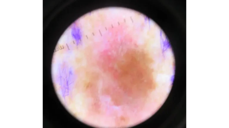
Step 3 — Book a dermatologist, not a GP first. For any ABCDE sign or urgent signal, request a dermatologist referral directly. Early-stage melanoma is best evaluated with dermoscopy — a specialised handheld lens that allows clinicians to examine lesion architecture invisible to the naked eye.
Step 4 — Prepare for a possible biopsy. If your dermatologist suspects melanoma, they will perform a punch or excisional biopsy — removing all or part of the lesion under local anaesthesia. This is a simple outpatient procedure. The tissue sample is examined in a laboratory to confirm diagnosis, depth, and staging.
Step 5 — Understand what staging means. If melanoma is confirmed, staging determines treatment. For a complete overview, read our pillar article on melanoma symptoms, stages, and treatment.
The NCI’s melanoma treatment resource provides full clinical detail on treatment options from surgery to immunotherapy. You can also explore our related guide on skin cancer warning signs and 12 symptoms for a broader skin cancer context.
What this means for you: Detection gives you control. Every melanoma warning sign in this article is visible. The biology works in your favour — if you act fast.
⚠️ Medical Disclaimer: This article is for educational purposes only and does not constitute medical advice, diagnosis, or treatment. Always consult a board-certified dermatologist or qualified healthcare provider for any concerns about your skin or health.
Frequently Asked Questions — Melanoma Warning Signs 2026
Q1. What are the first warning signs of melanoma?
The earliest warning signs include asymmetry in a mole, irregular or blurred borders, multiple colors within a single lesion, and a diameter exceeding 6mm. Any mole that is actively changing in any of these features is the most important early signal.
Q2. Can melanoma appear without a mole?
Yes. Up to 70–80% of melanomas develop on apparently normal skin — not from existing moles. Amelanotic melanomas appear pink or skin-colored and are frequently mistaken for rashes or pimples.
Q3. How fast does melanoma grow?
It depends on the type. Superficial spreading melanoma grows slowly over months or years. Nodular melanoma — the most dangerous subtype — can grow and deepen within 2–4 weeks. Any rapidly growing skin lesion should be evaluated urgently.
Q4. What does early-stage melanoma look like?
Early melanoma may look like an irregular mole with uneven color, ragged edges, or slight elevation. Some early lesions appear completely flat with subtle color variation. Review verified clinical photos at the NCI melanoma photo library.
Q5. Can melanoma appear on legs or feet?
Yes. The lower legs are the most common melanoma location in women. Melanoma also develops on the soles of feet and palms — particularly acral lentiginous melanoma, which is not caused by sun exposure.
Q6. Is a dark line under a nail always melanoma?
Not always — trauma can also cause nail streaks. However, a dark streak that doesn’t grow out with the nail, widens, or is accompanied by skin darkening around the nail (Hutchinson’s sign) requires immediate dermatological evaluation.
Q7. What is the ABCDE rule for melanoma?
ABCDE stands for: Asymmetry, Border irregularity, Color variation, Diameter over 6mm (or Dark), and Evolving change. This framework is endorsed by the Skin Cancer Foundation as the primary self-screening tool.
Q8. When should I see a doctor about a mole?
See a dermatologist within one week if a mole shows any ABCDE warning sign. Seek urgent care within 48–72 hours for a rapidly growing, bleeding, or non-healing lesion. Don’t wait for a scheduled annual exam. Read more in our ABCDE melanoma warning signs guide.
Q9. What is the survival rate for early-detected melanoma?
When melanoma is detected at Stage I (localised), the five-year survival rate reaches 99%. This drops to approximately 74% at the regional lymph node stage and 35% when it spreads to distant organs. The data is clear: early detection is the single most powerful survival factor.
Q10. Can melanoma be mistaken for a bruise or rash?
Yes — this is one of the most dangerous misdiagnoses. Amelanotic melanoma mimics rashes, and acral lentiginous melanoma is frequently confused with bruising under nails or on the soles. If a “bruise” doesn’t fully resolve within 2–3 weeks or a “rash” doesn’t respond to standard treatment, seek evaluation.
Q11. How often should I do a skin self-exam?
Dermatologists recommend a monthly head-to-toe self-exam and a professional skin check at least once per year — or every six months if you have risk factors such as a family history of melanoma, 50+ moles, or a history of tanning bed use. See full NCI SEER training data on melanoma signs for clinical reference.
For additional guidance on related conditions, explore basal cell cancer cure rates and treatment options and our comprehensive article on AI-assisted melanoma screening advances in 2026.
About this content
How this article was put together: researched from recognised health sources, drafted with the help of AI tools, and edited by hand, with sources linked throughout.

Sameer Patel is the founder and editor of My Medicine Advisor. He is not a doctor or medical professional — before starting this site he worked in banking,…
Medical disclaimer
The content on MyMedicineAdvisor is provided for general informational and educational purposes only and is not a substitute for professional medical advice, diagnosis, or treatment. Health information on this website should not be used to diagnose, treat, cure, or prevent any condition without guidance from a qualified healthcare professional. Always seek the advice of your doctor, physician, or another licensed healthcare provider with any questions you may have regarding a medical condition, symptoms, medications, or treatment decisions.