On This Page – Quick Medical Summary
Every year in the United States, over 100,000 Americans are diagnosed with melanoma — the deadliest form of skin cancer. The good news? When caught early, the 5-year survival rate reaches 99%. The ABCDE rule is the dermatologist-approved system to detect melanoma warning signs at home before they become life-threatening. This guide covers every sign, every hidden location, and the exact self-exam steps your doctor uses.
What Is the ABCDE Rule for Melanoma?
Most people don’t realize that skin cancer is the most common cancer in the United States — and melanoma is its most aggressive form. It spreads faster than basal cell or squamous cell carcinoma, making early detection critical.
The ABCDE rule was developed in 1985 by Dr. Darrell Rigel and colleagues at NYU as a practical memory tool to help both patients and clinicians identify suspicious moles. It remains the gold standard for self-examination used by dermatologists worldwide.
According to the American Cancer Society, an estimated 122,680 new cases of melanoma will be diagnosed in the U.S. in 2026 alone. Understanding melanoma warning signs isn’t optional — it’s survival knowledge.
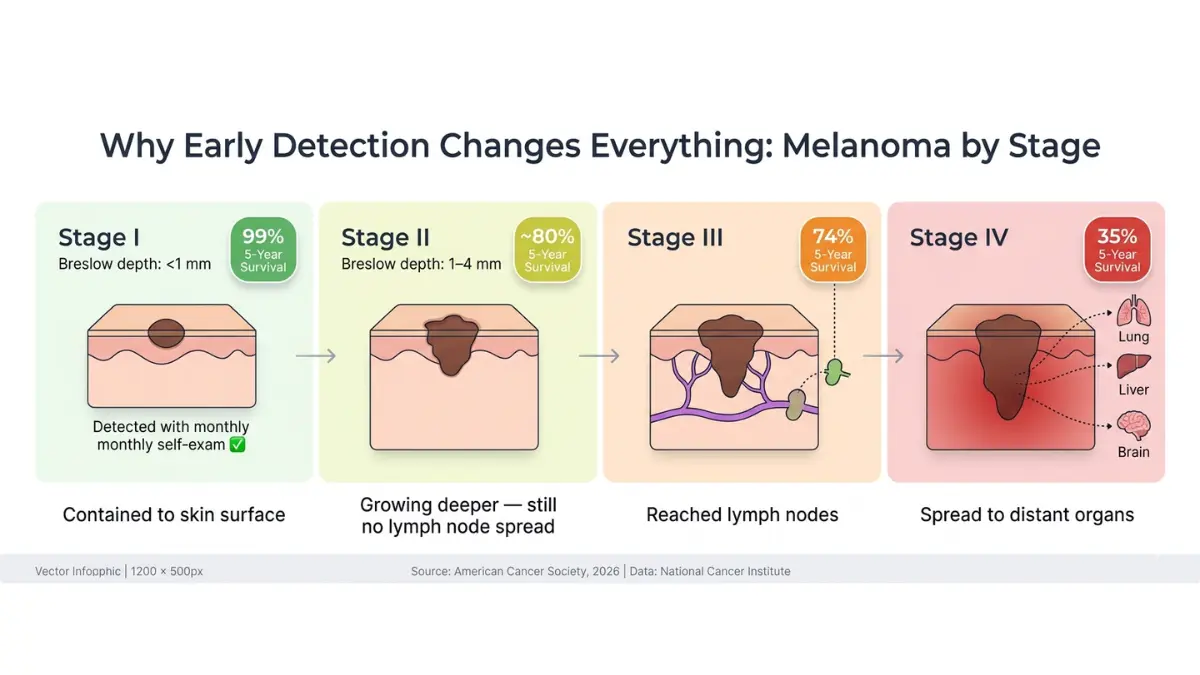
Why early detection changes everything:
| Stage at Detection | 5-Year Survival Rate |
|---|---|
| Localized (skin only) | 99% |
| Regional (lymph nodes involved) | 74% |
| Distant (spread to organs) | 35% |
Source: American Cancer Society, 2026
The difference between a 99% and 35% survival rate comes down to one thing: catching it early. That’s what the ABCDE rule does.
If you have a family history of melanoma or multiple atypical moles, use our Genetic Risk Assessment Tool to evaluate your personal inherited risk before your next dermatology appointment.
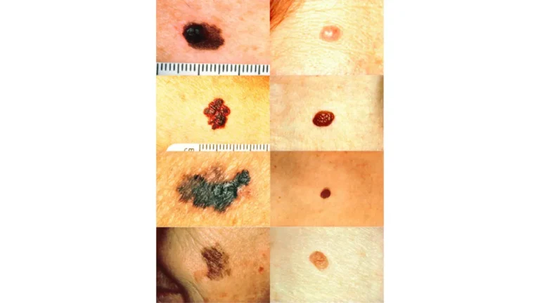
The 5 ABCDE Melanoma Warning Signs — A Precise Clinical Breakdown
This is the core of every skin self-exam. Each letter targets a specific visible feature that separates a harmless mole from a dangerous one. Here’s what competitors don’t explain thoroughly:
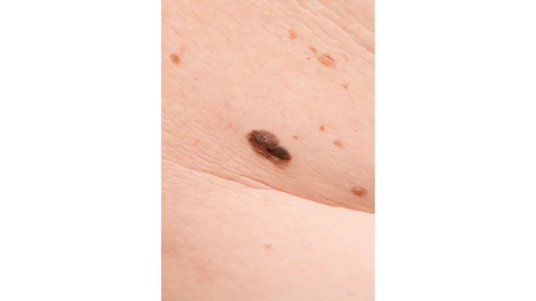
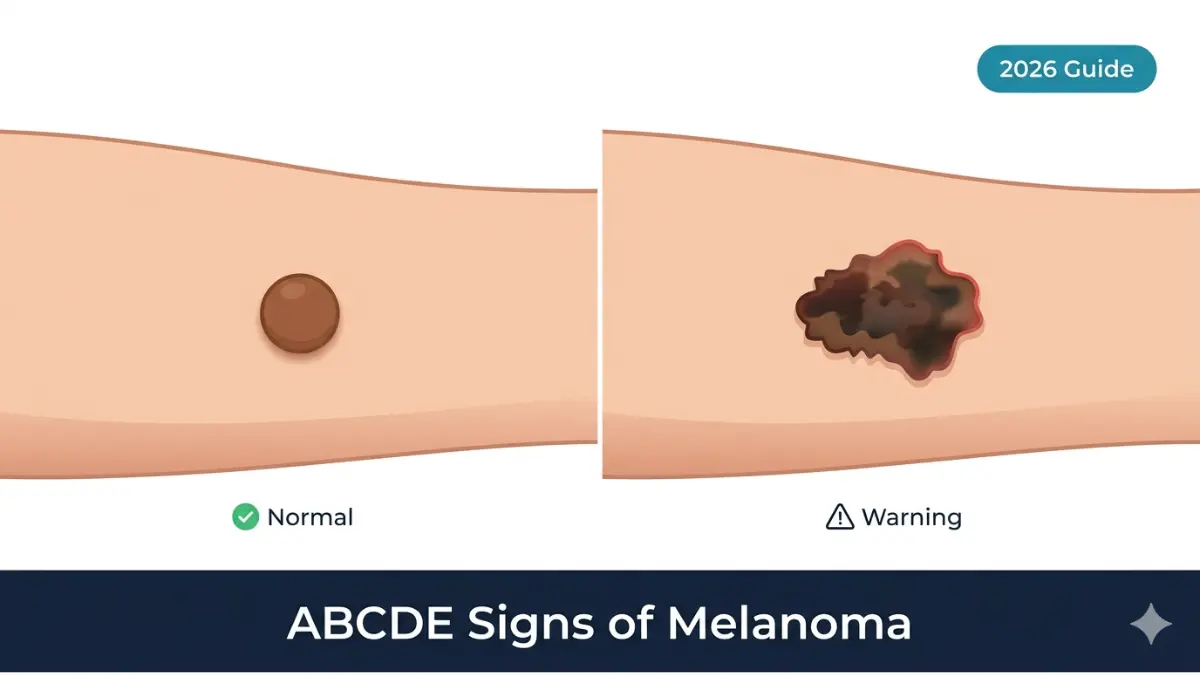
A — Asymmetry: The Mirror Test
What it means: Draw an imaginary line through the center of a mole. In a healthy mole, both halves mirror each other. In a melanoma warning sign, one half looks completely different from the other — a different shape, elevation, or size.
The mirror test: Hold a small mirror horizontally across the middle of the mole in a photo. If the reflection doesn’t match the other half, consider it suspicious.
Normal moles are round or oval and symmetrical. Most melanomas are not.
B — Border: Ragged, Notched, or Blurred Edges
What it means: A healthy mole has a smooth, clearly defined edge — like a circle drawn with a compass. A melanoma warning sign has irregular, scalloped, notched, or blurred borders that fade unevenly into surrounding skin.
According to the American Academy of Dermatology, borders that are poorly defined or that look like they’re “bleeding” into the skin are among the earliest and most reliable visual signals.
Key distinction:
- Normal: Clean, sharp, well-defined border
- Warning: Jagged, scalloped, notched, or ink-stained appearance
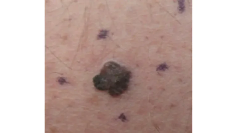
C — Color: Multiple Shades in One Mole
What it means: A benign mole is one uniform shade of brown. A melanoma warning sign often contains multiple colors within a single lesion — shades of tan, dark brown, black, and sometimes areas of red, white, or blue.
Critical gap all top competitors miss: Some melanomas are amelanotic — meaning they contain little to no pigment and appear pink, flesh-colored, or red. These are the most dangerous because they’re the easiest to dismiss as a pimple or scar. The National Cancer Institute specifically warns about late diagnosis of amelanotic melanoma due to its atypical appearance.
Color warning checklist:
- ✅ Multiple browns or blacks in one spot
- ✅ A pink or flesh-colored mole that is changing
- ✅ Areas of white (depigmentation) within a dark mole
- ✅ Any blue or red zones inside a pigmented lesion
D — Diameter: Larger Than a Pencil Eraser
What it means: Melanomas are typically larger than 6mm in diameter at detection — roughly the size of a pencil eraser (¼ inch). However, this is the most misunderstood rule: melanomas absolutely can and do present at smaller sizes.
The real clinical rule: Any mole — regardless of size — that shows the other ABCDE warning signs should be evaluated. The 6mm guideline is a threshold, not a clearance.
| Reference Size | Measurement |
|---|---|
| Pencil eraser | ~6 mm |
| Ballpoint pen tip | ~1 mm |
| Standard shirt button | ~15 mm |
Never dismiss a mole as “too small to be melanoma.” Size alone does not determine risk. For context on how melanoma detection has evolved with AI in 2026, read our guide on AI screening advances in melanoma detection.
E — Evolving: The Most Critical — and Most Overlooked — Sign
What it means: This is the single most important letter. Any change in a mole over time is a melanoma warning — whether in size, shape, color, elevation, or new symptoms like itching, bleeding, oozing, or crusting.
The Skin Cancer Foundation notes that 20–30% of melanomas develop within existing moles, while 70–80% arise on normal-looking skin. This means a mole you’ve had for years can become dangerous.
Evolution warning signs:
- A mole growing noticeably larger over weeks or months
- Color deepening or becoming patchy
- A flat mole starting to raise or develop a bump
- Spontaneous bleeding without injury
- Persistent itching, tenderness, or crusting
- Any mole that “just doesn’t feel right”
The Ugly Duckling Sign — The Unofficial 6th Check
No competitor explains this thoroughly. Most moles on your body share a family resemblance — they look similar to each other. A melanoma warning sign often stands out like an ugly duckling — noticeably larger, darker, lighter, or differently shaped than all surrounding moles.
If one mole looks like the odd one out, even if it doesn’t tick every ABCDE box, get it checked. Isolated single moles with no neighbors to compare against are also considered ugly ducklings by clinical guidelines.
For a broader look at how melanoma differs from other skin cancers, see our detailed breakdown: Melanoma vs. Skin Cancer: Key Differences Explained.
Quick-Reference ABCDE Table:
| Letter | What to Check | Normal | Warning Sign |
|---|---|---|---|
| A | Asymmetry | Both halves match | One half different |
| B | Border | Smooth, sharp | Ragged, blurred, notched |
| C | Color | Single shade of brown | Multiple colors, or pink/flesh |
| D | Diameter | Under 6mm, stable | Over 6mm, or growing |
| E | Evolving | Unchanged for years | Any recent change |

Melanoma Warning Signs on Dark Skin, Nails & Hidden Locations
This is the information gap that costs lives — and no top competitor covers it adequately.
Melanoma in People of Color
Acral lentiginous melanoma (ALM) is the most common form of melanoma in Black, Hispanic, and Asian patients. It does not follow the typical ABCDE pattern. The CDC’s skin cancer data shows that people of color are disproportionately diagnosed at later, less treatable stages — largely because of the false belief that darker skin protects against melanoma.
Marcus, a 48-year-old Black man in Atlanta, was told by a general practitioner for two years that the dark streak under his thumbnail was a bruise. It was ALM. His diagnosis came at Stage III.
Where ALM appears:
- Under fingernails or toenails — looks like a dark brown or black streak running lengthwise
- Palms of the hands — irregular dark patches on non-sun-exposed skin
- Soles of the feet — especially between toes and on the heel
Other Hidden Melanoma Locations
These locations are rarely mentioned — even by established medical publishers:
- Mucosal melanoma: Inside the mouth, nasal passages, or genitals — may appear as dark, irregular patches
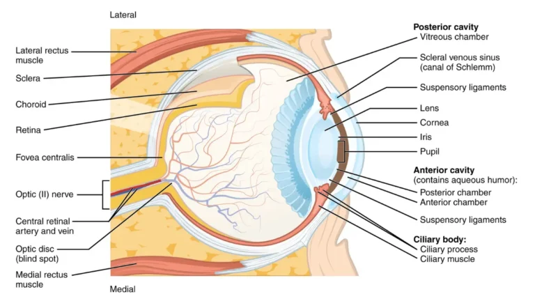
- Uveal (eye) melanoma: A dark spot appearing in the colored iris — detected during eye exams
- Scalp melanoma: Hidden under hair — requires a partner or mirror to examine
The ABCDE rule is less reliable in these locations. For nail and palm melanomas, look instead for: a new dark streak in a nail, a spot that isn’t healing, or a painless dark growth in an unusual area. Our skin cancer warning signs overview covers all skin cancer types including less common presentations.
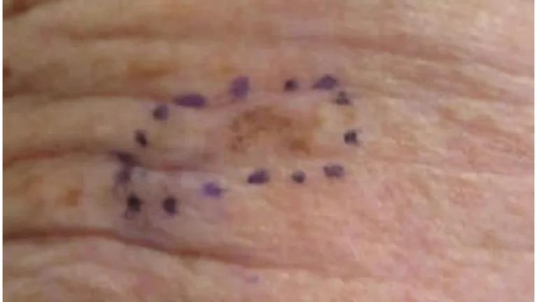
How to Check Your Moles at Home — A 7-Step Monthly Self-Exam
No competitor on the first page of Google provides a real, actionable self-exam protocol. This section is your complete guide.
What you need:
- Full-length mirror
- Small handheld mirror
- Bright overhead lighting
- Your smartphone camera (for mole mapping)
- A partner for hard-to-see areas (back, scalp)
When to do it: The first Sunday of every month — set a calendar reminder.
The 7-Step Full-Body Melanoma Check
Step 1 — Face, Ears & Scalp Examine your face front and sides in the mirror. Use a comb or hair dryer to part the hair in sections and inspect your scalp. Use the handheld mirror to check behind your ears and the back of your neck.
Step 2 — Neck, Chest & Abdomen Check the front and sides of your neck. Examine the full chest and stomach — including under the breasts for women.
Step 3 — Arms & Hands Raise both arms and check all sides — front, back, inner arm, and underarm. Don’t skip between the fingers and under your fingernails. Look for any new dark streaks in nails.
Step 4 — Back & Buttocks Use the handheld mirror with the full-length mirror to check your entire back — upper, middle, and lower. Ask a partner to help. Check the buttocks and between the cheeks.
Step 5 — Legs — Both Sides Fully Check the front, back, and inner thighs. Don’t skip the back of the knees — a common melanoma warning location.
Step 6 — Feet, Toes & Soles Sit down and lift each foot. Check the soles, between every toe, and under all toenails. These are the areas most often missed.
Step 7 — Genitals & Groin Use a mirror. Melanoma can develop anywhere, including in areas that never see the sun. Mucosal melanoma in this region is rare but real.
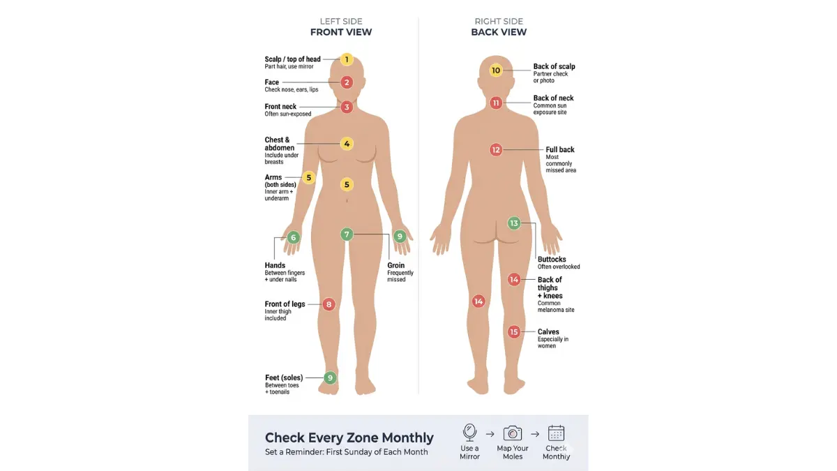
Mole mapping tip: Photograph any mole you’re monitoring and date the image. Comparing photos from 2–3 months apart is the most reliable way to detect change — which triggers the most important ABCDE letter: E for Evolving.
Not sure if what you’re seeing is worth worrying about? Use our Symptom Checker to assess your skin concern before your appointment.
The AAD’s free downloadable Body Mole Map is a printable tool to document and track every mole on your body.
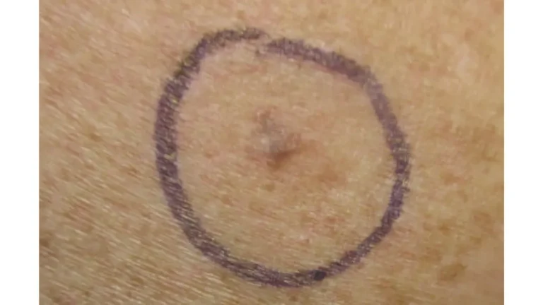
When to See a Dermatologist — The Melanoma Warning Checklist
Many people delay action because they’re afraid of overreacting. Dermatologists agree: there is no such thing as overreacting about a suspicious mole.
See a dermatologist within 2 weeks if you notice:
- ✅ Any mole meeting one or more ABCDE criteria
- ✅ A mole that itches, bleeds, or crusts without injury
- ✅ A new dark streak under a fingernail or toenail
- ✅ A sore on the skin that does not heal after 3 weeks
- ✅ A mole that “feels different” — even if it looks normal
- ✅ Any rapidly growing new skin growth
- ✅ A pink or skin-colored bump that bleeds easily
What to expect at a dermatologist skin check:
- Visual inspection — full-body exam under bright light
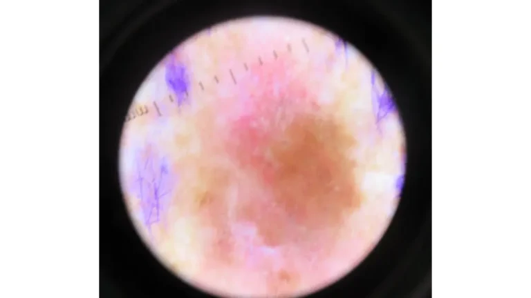
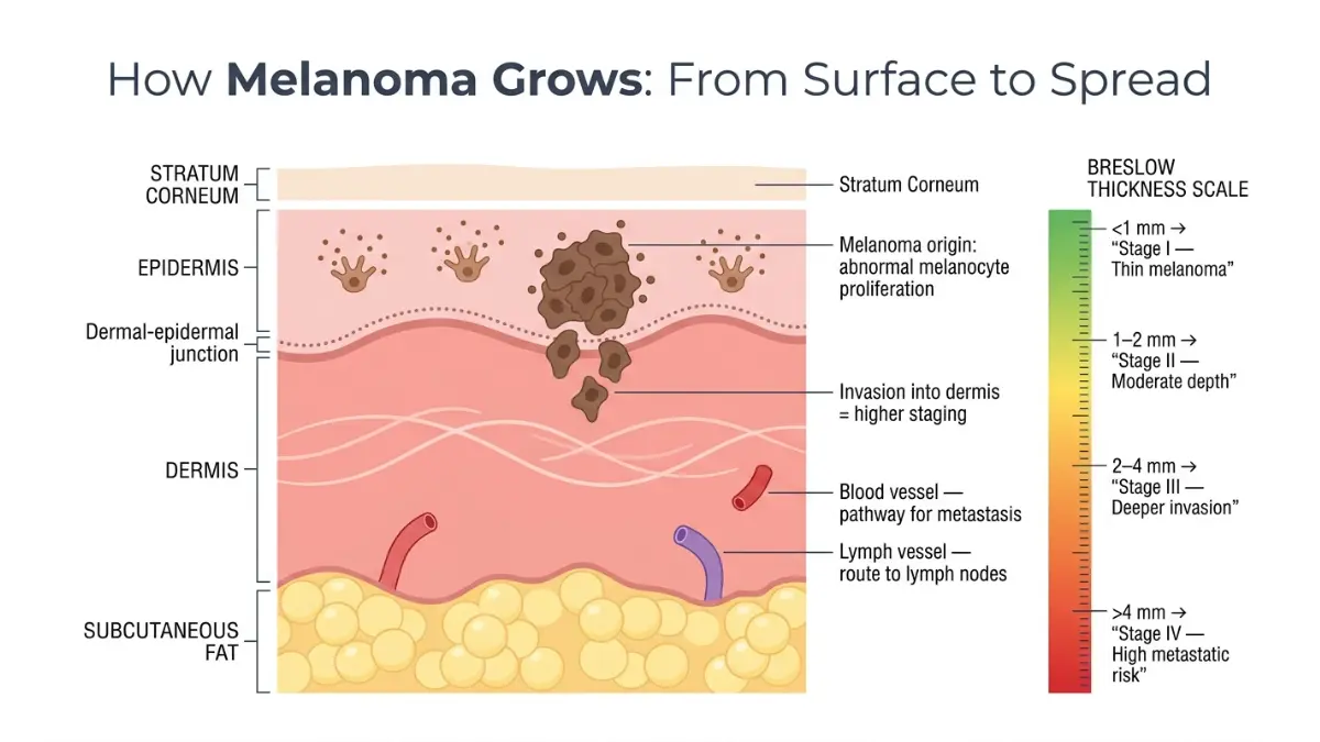
- Dermoscopy — a dermatoscope magnifies moles up to 10x, revealing subsurface pigment patterns invisible to the naked eye
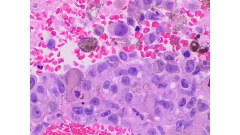
- Biopsy decision — if suspicious, a small tissue sample is removed under local anesthetic; results typically return within 5–10 business days
Understanding what a biopsy report means is important. Our guide on biopsy results and timelines walks you through every result type and what to do next.
Who is highest risk?
- Fair skin, light eyes, or red/blonde hair
- Personal or family history of melanoma
- 50+ moles on the body
- History of severe sunburns (especially before age 18)
- Tanning bed use — increases melanoma risk by 75% in users under 35, per WHO data
- Organ transplant recipients (immunosuppressive medications reduce tumor surveillance)
- Outdoor workers with daily unprotected UV exposure
If you’ve been diagnosed or are concerned about Stage 4 progression, our clinical guide to Stage 4 melanoma treatments provides a comprehensive breakdown of current therapy options.
Proven Melanoma Prevention — 7 Dermatologist-Backed Steps for 2026
Detecting melanoma warning signs early saves lives. Preventing the conditions that cause it saves even more.
1. Wear Broad-Spectrum SPF 30+ Sunscreen — Every Day Apply at least 1 ounce (a full shot glass) to all exposed skin 15 minutes before going outside. Reapply every 2 hours — or immediately after swimming or sweating. SPF 30 blocks approximately 97% of UVB rays; SPF 50 blocks 98%.
2. Avoid Peak UV Hours UV radiation peaks between 10am and 4pm. The CDC’s skin cancer prevention guidelines recommend seeking shade during these hours or wearing protective clothing.
3. Never Use Tanning Beds The WHO classifies tanning beds as Group 1 carcinogens — the same category as tobacco and asbestos. First use before age 35 increases melanoma risk by 75%.
4. Wear UV-Protective Clothing UPF-rated (Ultraviolet Protection Factor) clothing, wide-brim hats (3-inch brim minimum), and UV-blocking sunglasses protect areas where sunscreen is easily missed.
5. Get an Annual Professional Skin Check A board-certified dermatologist can identify melanoma warning signs invisible to the naked eye. Dermoscopy detects suspicious pigment patterns at a microscopic level. Annual full-body exams are recommended by the Skin Cancer Foundation for all adults over 40, and earlier for high-risk individuals.
6. Get Vitamin D Safely Adequate vitamin D is important for immune function and bone health — but sun exposure is not the safest source. Supplementation (1,000–2,000 IU/day) is a clinically safer alternative to UV exposure for maintaining levels. Discuss dosing with your physician.
7. Protect Immunosuppressed Patients More Aggressively Organ transplant recipients and patients on long-term immunosuppressive therapy face dramatically elevated skin cancer risk. More frequent dermatology checks (every 3–6 months) are recommended by transplant medicine guidelines.
For deeper reading on the full melanoma clinical picture — from early detection through treatment — visit our comprehensive melanoma symptoms, stages, and treatment guide.
FAQs: Melanoma Warning Signs & Mole Checks (2026)
Q1. What does melanoma look like in its early stages?
Early melanoma often appears as a mole or flat pigmented spot that has recently changed in color, size, shape, or texture. It may have uneven borders, multiple colors, or be slightly raised. Many early melanomas are painless.
Q2. Can melanoma be pink or flesh-colored?
Yes. Amelanotic melanoma lacks pigment and appears pink, skin-colored, or lightly red. It is frequently misidentified as a pimple, scar, or benign cyst — making it one of the most dangerous and late-diagnosed forms.
Q3. How do I know if a mole is cancerous or harmless?
Use the ABCDE rule: asymmetry, irregular border, multiple colors, diameter over 6mm, and evolving changes. If it meets any one of these criteria, schedule a dermatologist appointment. When in doubt, always get it checked — early-stage melanoma is highly curable.
Q4. What does the ABCDE rule stand for in melanoma?
ABCDE stands for: Asymmetry, Border irregularity, Color variation, Diameter over 6mm, and Evolving changes. Each letter represents a clinical warning feature that can indicate a mole may be developing into melanoma.
Q5. How fast does melanoma grow?
Superficial spreading melanoma — the most common type — can grow slowly over months to years. Nodular melanoma grows much faster, sometimes over weeks. This is why monthly self-exams and annual dermatology checks matter enormously.
Q6. Can melanoma appear on areas not exposed to the sun?
Yes. Melanoma can develop on the soles of the feet, palms, under nails, inside the mouth, in the eyes, and in the genitals — areas with little to no sun exposure. This is especially important for people of color, where acral lentiginous melanoma accounts for most diagnoses.
Q7. What is the survival rate for melanoma if caught early?
When detected while still localized to the skin, the 5-year survival rate is 99%. This drops to 74% if it reaches regional lymph nodes, and 35% if it spreads to distant organs. Early detection is the single most powerful factor in survival outcomes.
Q8. How often should I check my moles?
The American Academy of Dermatology recommends a full-body self-exam once a month. In addition, see a board-certified dermatologist for a professional full-body skin check at least once a year — or every 3–6 months if you are high-risk.
Q9. Is a mole that itches always a sign of melanoma?
Not always — dry skin or eczema can cause itching. However, persistent itching in a single mole, especially one that is also changing in appearance, is a recognized melanoma warning sign under the “E for Evolving” criterion. Don’t dismiss it.
Q10. What is the Ugly Duckling sign in melanoma detection?
The Ugly Duckling sign refers to a mole that looks distinctly different from all your other moles — larger, darker, lighter, or oddly shaped compared to nearby moles. It’s a clinical screening concept that works alongside the ABCDE rule and is highly effective at identifying melanoma that doesn’t meet every letter criterion.
Q11. When should I go to the ER vs. make a regular appointment for a suspicious mole?
A regular dermatology appointment is appropriate for a mole that looks suspicious but isn’t causing acute symptoms. Go to the ER or urgent care immediately if a mole is bleeding heavily and won’t stop, is rapidly growing over days, or is accompanied by fever or swollen lymph nodes — which may indicate active systemic spread.
For the latest melanoma research, 2026 incidence data, and current treatment options, read our Melanoma Statistics 2026 Report and our Melanoma vs. Skin Cancer Differences Guide.
⚠️ Medical Disclaimer: This content has been reviewed by a board-certified dermatologist and is intended for educational purposes only. It does not replace professional medical advice, diagnosis, or treatment. If you notice any melanoma warning signs, contact a licensed healthcare provider promptly.
About this content
How this article was put together: researched from recognised health sources, drafted with the help of AI tools, and edited by hand, with sources linked throughout.

Sameer Patel is the founder and editor of My Medicine Advisor. He is not a doctor or medical professional — before starting this site he worked in banking,…
Medical disclaimer
The content on MyMedicineAdvisor is provided for general informational and educational purposes only and is not a substitute for professional medical advice, diagnosis, or treatment. Health information on this website should not be used to diagnose, treat, cure, or prevent any condition without guidance from a qualified healthcare professional. Always seek the advice of your doctor, physician, or another licensed healthcare provider with any questions you may have regarding a medical condition, symptoms, medications, or treatment decisions.