On This Page – Quick Medical Summary
Melanoma in dark skin behaves differently from melanoma in lighter skin. It develops in sun-protected areas — not from sun damage — and is diagnosed at late stages in over 52% of Black patients. Knowing the exact symptoms and hidden locations can be lifesaving.
The Case That Changed Everything
In 1977, reggae legend Bob Marley noticed a dark mark forming beneath his toenail. He assumed it was a football injury. His doctors — unfamiliar with how melanoma on dark skin presents — were slow to act. By the time he was correctly diagnosed with acral lentiginous melanoma, the cancer had spread. Bob Marley died at 36.
His story is not an outlier. It is a pattern repeated thousands of times each year across America.
According to the American Association of Medical Colleges, Black patients are more than three times as likely to be diagnosed with melanoma at a late stage compared to non-Hispanic white patients. The five-year survival rate for Black patients sits at just 66–71%, versus 94% for white patients.
This gap is not biology. It is a failure of awareness — in patients, in doctors, and in medical education.
If you have dark skin — whether you’re Black, Hispanic, South Asian, Southeast Asian, Middle Eastern, or of mixed heritage — this guide gives you exactly what you need to know: where melanoma hides, what it looks like on your skin tone, why it gets missed, and what you can do today.
Use our Symptom Checker if you’ve recently noticed any unusual skin or nail changes.
Why Melanoma in Dark Skin Is Fundamentally Different
Melanoma Dark Skin Is Not a Sun Cancer — That’s Why It Gets Missed
Most skin cancer campaigns focus on UV radiation, sunburn, and tanning beds. For people with dark skin, this messaging is almost entirely irrelevant — and dangerously misleading.
Darker skin contains more melanin, which provides natural photoprotection. This is why melanoma dark skin cases are rarely caused by UV exposure. Instead, the most common type in people of color — acral lentiginous melanoma (ALM) — develops in sun-protected zones: nails, palms, and soles.
The National Cancer Institute confirms that ALM accounts for approximately 36% of melanomas in Black patients, compared to just 2–3% in white patients. It is biologically distinct, grows rapidly, and is discovered late because it appears in locations no one thinks to check.
Here is how melanoma types break down across skin tones:
| Melanoma Type | Most Common In | Primary Location |
|---|---|---|
| Superficial Spreading | White/light skin | Back, legs, arms |
| Acral Lentiginous (ALM) | Black, Asian, Hispanic skin | Palms, soles, under nails |
| Mucosal Melanoma | All skin types | Mouth, genitals, anal area |
| Nodular Melanoma | All skin types | Any body site |
| Amelanotic Melanoma | All skin types | Can appear anywhere, often pink/red |
Understanding that skin cancer on dark skin follows different rules is the first step toward survival. To understand the full picture of all melanoma types, read our guide on amelanotic melanoma — a colorless variant that even trained doctors miss on any skin tone.
A comprehensive PMC review of skin cancer in people of color confirms that symptoms in darker-skinned populations occur in locations rarely examined during routine medical appointments.
The 8 Hidden Locations Where Melanoma Hides on Dark Skin
8 Places Melanoma Hides on Dark Skin — Including Spots Your Doctor Never Checks
This is the section your competitors don’t have. Every standard melanoma article tells you to check your moles. But melanoma in dark skin doesn’t usually start on sun-exposed moles. It starts somewhere else entirely.
Know these 8 locations. Check them monthly.
1. Sole of the Foot (Plantar Region)
This is the most common melanoma site in Black and Asian patients. It looks like a bruise that never heals, a dark irregular patch, or a fading discolored area.
Between 30–40% of melanoma cases in people of color appear here, according to the Skin Cancer Foundation’s guide on skin of color. Check both feet monthly, especially along the heel and arch.
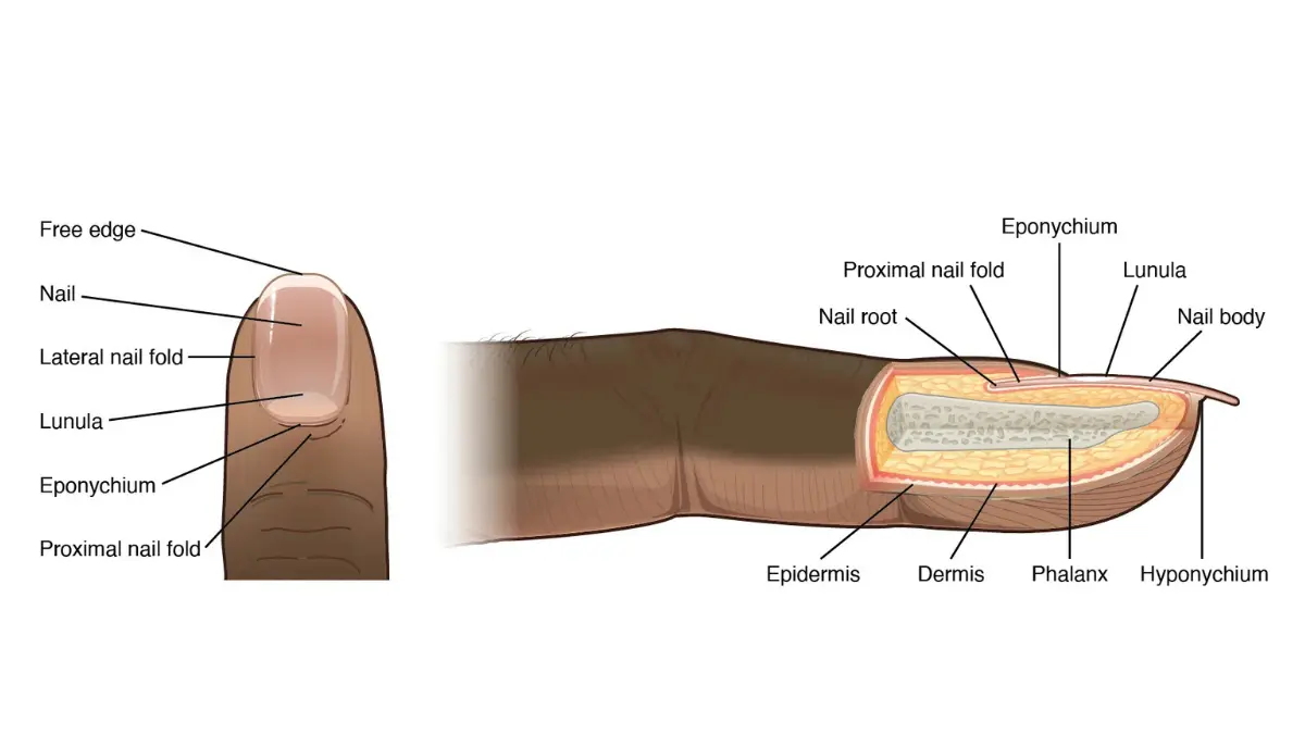
2. Under or Around Fingernails and Toenails
Look for a dark brown or black longitudinal streak running the length of the nail. This is called Hutchinson’s sign when the pigment spreads onto the surrounding skin — a recognized emergency warning.
⚠️ Callout: If you see dark pigment spreading from under your nail onto the surrounding skin or cuticle — see a dermatologist within one week. Do not wait.
Our detailed guide on subungual melanoma and nail melanoma covers exactly what to look for under each nail and when to act.
3. Palm of the Hand
An irregular dark patch, thickened area, or lesion that won’t heal. Often mistaken for a callus or pressure mark.
4. Inside the Mouth (Mucosal Melanoma)
Dark or brownish-black irregular patches on the gums, palate, or inner cheeks. The American Academy of Dermatology notes that mucosal sites are routinely overlooked in standard skin examinations.
5. Genital and Anal Area
Dark or pinkish-red irregular lesions in these areas are almost never spontaneously examined — by patients or providers. This leads to severely delayed diagnosis.
6. Between the Toes
A site frequently skipped even during thorough foot exams. Dark discoloration, nodules, or non-healing sores here warrant urgent dermatology review.
7. Under the Breast and in Skin Folds
Friction zones can harbor melanoma dark skin lesions that look like darkening of the skin or chronic irritation. These are almost never discussed in melanoma prevention literature.
8. Scalp and Behind the Ear
Hard to see without a two-mirror system. Many scalp melanomas in dark-skinned patients are first noticed by barbers or hairdressers — not doctors.
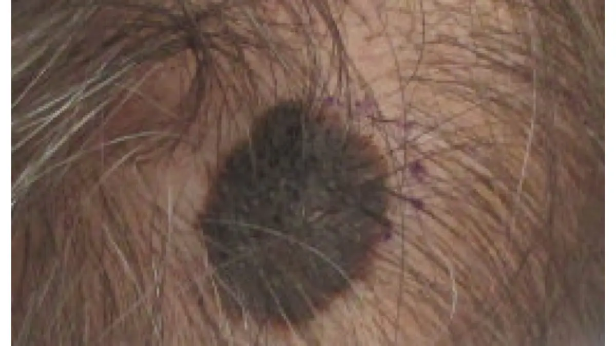
For a comprehensive map of every location melanoma appears across all skin types, see our guide on where melanoma appears on the body.
Use our Genetic Risk Assessment Tool to evaluate your personal baseline risk factors, including family history and skin type.
The Late Diagnosis Crisis — Why Dark Skin Patients Are Caught Too Late
Why Melanoma in People of Color Is Diagnosed Dangerously Late — and Who’s Responsible
The survival disparity in melanoma dark skin is not primarily biological. It is systemic. Five documented causes drive the late-diagnosis crisis in America.
1. The “Dark Skin Can’t Get Cancer” Myth
A 2022 study published in the Journal of the American Academy of Dermatology found that Black and Hispanic patients are significantly less likely to perform skin self-exams — partly because they’ve been told, or assumed, that darker skin is fully protected.
It is not. Melanin reduces UV risk. It does not eliminate ALM, mucosal melanoma, or nodular melanoma risk.
2. Medical Training Ignores Dark Skin
Dermatology textbooks and training curricula have historically featured melanoma almost exclusively on white skin. This creates a diagnostic blind spot when physicians encounter a dark streak beneath a nail or an irregular lesion on a brown palm.
3. Documented Provider Bias
Studies confirm that Black patients receive fewer thorough skin exams during primary care appointments. Concerns are more likely to be attributed to benign causes — “old bruise,” “pressure mark,” “fungal infection” — without biopsy.
4. Insurance and Access Gaps
According to the CDC’s research on melanoma in non-Hispanic Black Americans, uninsured and underinsured patients face longer delays between first symptom and confirmed diagnosis. Access to dermatology specialists is disproportionately limited in underserved communities.
5. Hidden Locations Are Never Examined
Nail beds, soles, and mucosal surfaces are not part of a routine annual physical. Without specific patient advocacy — asking the doctor to check these areas — they are left unexamined for years.
The numbers tell the full story:
| Group | 5-Year Melanoma Survival | Diagnosed at Late Stage |
|---|---|---|
| Non-Hispanic White | 94% | 16% |
| Hispanic | ~82% | 26% |
| Non-Hispanic Black | 66–71% | 52% |
Sources: CDC; Melanoma Research Alliance 2025
What This Means For You: If a doctor dismisses a dark, changing spot because you have dark skin — ask for a biopsy or a dermatology referral directly. You have an absolute right to advocate for your own health.
Our pillar resource, Melanoma: Symptoms, Stages & Treatment, covers what happens after a positive biopsy and what treatment options exist at every stage.
The Modified ABCDE System for Dark Skin
Standard ABCDE Doesn’t Work the Same on Dark Skin — Use This Updated System
Every competitor article lists the standard ABCDE checklist. None of them explain why it needs to be modified for people with darker skin tones.
On Fitzpatrick Type V–VI skin (deep brown to dark brown/black), color variations within a lesion are far harder to see. Borders appear less distinct. The standard system, designed for lighter skin, gives a false sense of security to people of color.
Here is the adapted framework:
| Standard | What to Know for Dark Skin |
|---|---|
| A – Asymmetry | Still applies; check if one side of a nail streak is wider than the other |
| B – Border | Borders may be subtle; focus on texture change, not just color edge |
| C – Color | May appear dark brown, grey, or black — not multicolored as in white skin |
| D – Diameter | Pencil eraser rule still applies; ALM lesions often grow larger before detection |
| E – Evolving | Most critical signal on dark skin — any change in shape, size, surface, or feel |
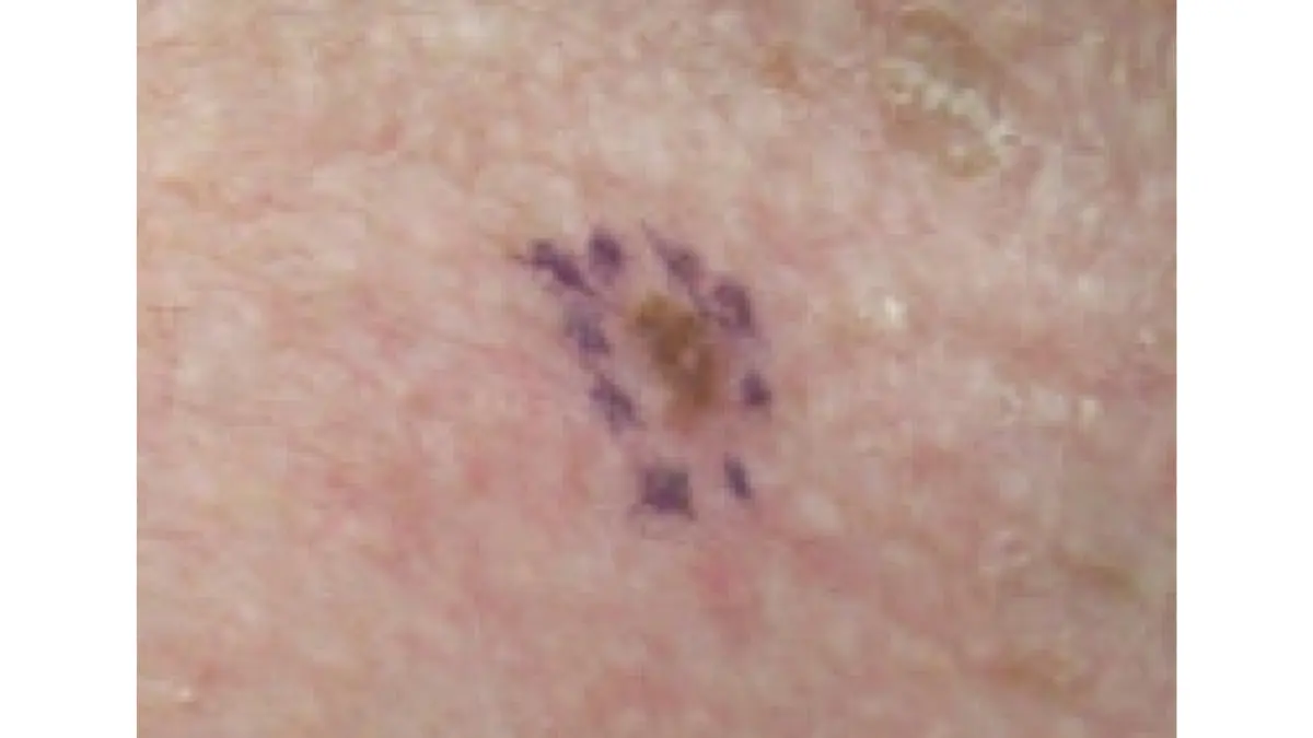
Add Two More Letters:
- F – Feel: Thickness, firmness, or a bump beneath a darkened area deserves immediate attention
- U – Ugly Duckling: A spot that looks different from all other marks on your skin — lighter, darker, raised, or textured differently
The American Cancer Society’s melanoma warning signs resource confirms that the ugly duckling sign is one of the most reliable early detection markers across all skin types.
Monthly self-check — 4 steps, under 5 minutes:
- Remove all nail polish once monthly and check each nail for streaks
- Examine both soles in full daylight using a hand mirror
- Ask a partner to check your scalp, behind the ears, and between shoulder blades
- Log any new or changed spot using our Symptom Checker to track changes over time
For the complete ABCDE guide with images and mole mapping instructions, see our dedicated resource on ABCDE warning signs and melanoma moles.
Prevention That Actually Works for Dark Skin Tones
Melanoma Prevention for Dark Skin: 7 Steps That Go Far Beyond Sunscreen
Most melanoma prevention campaigns are built for white skin. They focus almost entirely on sunscreen and sun avoidance — strategies that are largely irrelevant for acral lentiginous melanoma, which has no UV cause.
Here are 7 evidence-based prevention and early-detection steps that are actually relevant to melanoma in dark skin:
Step 1: Monthly Foot-and-Nail Inspection
Remove all nail polish — on fingers and toes — once per month. Check every nail for dark streaks, particularly any that are widening or have appeared recently.
Step 2: Request a Full-Body Skin Exam Annually
Do not assume your primary care doctor checks everywhere. Explicitly say: “I’d like a full skin exam today, including my scalp, nails, palms, soles, and between my toes.” Dermatologists are trained for this; general practitioners often are not.
Step 3: Know and Share Your Family History
ALM can have a genetic component. If any relative has had an unusual nail discoloration, foot lesion, or undiagnosed skin cancer, document it and share it with your doctor. Our Genetic Risk Assessment Tool helps you map this information before your appointment.
Step 4: Still Use Sunscreen — Especially for South Asian and Hispanic Skin
For Fitzpatrick Type III–IV skin — which includes many Hispanic, South Asian, and Mediterranean individuals — UV-driven melanoma risk is moderate and real. Use SPF 30+ broad-spectrum sunscreen on face, neck, and hands daily.
Read our dermatologist-curated guide on the best sunscreens for melanoma prevention to find formulas that don’t leave white cast on brown skin.
Step 5: Track Chronic Wounds That Don’t Heal
Any sore, wound, or darkened area on the foot or palm that persists beyond four weeks without healing should be biopsied, not treated as an injury.
Step 6: Advocate at Every Appointment
The MSK Cancer Center’s guide on melanoma on dark skin explicitly states that early detection requires patient self-advocacy. Use this exact phrase: “I’d like a biopsy referral” if a doctor dismisses a changing lesion.
Step 7: Learn the Full Prevention Landscape
For a complete, evidence-based prevention framework, read our guide on how to prevent melanoma — including lifestyle factors, genetic screening, and when to get a sentinel lymph node biopsy.
The NIH’s research on melanoma survival disparities confirms that improved public education — not new treatments — is the most impactful lever for reducing the mortality gap in dark-skinned populations.
Your 2026 Action Plan — What to Do Right Now
Melanoma Dark Skin — 2026 Action Plan (Dermatologist-Reviewed)
You don’t need to wait for symptoms to start protecting yourself. Here is your immediate action plan:
5 Non-Negotiable Steps:
- Remove nail polish monthly and inspect every nail for new dark streaks
- Book a full-body dermatology exam — specify palms, soles, scalp, and mucosal areas
- Use our Symptom Checker to document and track any changing spot from today
- Learn your survival odds at every stage — our melanoma survival rate by stage guide breaks down exactly what early versus late diagnosis means for your prognosis
- Share this article — the most likely person to catch your melanoma early is someone who knows you well enough to notice a change
See a Dermatologist Urgently If You Notice:
- A dark streak under a nail that is widening or appeared suddenly
- A sore on the sole of your foot that hasn’t healed in 4+ weeks
- Any dark spot in your mouth, on your gums, or on the inner lining of your cheek
- A mole or dark mark that has changed in feel, size, or shape in the past 3 months
For a complete foundation of knowledge on this disease, bookmark our pillar resource: Melanoma: Symptoms, Stages & Treatment — 2026 Complete Guide.
⚕️ Medical Disclaimer: This article is for educational and informational purposes only. It does not constitute medical advice, diagnosis, or treatment. Always consult a board-certified dermatologist or qualified healthcare provider regarding any skin changes or medical concerns. If you notice a rapidly changing skin lesion, seek medical evaluation promptly.
Frequently Asked Questions — Melanoma Dark Skin
1. What does melanoma look like on dark skin?
It often appears as a dark brown or black streak under a nail, an irregular patch on the sole of the foot, or a non-healing sore on the palm. Color variation is less obvious than on lighter skin.
2. Can Black people get melanoma?
Yes. While the lifetime risk is lower (1 in 1,000 for Black individuals vs. 1 in 33 for white individuals), Black patients are diagnosed at far later, more deadly stages.
3. Where does melanoma usually appear on dark skin?
The sole of the foot, under fingernails and toenails, the palm of the hand, inside the mouth, and in the genital or anal area are the most common sites.
4. What is acral lentiginous melanoma?
It is the most common type of melanoma in Black, Asian, and Hispanic patients. It grows in sun-protected areas — particularly palms, soles, and nail beds — and has no UV cause.
5. Why is melanoma diagnosed late in people of color?
Five documented reasons: the “dark skin is protected” myth, undertrained providers, implicit provider bias, insurance access gaps, and hidden anatomical locations never examined routinely.
6. What is Hutchinson’s sign?
It is the spread of dark pigment from under a nail onto the surrounding skin or cuticle. It is a recognized emergency warning sign for nail melanoma.
7. Does sunscreen prevent melanoma on dark skin?
For UV-driven melanoma — yes, partially. For acral lentiginous melanoma — no, because it has no UV cause. Foot and nail inspection is more important for dark-skinned individuals.
8. What is the 5-year survival rate for melanoma in Black patients?
Approximately 66–71%, compared to 94% in white patients. This gap is driven by late-stage diagnosis, not biology.
9. How often should people with dark skin check for skin cancer?
Monthly self-exams (including nail inspection with polish removed) and an annual professional skin exam with a dermatologist.
10. Can melanoma appear inside the mouth on dark skin?
Yes. Mucosal melanoma develops on the inner lining of the mouth, gums, and palate. Any persistent dark patch inside the mouth should be evaluated by a doctor.
11. What if my doctor dismisses my skin concern?
Request a biopsy referral directly. Say: “I would like a referral to a dermatologist for a biopsy.” You are entitled to a second opinion. Dismissed concerns are a documented contributor to late-stage diagnosis in people of color.
Explore more dermatology, cancer, and skin health guides at mymedicineadvisor.com/health/.
Medical disclaimer
The content on MyMedicineAdvisor is provided for general informational and educational purposes only and is not a substitute for professional medical advice, diagnosis, or treatment. Health information on this website should not be used to diagnose, treat, cure, or prevent any condition without guidance from a qualified healthcare professional. Always seek the advice of your doctor, physician, or another licensed healthcare provider with any questions you may have regarding a medical condition, symptoms, medications, or treatment decisions.