
First Signs of Breast Cancer: 12 Early Symptoms + Photos
Sarah Chen, a 38-year-old marketing executive from Boston, discovered a small, painless lump while applying body lotion after her evening shower. She almost dismissed it as nothing serious—no pain, no visible changes. Three weeks later, her doctor confirmed what that subtle change meant: stage 1 breast cancer. Sarah’s story mirrors thousands of women who catch breast cancer early simply by recognizing warning signs their bodies communicate.
The first signs of breast cancer include a painless breast lump in or near the breast tissue, nipple discharge that isn’t breast milk, skin dimpling resembling an orange peel, nipple inversion or changes in direction, breast swelling without a detectable lump, redness or flaky skin on the breast or nipple area, armpit lumps from swollen lymph nodes, unexplained breast pain, thickening of breast tissue, changes in breast size or shape, areola discoloration, and persistent breast irritation. Early detection through recognizing these breast cancer symptoms significantly improves survival rates, with women receiving regular screenings showing a 26% lower breast cancer death rate compared to those who don’t get screened.
Why Early Detection Saves Lives
In 2025, an estimated 316,950 women will be diagnosed with invasive breast cancer in the United States, with an additional 59,080 cases of non-invasive breast cancer expected. The encouraging reality: breast cancer death rates have declined 44% since 1989, primarily due to improved early detection methods and increased awareness of breast cancer warning signs. According to the National Cancer Institute’s breast cancer research, detecting breast cancer before symptoms appear through screening mammography allows treatment to begin earlier when cancer is most treatable.
Understanding these early signs of breast cancer empowers you to take action. Many women use digital tools to track symptoms and assess their risk—our Symptom Checker helps identify potential concerns that warrant medical evaluation, while the Genetic Risk Assessment Tool evaluates your personal breast cancer risk based on family history and other factors.
12 Early Warning Signs: Quick Reference Guide
Most breast lumps are benign, but any new breast abnormalities require professional evaluation. The Centers for Disease Control and Prevention emphasizes that recognizing these breast cancer symptoms early enables timely diagnosis and significantly better treatment outcomes.
⚠️ Medical Disclaimer: This article is for educational purposes only and does not constitute medical advice. Always consult with a qualified healthcare provider for personalized guidance regarding breast cancer symptoms, screening, and diagnosis.
The 12 Early Symptoms – Comprehensive Breakdown
12 Early Warning Signs of Breast Cancer You Must Never Ignore
Understanding breast cancer symptoms requires careful attention to subtle body changes. These twelve early signs represent the most common breast cancer warning signs documented across international medical research and clinical practice.
1. Breast Lumps: The Most Common First Sign
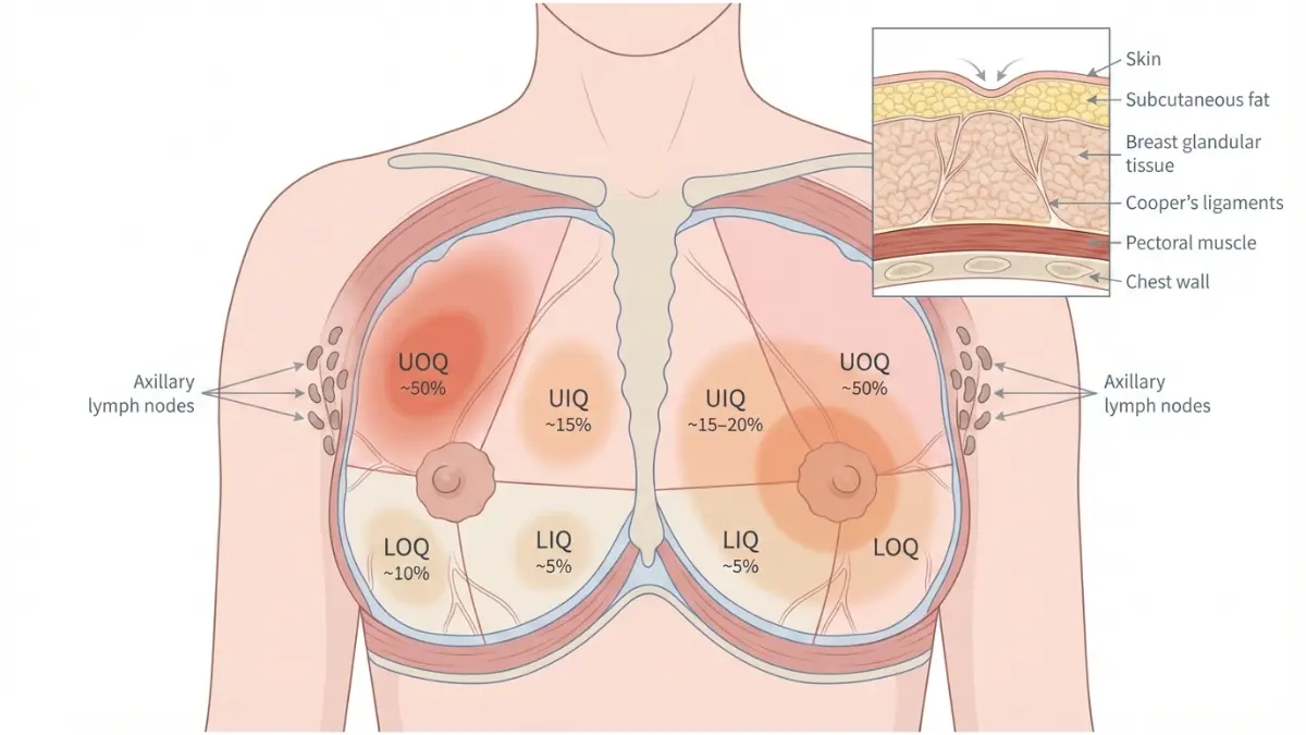
Breast lumps remain the most frequently reported initial breast cancer symptom, accounting for the majority of cancer diagnoses. Malignant breast lumps typically feel hard, irregular in shape, and fixed to surrounding tissue, unlike benign lumps that move freely and feel smooth. The upper outer quadrant of the breast (near the armpit) contains the highest concentration of breast tissue and represents the location where approximately 50% of breast cancers develop. Cancerous lumps rarely cause pain initially, which is why women often delay seeking evaluation.
💡 What This Means For You: Perform monthly self-examinations to establish your breast’s normal texture. If you detect any breast lump larger than a pea, schedule an appointment within one week. Use our Symptom Checker to document when you first noticed the change and accompanying symptoms before your medical visit.
2. Nipple Discharge: When to Worry
Nipple discharge that occurs without squeezing the nipple and affects only one breast warrants immediate medical evaluation. While discharge from both nipples often indicates benign hormonal changes, spontaneous unilateral discharge—especially if bloody, clear, or occurring in non-lactating women—can signal ductal carcinoma in situ (DCIS), an early form of breast cancer confined to milk ducts. Paget’s disease of the breast, which affects the nipple and surrounding areola, frequently presents with nipple discharge alongside crusting and scaling. According to research published in the National Institutes of Health database, 5-15% of women with pathologic nipple discharge receive a breast cancer diagnosis.
💡 What This Means For You: Document the discharge color, frequency, and whether it occurs spontaneously or only with pressure. Bloody or clear discharge from a single duct requires urgent evaluation. International oncologist Dr. Maria Gonzalez from Barcelona’s Vall d’Hebron Hospital emphasizes: “Any spontaneous nipple discharge in non-pregnant, non-breastfeeding women deserves investigation regardless of color.”
3. Breast Skin Changes and Dimpling
Breast dimpling, medically termed “peau d’orange” (French for “orange peel”), creates a dimpled, pitted appearance caused by cancer cells blocking lymphatic drainage in the skin. This breast skin change represents a serious warning sign that shouldn’t be ignored. Puckering occurs when tumors pull on Cooper’s ligaments (connective tissue supporting the breast), creating visible indentations. Unlike temporary dimpling from underwire bras or sleeping positions, cancerous dimpling persists and often worsens. Skin retraction—when the skin pulls inward—accompanies deeper tumors attached to chest wall muscles.
💡 What This Means For You: Stand before a mirror in good lighting with arms at your sides, then raised overhead. Look for any areas where skin appears tethered or dimpled. Photograph any changes to track progression. These breast abnormalities often indicate inflammatory breast cancer, a rapidly progressing form requiring immediate intervention.
4. Nipple Changes and Inversion
Nipple inversion occurs when the nipple turns inward instead of pointing outward. While some women have naturally inverted nipples since puberty, sudden nipple inversion or a change in nipple direction represents a significant breast cancer warning sign. Paget’s disease of the breast affects the nipple area and may cause the nipple to flatten, invert, or change angle. Tumors located directly beneath the nipple pull on milk ducts, causing retraction. Additional concerning nipple changes include scaling, crusting, flaking skin on the nipple or areola, redness, or the development of sores that don’t heal within two weeks—all potential indicators of breast cancer symptoms requiring evaluation.
💡 What This Means For You: Compare current photos of your breasts to photos from six months ago. If your previously normal nipple has inverted or shifted direction, schedule evaluation within one week. This change often accompanies an underlying mass.
5. Breast Swelling and Size Changes
Unilateral breast swelling—when one breast becomes noticeably larger, heavier, or swollen compared to the other—represents a critical early sign of breast cancer. Inflammatory breast cancer (IBC) causes rapid breast swelling, warmth, and heaviness within weeks or even days, making it one of the most aggressive forms. Unlike hormonal breast swelling that affects both breasts symmetrically before menstruation, cancerous swelling remains persistent and typically worsens.
The affected breast may feel firm, warm to touch, and appear red or discolored. Women sometimes mistake IBC symptoms for breast infection (mastitis), but mastitis typically includes fever and responds to antibiotics within 48-72 hours, while IBC symptoms do not improve with antibiotics.
💡 What This Means For You: Measure your breast size if you notice swelling. If one breast increases more than one cup size within a month, or if swelling accompanies redness and warmth, seek immediate medical evaluation. Monitor your overall health using our BMI Calculator as obesity increases breast cancer risk.
6. Armpit Lumps and Lymph Node Swelling
Swollen lymph nodes in the armpit (axillary lymph nodes) may indicate that breast cancer has spread beyond the breast tissue into the lymphatic system, a process called lymph node metastasis. The lymphatic system serves as the body’s drainage network, and breast tissue drains primarily into axillary lymph nodes located in the armpit. When cancer cells travel through lymph vessels, they often lodge in these nodes first. Cancerous lymph nodes typically feel hard, fixed (non-movable), and painless, unlike swollen nodes from infection which feel tender, soft, and mobile. Research published in the NIH database shows that the presence of metastatic breast cells in axillary lymph nodes significantly impacts staging and treatment decisions.
💡 What This Means For You: Palpate your armpits monthly while lying down with your arm raised. Normal lymph nodes feel soft and movable. Hard, fixed lumps larger than one centimeter require evaluation within one week. Dr. Kenji Tanaka from Tokyo’s Cancer Institute emphasizes: “Axillary lymphadenopathy deserves investigation even without detectable breast mass.”
7. Breast Redness and Inflammation
Breast redness (erythema) covering more than one-third of the breast surface represents a hallmark inflammatory breast cancer symptom. IBC presents with rapid onset of diffuse redness, warmth, edema, and peau d’orange skin changes caused by cancer cells blocking dermal lymphatic vessels. The affected breast may appear bruised, pink, red, or purple—particularly visible in lighter skin tones. Women with darker skin should look for areas that appear darker, warm to touch, or have texture changes, as breast redness may be less visible.
Unlike breast infections that develop suddenly with fever and flu-like symptoms, inflammatory breast cancer symptoms progress rapidly over weeks without systemic infection signs. This aggressive form accounts for 1-5% of all breast cancer cases but disproportionately affects younger women under 40.

💡 What This Means For You: If breast redness, warmth, and swelling don’t improve after 72 hours of antibiotics prescribed for suspected infection, insist on immediate mammography or ultrasound. IBC requires urgent diagnosis and treatment initiation.
8. Breast Pain and Tenderness Patterns
While most breast cancers don’t cause pain initially, persistent non-cyclical breast pain unrelated to menstrual cycles can occasionally signal malignancy. Cyclical breast pain that corresponds with hormonal fluctuations typically indicates benign conditions, but new, persistent, localized pain in one specific breast area deserves evaluation. Inflammatory breast cancer frequently causes breast tenderness along with other symptoms. Sharp, stabbing pain confined to one breast quadrant, especially when accompanied by a palpable mass, requires assessment.
Approximately 5-10% of breast cancer patients report breast pain as an initial symptom. Musculoskeletal chest pain often worsens with arm movement, while pain from breast tissue remains constant regardless of movement.
💡 What This Means For You: Track pain patterns in a journal for one month. Note timing relative to menstrual cycle, severity on a 1-10 scale, location, and aggravating factors. If pain persists beyond two cycles or intensifies, schedule evaluation. Meanwhile, assess your overall health metrics with our Body Fat Calculator since higher body fat percentage correlates with increased breast cancer risk.
9. Breast Texture Changes and Thickening
Breast tissue thickening feels different from a distinct lump—it’s a firm, dense area that lacks clear borders. Dense breast tissue on mammography can mask tumors, making detection more challenging and highlighting the importance of clinical breast examination. When you palpate your breasts, thickening feels like pressing on the heel of your hand rather than your palm. This breast texture change may indicate cancerous tissue infiltrating normal breast tissue.
Unlike premenstrual breast changes that affect both breasts diffusely and resolve after menstruation, cancerous thickening remains localized, persistent, and unilateral. Women with naturally dense breast tissue face 1.5 to 2 times higher breast cancer risk, making regular professional screening essential.
💡 What This Means For You: Establish your breast’s normal texture through monthly self-examination at the same point in your menstrual cycle (5-7 days after period starts). New thickening persisting more than one cycle requires evaluation. Track your health metrics using our Water Intake Calculator to support overall wellness during screening.
10. Breast Abnormalities in Shape
Sudden breast asymmetry or shape distortion can signal underlying breast cancer. While most women have slight natural breast asymmetry, rapid changes—such as one breast becoming noticeably larger, developing an unusual contour, or changing profile when viewed from the side—warrant investigation. Cancerous changes pull breast tissue inward, creating visible distortion. Breast contour changes appear most obvious when raising arms overhead or leaning forward.
Tumors attached to chest wall muscles cause visible retraction when contracting pectoral muscles (pressing palms together). Compare your breasts in multiple positions: arms at sides, arms overhead, hands on hips pressing inward, and leaning forward. Any new asymmetry in size, shape, or contour requires documentation and evaluation.
💡 What This Means For You: Photograph your breasts monthly from multiple angles to track subtle changes. Early detection through shape monitoring saves lives. International guidelines recommend clinical breast examination annually for women over 40 and those with elevated risk factors assessed through tools like our Genetic Risk Assessment Tool.
11. Areola Changes and Discoloration
The areola (pigmented skin surrounding the nipple) can develop concerning changes indicating Paget’s disease of the breast, a rare breast cancer affecting the nipple and areola. Warning signs include scaliness, crusting, flaking, thickening, redness, or oozing that persists beyond two weeks. Many women initially mistake these symptoms for eczema or dermatitis, but unlike these benign skin conditions, Paget’s disease doesn’t improve with topical steroids. The affected areola may appear redder, darker, or lighter than normal. Persistent itching, burning, or tingling sensations accompany visible changes. Approximately 1-3% of breast cancers are Paget’s disease, which usually coexists with underlying ductal carcinoma in situ or invasive breast cancer.
💡 What This Means For You: If areola changes don’t resolve within two weeks of over-the-counter eczema treatment, schedule evaluation. Request biopsy if skin changes persist—Paget’s disease requires definitive diagnosis. Dr. Sophie Laurent from Paris’s Institut Curie notes: “Persistent unilateral nipple eczema is Paget’s disease until proven otherwise.”
12. Persistent Breast Irritation
Ongoing breast irritation—including itching, tingling, burning sensations, or discomfort that persists for weeks—can indicate inflammatory breast cancer or other serious conditions. While occasional breast itching from dry skin, allergies, or ill-fitting bras is normal, persistent irritation localized to one breast requires attention. Inflammatory breast cancer causes itching due to lymphatic vessel blockage. Some women describe a “sunburned” feeling on breast skin. Irritation accompanied by visible changes (redness, swelling, texture changes) requires urgent evaluation. Even without visible changes, persistent discomfort lasting more than four weeks deserves assessment. Document symptom duration, severity, location, and any factors that improve or worsen symptoms.
💡 What This Means For You: If breast irritation persists beyond two weeks despite eliminating potential irritants (new detergents, fabrics, body products), schedule evaluation. Maintain optimal health during screening with our comprehensive Health Tips resources covering nutrition, exercise, and wellness strategies.
Key Takeaway: These twelve early signs of breast cancer represent the most common breast cancer symptoms reported by patients and documented in medical research. While many of these changes result from benign conditions, only medical evaluation can definitively distinguish normal variations from serious breast abnormalities. The National Cancer Institute emphasizes that early detection through symptom awareness and regular screening significantly improves breast cancer treatment outcomes.
Self-examination + Early Detection Methods
How to Perform Breast Cancer Self-Examination: Expert-Guided Steps
While clinical breast examinations and mammography remain the primary screening methods, breast self-awareness helps women recognize changes that warrant medical evaluation. Understanding your breast’s normal appearance and texture enables early identification of breast cancer symptoms.
Monthly Self-Examination Protocol
Breast self-examination should be performed monthly, ideally 5-7 days after menstruation begins when breast tissue is least tender and lumpy. The National Institutes of Health recommends a systematic approach using visual inspection and manual palpation to detect breast lumps, skin changes, or other abnormalities. Postmenopausal women should choose a consistent date each month.
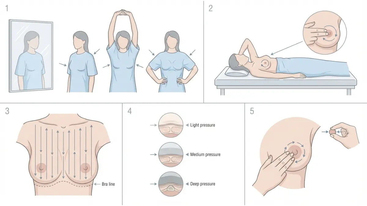
Visual Inspection (2-3 minutes):
- Stand shirtless before a well-lit mirror with arms at sides
- Examine for asymmetry, breast dimpling, nipple changes, skin discoloration, or visible lumps
- Raise arms overhead and observe for breast skin changes or contour distortions
- Place hands on hips, press inward to flex chest muscles, and check for retraction
- Lean forward to assess whether breasts hang evenly
Manual Palpation (5-7 minutes per breast):
- Lie flat with a pillow under your shoulder on the examination side
- Use the pads of your three middle fingers (not fingertips) in small circular motions
- Apply three pressure levels: light (skin surface), medium (mid-breast tissue), and deep (near chest wall)
- Follow vertical strip pattern from collarbone to bra line, armpit to center
- Examine the entire breast including armpit area where axillary lymph nodes reside
- Squeeze each nipple gently to check for nipple discharge
Women tracking their breast health can document findings using our Symptom Checker before medical appointments. Those with elevated risk factors should utilize our Genetic Risk Assessment Tool to understand when enhanced screening becomes necessary.
💡 What This Means For You: Breast cancer self-examination supplements but doesn’t replace professional screening. Report any persistent breast abnormalities—including breast lumps, breast thickening, nipple changes, or breast redness—to your healthcare provider within one week of detection.
Clinical Detection Methods Comparison
The U.S. Preventive Services Task Force recommends biennial mammography screening for women aged 40-74 at average risk, with the option for annual screening based on individual preference. Updated 2024 guidelines lowered the screening age from 50 to 40 years due to increasing breast cancer incidence in younger women.
Research published in the NIH database shows that combining mammography with ultrasound increases breast cancer detection rates by 97.3% compared to mammography alone in women with dense breast tissue. However, supplemental screening also increases false-positive rates and biopsies for benign conditions.
💡 What This Means For You: Women with dense breast tissue, family history, or genetic mutations benefit from enhanced screening beyond standard mammography. The American College of Radiology provides guidelines for supplemental screening based on risk factors. Discuss your breast cancer risk profile with your physician to determine optimal screening intervals. Track your overall health metrics with our BMI Calculator since obesity increases breast cancer risk and can affect mammography effectiveness.
Insurance Coverage Considerations:
- The Affordable Care Act mandates coverage for biennial mammography for women 40+ without copays
- Many states require insurance coverage for supplemental screening in women with dense breasts
- High-risk screening (MRI, genetic testing) requires medical necessity documentation
- Diagnostic mammograms (following abnormal findings) may have different coverage than screening mammograms
Digital Health Tools for Monitoring
Modern technology enhances breast cancer early detection through AI-powered risk assessment, symptom tracking applications, and genetic testing platforms. Women with BRCA1 or BRCA2 mutations face 45-65% lifetime breast cancer risk, making genetic risk stratification essential for screening decisions. Our Genetic Risk Assessment Tool evaluates personal and family history to identify women requiring enhanced surveillance protocols including annual breast MRI starting at age 25-30.
Digital symptom diaries enable women to document breast changes systematically—recording lump characteristics, nipple discharge color and frequency, breast pain patterns, and skin texture changes. Patient testimonial: “Using a symptom tracker helped me notice my nipple inversion was progressively worsening over three months, not just a temporary change. That documentation convinced my doctor to order immediate imaging, catching my ductal carcinoma in situ early.” — Maria S., 47, Atlanta
💡 What This Means For You: Leverage technology to enhance breast cancer awareness. Photograph your breasts monthly from multiple angles to track subtle changes in breast size, shape, and skin appearance. Maintain optimal wellness during screening with evidence-based lifestyle strategies from our Health Tips section covering nutrition, exercise, and weight management—factors that significantly influence breast cancer risk.
Risk Factors + Who’s At Highest Risk
Understanding Your Breast Cancer Risk: Factors You Can and Cannot Control
Breast cancer risk results from complex interactions between genetic predisposition, hormonal factors, and lifestyle choices. Women who inherit harmful BRCA1 or BRCA2 gene mutations face a 60% or greater lifetime risk of developing breast cancer, compared to approximately 13% in the general population. Understanding both modifiable and non-modifiable risk factors empowers women to make informed decisions about screening and prevention strategies.
Non-Modifiable Risk Factors
Age remains the strongest breast cancer risk factor, with incidence increasing significantly after age 50. According to the Centers for Disease Control and Prevention’s breast cancer surveillance data, women aged 50-74 represent the highest-risk age group for breast cancer diagnosis. Family history amplifies risk considerably—having a first-degree relative (mother, sister, or daughter) with breast cancer doubles a woman’s risk, while multiple affected relatives increase risk further.
Genetic mutations beyond BRCA1/BRCA2 include rare variants in PALB2 (conferring 2.3-fold risk), CHEK2 (approximately twofold risk), ATM, and TP53 genes that collectively account for 5-10% of hereditary breast cancers. Women should utilize our Genetic Risk Assessment Tool to evaluate whether genetic testing is appropriate based on family history patterns.
Race and ethnicity influence breast cancer patterns, with white women having slightly higher incidence than Black women, but Black women experiencing higher mortality rates due to more aggressive tumor subtypes and healthcare access disparities.
Modifiable Lifestyle Factors
The National Cancer Institute identifies several modifiable breast cancer risk factors that women can address through lifestyle changes. Physical inactivity increases postmenopausal breast cancer risk, while regular exercise (150 minutes of moderate activity weekly) reduces risk by 10-20%. Women can track their fitness progress using our Heart Rate Zone Calculator to optimize cardiovascular exercise intensity.
Obesity significantly elevates breast cancer risk, particularly after menopause, through multiple mechanisms including increased estrogen production by adipose tissue. Maintaining healthy body weight through balanced nutrition reduces risk substantially. Calculate your current status with our BMI Calculator and create personalized weight management strategies using our Weight Loss Calculator and Calorie Deficit Calculator.
Alcohol consumption demonstrates a clear dose-response relationship with breast cancer—even one drink daily increases risk by 7-10%, while 2-3 drinks daily raise risk approximately 20% compared to non-drinkers. Research in the NIH database confirms that every 10 grams of daily alcohol consumption (approximately one drink) correlates with a 10% breast cancer risk increase.
Postmenopausal hormone therapy (HRT), particularly estrogen-progestin combinations, increases breast density and breast cancer risk. Women considering HRT should discuss individual risk-benefit profiles with healthcare providers, especially those with dense breast tissue where the association between HRT and breast cancer appears strongest.
💡 What This Means For You: Focus on factors within your control. Dr. Amara Okafor from Nigeria’s Lagos University Teaching Hospital advises: “Women cannot change their genetics or age, but lifestyle modifications—maintaining healthy weight, limiting alcohol, and exercising regularly—reduce breast cancer risk by 20-40% in epidemiological studies.” Optimize your nutrition with our Macro Calculator to support healthy body composition.
High-Risk Groups Requiring Enhanced Screening
Women at substantially elevated risk require intensified surveillance starting earlier than average-risk populations, typically beginning at age 30 with annual mammography plus breast MRI. High-risk categories include:
- BRCA1/BRCA2 mutation carriers and their first-degree untested relatives
- Lifetime risk ≥20-25% calculated by validated risk models
- Personal history of chest radiation between ages 10-30 (Hodgkin lymphoma survivors)
- Previous diagnosis of atypical hyperplasia, lobular carcinoma in situ, or ductal carcinoma in situ
Enhanced screening protocols significantly improve early detection of breast cancer symptoms in high-risk women, enabling treatment when cancers are smaller and more treatable.
When To See A Doctor + Diagnosis Process
Taking Action: When to Seek Medical Evaluation for Breast Cancer Symptoms
Recognizing when breast cancer symptoms require urgent medical attention can dramatically impact treatment outcomes. Any new breast lump, persistent nipple discharge, or inflammatory changes warrant evaluation within one week, as early breast cancer diagnosis significantly improves survival rates.
Red Flag Symptoms Requiring Immediate Attention
Seek medical evaluation within 24-48 hours if you experience:
- Rapidly progressing breast redness, warmth, and swelling—potential inflammatory breast cancer
- Bloody or spontaneous nipple discharge from one breast
- Breast dimpling or “orange peel” skin texture appearing suddenly
- New breast lump with accompanying skin changes or nipple retraction
- Persistent breast pain with palpable mass or skin changes
- Armpit lumps that feel hard, fixed, and painless
Patient story: Maria’s decisive action saved her life. When she noticed her left breast becoming increasingly red and tender over five days—initially dismissed as mastitis—her condition didn’t improve with antibiotics. Maria insisted on immediate imaging after 72 hours. Diagnostic tests confirmed inflammatory breast cancer symptoms, and urgent treatment began within one week. “Don’t wait if something feels wrong,” Maria emphasizes. “Inflammatory breast cancer progresses rapidly, and every day matters.”
Use our Symptom Checker to document symptom onset, progression, and characteristics before your appointment, providing your physician with detailed information for accurate breast cancer diagnosis.
The Diagnostic Journey: What to Expect
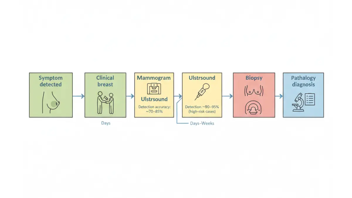
The breast cancer diagnosis process typically begins with clinical breast examination followed by imaging studies, progressing to biopsy when suspicious findings emerge. Understanding this timeline reduces anxiety and helps women advocate for timely evaluation.
Typical Diagnostic Timeline:
| Stage | Procedure | Timeline | Purpose |
|---|---|---|---|
| Initial Visit | Clinical breast exam, history | Day 1 | Assess breast abnormalities, risk factors |
| Imaging | Diagnostic mammogram, ultrasound | 1-7 days | Characterize mass location, size |
| Additional Imaging | Breast MRI (if needed) | 1-2 weeks | Evaluate extent, multifocal disease |
| Biopsy | Core needle or fine-needle aspiration | 1-2 weeks | Obtain tissue for definitive diagnosis |
| Pathology Results | Microscopic tissue analysis | 3-7 days | Confirm breast cancer, determine subtype |
| Staging | Additional tests if cancer confirmed | 1-2 weeks | Determine clinical prognostic stage |
Research published in the NIH database shows that diagnostic mammography combined with breast ultrasound achieves 95% accuracy in evaluating clinical breast concerns in women under 50 years. Biopsy remains the only definitive method to diagnose breast cancer—imaging findings alone cannot distinguish benign from malignant masses with certainty.
💡 What This Means For You: Most breast cancer diagnoses occur within 2-4 weeks from initial evaluation to biopsy results. If your healthcare provider recommends “watchful waiting” for a new, persistent breast lump or concerning breast skin changes, request immediate imaging. International oncologist Dr. Hiroshi Yamamoto from Japan’s National Cancer Center advises: “New breast abnormalities in women over 30 deserve imaging evaluation, not observation alone.” Maintain optimal health during diagnostic evaluation with our BMI Calculator and Body Fat Calculator to track wellness metrics.
Questions to Ask Your Doctor
Before diagnostic mammogram:
- Do I need diagnostic or screening mammogram?
- Should ultrasound be performed simultaneously?
- Will my breast density affect mammogram accuracy?
If biopsy is recommended:
- What type of biopsy do you recommend (fine-needle, core-needle, surgical)?
- How accurate is this biopsy method?
- When will I receive results?
- What happens if biopsy shows breast cancer?
If breast cancer is diagnosed:
- What is my breast cancer stage and subtype?
- What additional tests determine treatment planning?
- What are my treatment options?
- Should I seek second opinion at comprehensive cancer center?
Download this question checklist by bookmarking our Health Tips section for comprehensive patient resources supporting informed decision-making throughout your diagnostic journey.
Lifestyle, Prevention + Support Resources
Beyond Detection: Prevention Strategies and Support Systems
Evidence-Based Prevention Strategies
Research published by the CDC demonstrates that physical activity significantly reduces breast cancer risk, with women engaging in regular vigorous activity experiencing approximately 40% lower risk compared to sedentary women. Adults should complete 150-300 minutes of moderate-intensity exercise weekly—optimize your cardiovascular workouts using our Heart Rate Zone Calculator to ensure proper training intensity. NIH studies confirm that Mediterranean dietary patterns rich in vegetables, fruits, whole grains, and lean proteins reduce breast cancer recurrence and improve survival outcomes in women previously diagnosed.
Maintain healthy body weight through balanced nutrition—our Macro Calculator provides personalized protein, carbohydrate, and fat targets supporting optimal body composition. Women struggling with weight management benefit from our Weight Loss Calculator combined with Calorie Deficit Calculator for science-based weight reduction strategies. Calculate your baseline metabolic needs with our BMR Calculator and track hydration using our Water Intake Calculator since adequate hydration supports metabolic health.
Support Resources and Clinical Trial Opportunities
The National Cancer Institute connects breast cancer patients with ongoing clinical trials investigating innovative treatments including immunotherapy, targeted therapies, and precision medicine approaches. Participation in NCI-supported breast cancer research advances scientific understanding while potentially providing access to cutting-edge treatments.
Comprehensive patient support includes:
- Cancer support groups connecting patients with survivors who understand the emotional journey
- Mental health counseling addressing anxiety, depression, and treatment-related stress
- Financial assistance programs covering treatment costs, medications, and transportation
- Nutritional counseling optimizing diet during treatment—track your protein needs with our Protein Intake Calculator to maintain muscle mass during chemotherapy
Take Action Today
Empower yourself through awareness and action. Bookmark our Symptom Checker for monthly breast self-examinations and utilize our Genetic Risk Assessment Tool to evaluate your personal breast cancer risk. Share these early signs of breast cancer with family members and friends—early detection through symptom recognition saves lives. Schedule your mammogram screening if you’re 40 or older, and explore our comprehensive Health Tips section for evidence-based wellness strategies supporting breast cancer prevention.
Frequently Asked Questions About Breast Cancer Symptoms
1. What are the first signs of breast cancer?
The first signs of breast cancer typically include a painless lump in the breast or underarm, nipple discharge (bloody or clear), skin dimpling or “orange peel” texture, nipple inversion, breast swelling, redness, and persistent changes in breast size or shape. Most women first notice a hard, irregular lump that doesn’t move freely.
2. Are all breast lumps cancerous?
No, approximately 80% of breast lumps are benign (non-cancerous) and caused by cysts, fibroadenomas, or hormonal changes. However, any new breast lump requires medical evaluation within one week since only imaging and biopsy can definitively distinguish benign from malignant lumps.
3. Does breast cancer always cause pain?
No, most breast cancers are painless initially, which is why women often delay seeking evaluation. However, inflammatory breast cancer and some advanced tumors can cause persistent, non-cyclical breast pain accompanied by other warning signs like redness, swelling, or skin changes.
4. How often should I perform breast self-examination?
Perform breast self-examination monthly, ideally 5-7 days after menstruation begins when breast tissue is least tender. Postmenopausal women should choose a consistent date each month to check for lumps, skin changes, nipple discharge, or other abnormalities.
5. When should I get my first mammogram?
The U.S. Preventive Services Task Force recommends starting mammography screening at age 40 for women at average risk, with biennial screening through age 74. Women with elevated risk factors (family history, genetic mutations, dense breasts) should begin screening earlier, typically at age 30.
6. What does inflammatory breast cancer look like?
Inflammatory breast cancer presents with rapid-onset breast redness, warmth, swelling, and “orange peel” skin texture covering at least one-third of the breast. The affected breast may appear bruised, feel heavy, and show dimpling without a distinct palpable lump—requiring immediate medical evaluation.
7. Can younger women get breast cancer?
Yes, while breast cancer risk increases significantly after age 50, approximately 11% of all cases occur in women under 45 years. Younger women with family history, BRCA mutations, or dense breast tissue face higher risk and should discuss early screening with their physicians.
8. What is nipple discharge and when is it concerning?
Nipple discharge is fluid leaking from the nipple. Spontaneous discharge from one breast that is bloody, clear, or occurs without squeezing warrants immediate evaluation, as it may indicate ductal carcinoma or Paget’s disease. Milky discharge from both breasts is usually benign.
9. How accurate is mammography for detecting breast cancer?
Digital mammography has 54-85% sensitivity depending on breast density, with 3D mammography (digital breast tomosynthesis) showing 15% higher detection rates. Dense breast tissue reduces mammography accuracy, making supplemental ultrasound or MRI necessary for complete evaluation in high-risk women.
10. Can lifestyle changes reduce breast cancer risk?
Yes, maintaining healthy weight, exercising 150+ minutes weekly, limiting alcohol to less than one drink daily, and avoiding postmenopausal hormone therapy reduce breast cancer risk by 20-40%. While you cannot change genetic factors, lifestyle modifications significantly impact overall risk.
11. What should I do if I find a breast lump?
Schedule an appointment with your healthcare provider within one week if you detect a new breast lump. Document when you first noticed it, its characteristics (size, texture, mobility), and any accompanying symptoms using a symptom tracker. Do not panic—most lumps are benign, but timely evaluation ensures early diagnosis if cancer is present.
About this content
How this article was put together: researched from recognised health sources, drafted with the help of AI tools, and edited by hand, with sources linked throughout.

Sameer Patel is the founder and editor of My Medicine Advisor. He is not a doctor or medical professional — before starting this site he worked in banking,…
Medical disclaimer
The content on MyMedicineAdvisor is provided for general informational and educational purposes only and is not a substitute for professional medical advice, diagnosis, or treatment. Health information on this website should not be used to diagnose, treat, cure, or prevent any condition without guidance from a qualified healthcare professional. Always seek the advice of your doctor, physician, or another licensed healthcare provider with any questions you may have regarding a medical condition, symptoms, medications, or treatment decisions.