On This Page – Quick Medical Summary
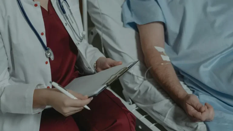
When 61-year-old David Chen received his CT scan report showing a 1.8 cm shadow near his right upper lobe, his pulmonologist said four words he didn’t expect: “You need a lung biopsy.”
His first question wasn’t about the nodule. It was: “What exactly is going to happen to me?”
A lung biopsy is a minimally invasive medical procedure in which a small sample of lung tissue is removed and examined under a microscope to confirm or rule out cancer. It is the only definitive method to diagnose lung cancer — imaging scans alone cannot confirm malignancy. For most patients, it is a same-day procedure with local or sedation anesthesia, and recovery takes just days.
This guide covers every type of lung biopsy, real risk rates, a step-by-step walkthrough of the procedure, and what the 2026 breakthroughs in robotic and liquid biopsy mean for patients today.
Disclaimer: This article is for educational purposes only and does not constitute medical advice. Always consult a qualified physician for diagnosis and treatment decisions.
What Is a Lung Biopsy and Why Do Doctors Order One?
The Only Test That Confirms Lung Cancer
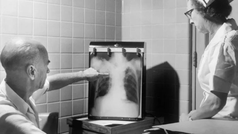
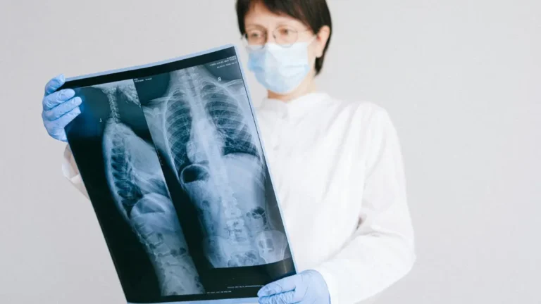
A CT scan, PET scan, or chest X-ray can detect abnormalities in your lungs. But none of them can tell a pathologist what type of cells are growing there. That is the job of a lung biopsy.
During a lung biopsy, a physician removes a small piece of tissue from the suspicious area. A pathologist then examines it under a microscope to determine whether cancer cells are present — and if so, which type. According to the National Cancer Institute, accurate tissue diagnosis is essential before any lung cancer treatment can begin.
Key fact: Only 3 to 4 in every 100 lung nodules detected on CT scans are cancerous. A lung biopsy procedure is what separates a confirmed diagnosis from an unfounded fear.
If you’ve already had a CT scan and are unsure what your results mean, our guide on lung cancer CT scan results explains exactly what each finding indicates.
When Does a Doctor Recommend a Lung Biopsy?
Your physician may recommend a lung tissue biopsy in the following situations:
- A suspicious nodule or mass found on CT, PET, or MRI imaging
- A lung abnormality that has grown over time on serial imaging
- Unexplained fluid around the lungs (pleural effusion)
- Unresolved infection or inflammation that doesn’t respond to antibiotics
- Known cancer elsewhere in the body with possible lung spread
- Confirming lung cancer type and stage before starting treatment
Understanding how lung cancer is diagnosed involves multiple steps — a lung biopsy is typically the definitive final step in that diagnostic pathway.
If you’re unsure whether your symptoms warrant further evaluation, use our free Symptom Checker to track and assess your concerns before your appointment.
The 5 Types of Lung Biopsy — Full Comparison
Not all lung biopsies are the same. The type recommended depends on the nodule’s location, size, your overall health, and your care team’s expertise. Here is the complete breakdown.
Master Comparison Table: All 5 Lung Biopsy Types
| Biopsy Type | Invasiveness | Anesthesia | Duration | Typical Recovery | Best Used For |
|---|---|---|---|---|---|
| CT-Guided Needle (Percutaneous) | Minimal | Local | 30–45 min | Same day | Peripheral nodules near chest wall |
| Bronchoscopy (Transbronchial) | Minimal | Sedation | 30–60 min | Same day | Central airway lesions |
| EBUS-TBNA | Minimal | Sedation | 30–60 min | Same day | Mediastinal lymph node staging |
| VATS (Thoracoscopic) | Moderate | General | 1–2 hrs | 2–5 days hospital | Hard-to-reach peripheral lesions |
| Open Lung Biopsy | Highest | General | 2–3 hrs | 5–7 days hospital | Last resort only |
Adapted from Wikimedia Commons Biopsie_Lunge_Computertomographie_BC.png, licensed under CC BY-SA 4.0.
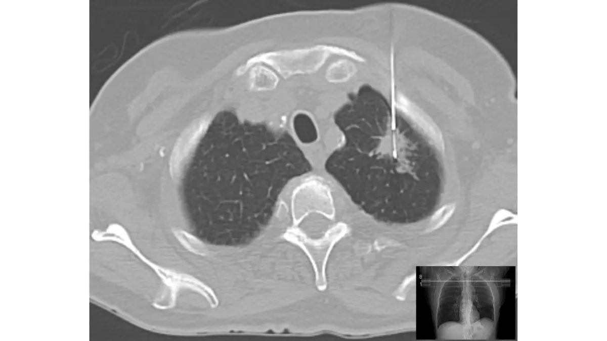
CT-Guided Needle Biopsy (Percutaneous Lung Biopsy)
This is the most commonly performed lung biopsy in the United States. A radiologist inserts a thin needle through the chest wall, guided in real time by CT imaging, to reach the nodule.
It uses only local anesthesia — you are awake but numb. Most patients go home the same day. It is best suited for nodules located near the outer edges of the lung. The American Lung Association confirms this is the most widely used approach for peripheral lung lesions.
Bronchoscopy Biopsy (Transbronchial Biopsy)
A bronchoscope — a thin, flexible tube with a tiny camera — is passed through the nose or mouth, down the windpipe, and into the airways. Forceps at the tip collect tissue from the target area.
You receive sedation (not full general anesthesia). This lung biopsy procedure works best when the nodule is near a central airway. Duration: 30 to 60 minutes.
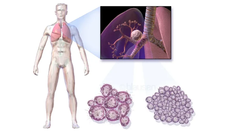
EBUS-TBNA (Endobronchial Ultrasound)
This is one of the most underexplained procedures in all of pulmonary medicine — and it deserves a plain-language explanation.
EBUS combines a bronchoscope with an ultrasound probe that can see through the airway walls. It allows physicians to biopsy lymph nodes in the chest without a single skin incision. It is the gold standard for staging whether lung cancer has spread to mediastinal lymph nodes — a critical piece of information that determines your treatment plan.
According to Johns Hopkins Medicine, EBUS-TBNA is particularly valuable because it can simultaneously diagnose and stage lung cancer in one procedure.
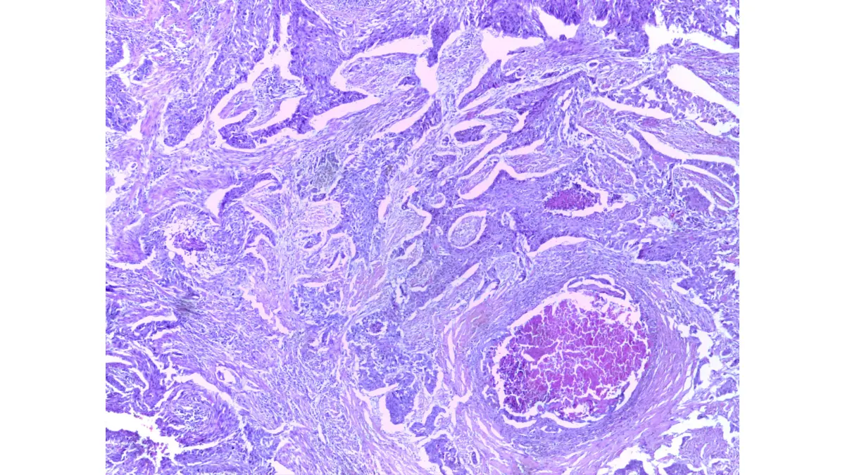
Adapted from Wikimedia Commons Squamous_Cell_Carcinoma_Lung_4x.jpg, licensed under CC BY-SA 4.0.
VATS (Video-Assisted Thoracoscopic Surgery)
A surgeon makes three small incisions between the ribs and inserts a thoracoscope with a camera. Instruments remove tissue through the other incisions. You are under general anesthesia.
VATS is more invasive than needle or bronchoscopic biopsy but allows the surgeon to view and sample areas that a needle cannot safely reach. Most patients stay 2–5 days post-procedure.
Open Lung Biopsy
This is the most invasive option — a full incision in the chest wall under general anesthesia. It is only recommended when all other biopsy methods have failed to produce a diagnosis. Open biopsies are rare in 2026 given the advancement of minimally invasive techniques.
To understand how biopsy type affects the staging of your cancer, read our detailed guide on lung cancer stages explained.
Step-by-Step — Exactly What Happens During a Lung Biopsy
Before Your Lung Biopsy (Days 1–3 Prior)
Your care team will prepare you thoroughly. Here is what to expect:
- Blood tests to assess clotting function and kidney health
- Pre-procedure imaging (CT scan) to map the target site precisely
- Medication review: Stop blood thinners such as aspirin, warfarin, or clopidogrel as directed — these raise lung biopsy bleeding risk
- Fasting: No food or drink for 4–6 hours before the procedure
- Consent form: Read carefully and ask every question you have
Important: Tell your care team about all supplements, including fish oil, vitamin E, and herbal products. These can increase bleeding risk just as pharmaceutical blood thinners do.
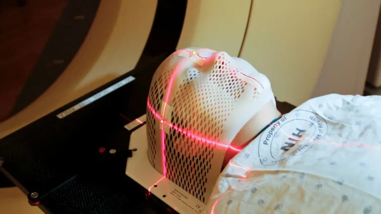
During the Procedure — Minute by Minute (Needle Biopsy Example)
Here is what a standard CT-guided lung biopsy looks like:
- You change into a hospital gown and lie on the CT table
- An IV line is placed in your arm or hand
- CT imaging confirms the exact biopsy site on your chest
- The skin is cleaned with antiseptic solution
- Local anesthetic is injected — you feel a brief sting, then numbness
- The biopsy needle is advanced toward the nodule under CT guidance
- You are asked to hold your breath briefly during needle insertion
- Tissue is collected — you may feel mild pressure but no sharp pain
- The needle is removed and gentle pressure is applied
- An immediate chest X-ray confirms your lung has not collapsed
Total time: 30 to 45 minutes for most patients.
Anxiety note: The majority of patients describe a CT-guided lung needle biopsy as far less painful or frightening than they anticipated. The preparation phase often causes more anxiety than the procedure itself.
For a bronchoscopy biopsy, a throat-numbing spray is applied first. The bronchoscope passes through your airway while you are sedated — you will not be able to speak or swallow during this time, but you will not feel pain.
The Cleveland Clinic’s lung biopsy procedure guide confirms that for most types, patients are monitored 2–4 hours after the procedure before discharge.
Lung Biopsy Risks, Complications & the Real Safety Numbers
Most Common Complications — With Actual Rates
Competitors list risks vaguely. Here are the actual complication rates from published clinical data:
| Complication | How Often | Severity | Typical Outcome |
|---|---|---|---|
| Pneumothorax (partial lung collapse) | 15–25% (CT-guided needle) | Mild in most cases | Self-resolves in hours; chest tube needed in ~5% |
| Minor bleeding / blood-tinged cough | 5–10% | Mild | Resolves within 5–7 days |
| Significant bleeding | <2% | Moderate | Managed medically |
| Infection | <1% | Mild–moderate | Treated with antibiotics |
| Air embolism | <0.1% | Potentially serious | Treatable with oxygen |
| Tumor seeding | <0.01% (extremely rare) | Varies | Addressed with targeted radiation if occurs |
Data sourced from StatPearls/NCBI — Percutaneous Lung Lesion Biopsy, updated 2025.
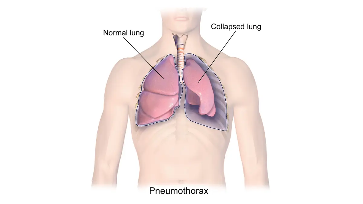
Adapted from Wikimedia Commons Blausen_0742_Pneumothorax.png, licensed under CC BY 3.0.
Does a Lung Biopsy Spread Cancer?
This is the most-searched fear around any lung cancer biopsy, and it deserves a direct, evidence-based answer.
Tumor seeding — where a small number of cancer cells track along the needle path — is a real but extraordinarily rare event occurring in fewer than 1 in 10,000 cases. The American Cancer Society confirms that the diagnostic benefit of a biopsy overwhelmingly outweighs this theoretical risk in virtually every clinical scenario.
Modern CT-guided techniques, coaxial needle systems, and immediate post-biopsy imaging have reduced even this already-rare risk further in 2026.
Who Faces a Higher Complication Risk?
Your physician will assess your individual profile. Higher-risk patients include those with:
- Severe COPD or emphysema — higher pneumothorax risk post-biopsy
- Blood clotting disorders — increased bleeding risk
- Single functioning lung — extra caution warranted
- Nodules located near major blood vessels — requires highly experienced interventional radiologist
- Advanced age with multiple comorbidities — assessed case by case
Key takeaway: Your physician does not recommend a lung biopsy without first evaluating whether the specific benefits outweigh your personal risk profile. Always ask your doctor which complications are most relevant to your situation.
To understand your genetic factors and hereditary cancer risk, our free Genetic Risk Assessment Tool can help you map your family history before your consultation.
Recovery & Results — Your Complete Post-Biopsy Timeline
Day-by-Day Recovery Guide
| Timeframe | CT-Guided Needle | Bronchoscopy | VATS |
|---|---|---|---|
| Day 0 | Home after 2–4 hr monitoring; post-biopsy chest X-ray clear | Home after 2 hr; mild sore throat normal | Hospital overnight; pain managed with IV medication |
| Days 1–2 | Rest; avoid lifting >5 lbs; no air travel or scuba diving | Light activity; throat soreness resolves | Walking encouraged; oral pain medication |
| Days 3–5 | Most patients return to desk work | Full normal activity | Gradual activity increase; drain removed if placed |
| Days 5–7 | Full recovery in most cases | — | Most patients discharged; return visit scheduled |
| Day 7+ | — | — | Resume light activity; no strenuous exercise for 2 weeks |
Do not fly or scuba dive after a lung biopsy. Changes in air pressure can trigger a pneumothorax even after an apparently uncomplicated procedure. Ask your physician specifically when air travel is safe for you.
When Will You Get Your Lung Biopsy Results?
- Standard pathology: 3–7 business days
- Molecular and biomarker testing (for targeted therapy eligibility): 2–3 weeks
- Complex cases (rare cancer types): Up to 4 weeks
Do not assume no news is good news. Follow up proactively with your care team if you haven’t received results within 10 business days.
For a complete guide on reading and understanding your pathology report, our article on biopsy results timeline and report guide walks through every section of the report in plain language.
Understanding Your Lung Biopsy Results
Positive result (cancer confirmed):
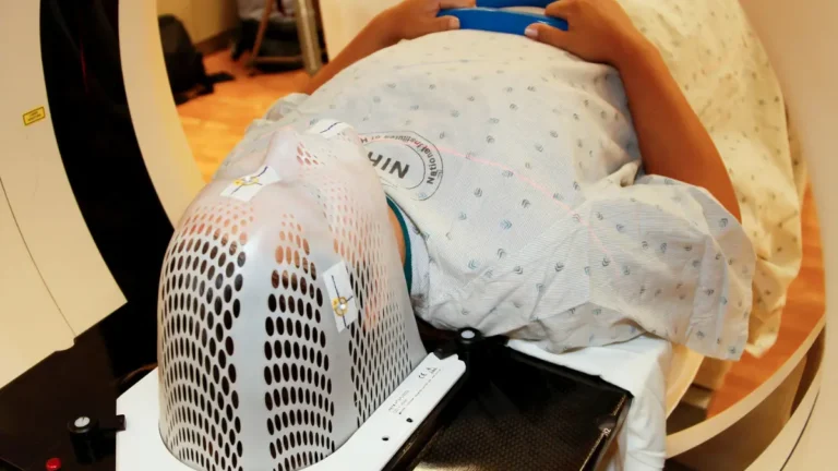
- Your physician will order staging tests: PET scan, brain MRI, bone scan
- Treatment planning begins — surgery, chemotherapy, radiation, targeted therapy, or immunotherapy depending on cancer type and stage
- See our guide on lung cancer FDA-approved drugs and treatments in 2026
Negative result (no cancer found):
- Does not always mean cancer is absent — it means no cancer was found in the sample collected
- If clinical suspicion remains high, your physician may recommend a repeat biopsy or surgical excision
Inconclusive result:
- Occurs when insufficient tissue was obtained or the sample was non-diagnostic
- A repeat lung biopsy or surgical approach (VATS) is typically recommended
Biomarker testing note: In 2026, most confirmed lung cancers undergo comprehensive molecular profiling — including EGFR, ALK, ROS1, KRAS, PD-L1, and BRAF mutations. These results determine eligibility for targeted therapies and immunotherapy. This testing is done on the same biopsy tissue and is standard of care.
To understand survival outcomes after a confirmed diagnosis, our evidence-based guide on stage 1 lung cancer survival rates and lung cancer statistics and survival rates provide detailed data by stage.
For help decoding your full pathology report after biopsy, our pathology result decoded guide breaks down every term physicians use.
2026 Breakthrough — Robotic, AI-Guided & Liquid Lung Biopsies
This is the section that no competitor has yet published in a patient-facing format. These are not experimental concepts — they are active clinical realities transforming how lung biopsies are performed in 2026.
Robotic Bronchoscopy — Biopsy Without a Chest Wall Incision
Ion Robotic Bronchoscopy uses a catheter guided by a robotic arm to navigate deep into the airways like a GPS system. It can reach peripheral lung nodules that were previously inaccessible via conventional bronchoscopy — without a single incision in the chest wall.
Key advantages:
- Same-day discharge in most cases
- No pneumothorax risk from chest wall puncture
- Suitable for nodules as small as 5–8 mm
- AI-assisted navigation maps the airway in 3D before the procedure begins
As Hackensack Meridian Health reported in January 2026, robotic bronchoscopy allows thoracic surgeons to both localize and biopsy small lung nodules without any external incisions — with patients going home the same day.
The technology is now expanding beyond its initial US centers into major academic hospitals in the UK, Canada, and Australia.
AI-Guided Biopsy Navigation — GPS for Your Lungs
Artificial intelligence is now embedded into CT-guided biopsy planning systems. Before the needle ever touches the skin, AI software:
- Identifies the safest needle trajectory to the nodule
- Maps nearby blood vessels to avoid
- Calculates the optimal entry angle to minimize pneumothorax risk
- Flags nodules on CT that radiologists may have initially missed
“AI doesn’t replace your surgeon — it gives them a GPS for your lungs.”
This technology is reducing the rate of inconclusive biopsies by improving targeting precision, particularly for sub-centimeter nodules.
Liquid Biopsy — A Blood Test That Reads Your Tumor
A liquid biopsy for lung cancer analyzes circulating tumor DNA (ctDNA) — tiny fragments of cancer DNA shed by tumors into the bloodstream. It requires only a standard blood draw.
Current status in 2026:
| Use Case | Standard of Care? |
|---|---|
| First-line lung cancer diagnosis | Not yet — tissue biopsy remains required |
| Monitoring treatment response | ✅ Yes — widely used |
| Detecting early recurrence | ✅ Yes — expanding rapidly |
| Identifying resistance mutations | ✅ Yes — standard in advanced NSCLC |
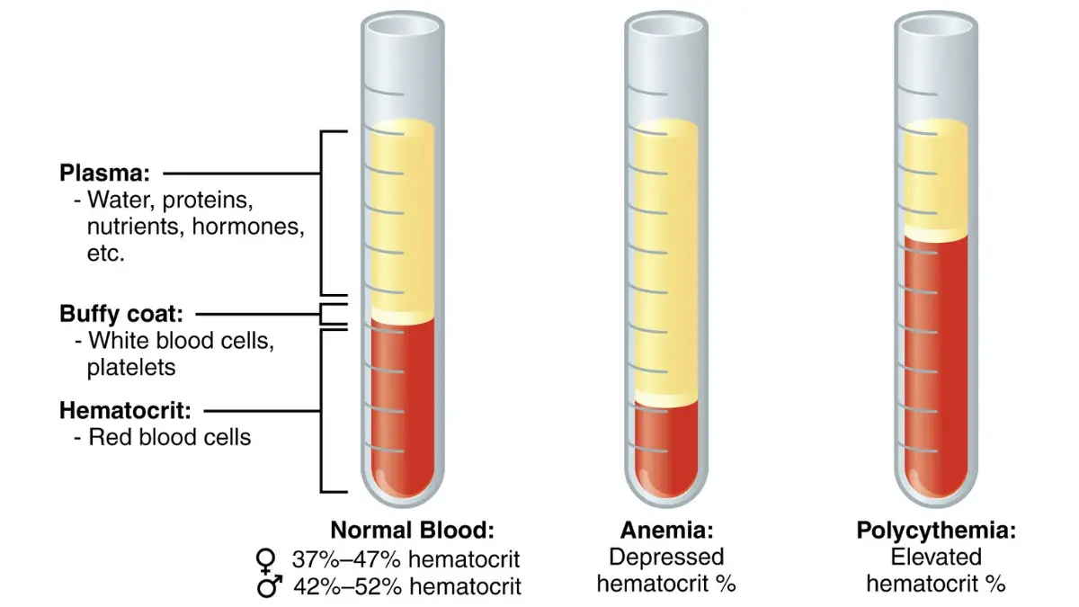
Adapted from OpenStax Anatomy and Physiology 2e, Chapter 18.1 An Overview of Blood, licensed under CC BY 4.0.
The NCI’s liquid biopsy research program confirms that liquid biopsy is a rapidly advancing field, with multiple FDA-authorized platforms now available for molecular monitoring.
Companies like Foundation Medicine and GRAIL are actively expanding liquid biopsy eligibility through ongoing trials. Within the next 2–3 years, liquid biopsy may partially replace repeat tissue biopsies for patients already under treatment.
FAQs: Lung Biopsy Questions Answered
1. How long does a lung biopsy take?
A needle or bronchoscopy biopsy takes 30–60 minutes. VATS takes 1–2 hours under general anesthesia.
2. Is a lung biopsy painful?
Most patients feel pressure but not pain. Local anesthesia or sedation is used for all types. Post-procedure soreness is mild and typically resolves within 48 hours.
3. Can a lung biopsy spread cancer?
Tumor seeding — cancer spreading along the biopsy needle track — occurs in fewer than 1 in 10,000 cases. The American Cancer Society confirms benefits far outweigh this risk for virtually all patients.
4. How long until lung biopsy results are ready?
Standard pathology: 3–7 business days. Molecular/biomarker testing: up to 3 weeks.
5. What does a positive lung biopsy result mean?
Cancer cells were confirmed in the tissue sample. Staging tests (PET, MRI) and treatment planning begin immediately.
6. Can I go home the same day?
Yes — for CT-guided needle biopsy and bronchoscopy. VATS requires 2–5 days in hospital. Open biopsy requires 5–7 days.
7. What is the most common complication?
Pneumothorax (partial lung collapse), occurring in 15–25% of CT-guided needle biopsies. Most cases resolve on their own within hours and require no intervention.
8. What is EBUS-TBNA?
A bronchoscopy technique combining ultrasound to biopsy mediastinal lymph nodes without any skin incision. It simultaneously diagnoses and stages lung cancer in one procedure.
9. Is a CT-guided biopsy the same as a needle biopsy?
Yes — CT-guided refers to the imaging used to direct the needle. The procedure is also called a percutaneous or transthoracic needle biopsy.
10. What happens if my lung biopsy is inconclusive?
Insufficient tissue or a non-diagnostic result means a repeat biopsy or VATS surgical biopsy will likely be recommended by your care team.
11. What is a liquid biopsy for lung cancer?
A blood test analyzing circulating tumor DNA. In 2026, it is used for monitoring treatment response and detecting recurrence — not yet as first-line diagnosis.
Final Summary: What Every Patient Should Know
A lung biopsy is the most accurate diagnostic tool available for lung cancer — with 95%+ diagnostic accuracy when performed by experienced specialists. It is not the frightening ordeal most patients fear. The majority of procedures are minimally invasive, same-day, and well-tolerated.
The 2026 landscape has fundamentally changed what is possible. Robotic bronchoscopy eliminates chest wall puncture entirely for many patients. AI-guided systems make needle biopsies safer and more precise. Liquid biopsy is transforming how physicians monitor treatment without repeat procedures.
What to do next:
- If you’ve been referred for a lung biopsy, ask your physician which type is recommended and why
- Request comprehensive biomarker and molecular testing be run on your biopsy sample — it is standard of care for NSCLC in 2026
- Read our full guide on what is lung cancer to understand your complete diagnostic and treatment pathway
Sources & References:
- National Cancer Institute — Lung Cancer
- American Lung Association — Lung Biopsy
- Cleveland Clinic — Lung Biopsy Procedure Guide
- Johns Hopkins Medicine — Lung Biopsy
- American Cancer Society — What Is a Biopsy?
- StatPearls/NCBI — Percutaneous Lung Lesion Biopsy
- Hackensack Meridian Health — Robotic Bronchoscopy 2026
- NCI — Liquid Biopsy Research
This article is for educational purposes only and does not constitute medical advice. Always consult a qualified physician for personalized diagnosis and treatment decisions.
About this content
This medical content is prepared through a structured publishing workflow with expert writing, clinical review and editorial quality checks.

Board Certifications: Internal Medicine (2005); Medical Oncology (2008); Hematology (2009) Experience: 20 years | Location: Houston, Texas Education: BS Biology, Duke University (1999); MD, Baylor College of Medicine…

Board Certifications: Anatomic Pathology (2006); Clinical Pathology (2006); Molecular Genetic Pathology (2010) Experience: 19 years | Location: Baltimore, Maryland Education: BS Biochemistry, Johns Hopkins University (1998); MD, Johns…

Board Certifications: Diagnostic Radiology (2009); Neuroradiology (2011); Breast Imaging (2013) Experience: 16 years | Location: Rochester, Minnesota Education: BS Physics, MIT (2000); MD, Mayo Clinic Alix School of…
Medical disclaimer
The content on MyMedicineAdvisor is provided for general informational and educational purposes only and is not a substitute for professional medical advice, diagnosis, or treatment. Health information on this website should not be used to diagnose, treat, cure, or prevent any condition without guidance from a qualified healthcare professional. Always seek the advice of your doctor, physician, or another licensed healthcare provider with any questions you may have regarding a medical condition, symptoms, medications, or treatment decisions.