On This Page – Quick Medical Summary
When Rachel, 31, was diagnosed with Stage II melanoma on her upper back in 2024, her dermatologist’s first question was: “Did you ever use tanning beds?” She had — just 12 sessions over three years in college. Tanning beds directly increase melanoma risk by up to 75%, according to the World Health Organization. If you’ve used them — even once — this research-backed guide covers exactly what that means for you today.
⚠️ This article is for educational purposes only and does not constitute medical advice. Always consult a licensed healthcare provider for personal medical decisions.
What the Science Actually Says About Tanning Beds and Melanoma Risk
IARC’s Classification: Why Tanning Beds Belong in the Same Category as Tobacco
The International Agency for Research on Cancer (IARC), part of the World Health Organization, classified tanning beds as Group 1 carcinogens in 2009 — reaffirmed through updated reviews in 2024. Group 1 is the highest-risk cancer classification. It places tanning beds alongside tobacco smoke, asbestos, and benzene.
This is not a precautionary label. It is settled, consensus-level science based on decades of population data.
Key facts:
- Tanning beds carry no scientifically recognized health benefit that cannot be achieved more safely
- There is no documented “safe dose” of tanning bed UV radiation for melanocyte DNA
- The classification applies to all commercial tanning beds, regardless of brand or lamp type
The Numbers That Changed Dermatology Forever
According to National Cancer Institute research, individuals who use tanning beds before age 35 face a 75% higher melanoma risk than those who never used them.
Additional data points:
- First use before age 25 raises risk by over 87% (Journal of the National Cancer Institute, 2022)
- Each additional 10 sessions increases melanoma risk by approximately 16–20%
- Tanning bed UV-A intensity is 10–15× higher than midday summer sun
Our melanoma statistics 2026 guide covers the full US incidence, mortality, and demographic breakdown.
Risk Table: Sessions × Skin Type × Age of First Use
| Sessions Per Year | Fitzpatrick Type I–II (Fair) | Fitzpatrick Type III–IV | Age of First Use |
|---|---|---|---|
| 1–5 | Moderate (+35%) | Low-moderate (+18%) | Any age |
| 6–15 | High (+60%) | Moderate (+35%) | Under 35 |
| 16+ | Very high (+75–87%) | High (+55%) | Under 25 |
What This Means For You: Even occasional tanning bed use carries documented, compounding risk. If you have fair skin, light hair, or a known family history of melanoma, your baseline risk is already elevated — tanning bed exposure multiplies it. Use our Genetic Risk Assessment Tool to evaluate your personal melanoma risk profile.
How Tanning Beds Damage Skin at the Cellular Level — Not Just a Tan
UV-A vs. UV-B: Which Wavelength Is More Dangerous in a Tanning Bed?
Most people assume UV-B is the bigger threat because it causes sunburn. This is incorrect in the context of tanning beds.
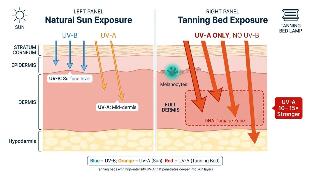
Tanning beds primarily emit UV-A radiation, which penetrates far deeper into the dermis and causes the most lasting cellular damage — silently, with no visible warning.
| Factor | UV-B | UV-A |
|---|---|---|
| Skin penetration | Outer epidermis | Deep dermis |
| Causes sunburn? | Yes | No |
| Drives tanning? | Partial | Primary driver |
| DNA mutation type | Direct CPD formation | Oxidative DNA damage |
| Tanning bed dose vs. sun | Lower | 10–15× higher |
According to the FDA’s tanning radiation safety guidance, the UV-A output of modern commercial tanning beds exceeds the equivalent midday sun dose by a factor of 10 to 15 within a typical 10-minute session.
What Happens to Your Melanocytes During One Session
This is the biological sequence that takes place every time a tanning bed lamp activates:
- UV-A photons penetrate through the epidermis to living melanocytes in the basal layer
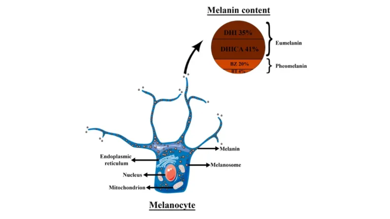
- Melanocytes produce melanin in response — the visible “tan”
- Simultaneously, UV-A creates cyclobutane pyrimidine dimers (CPDs) — DNA helix distortions that impair cell replication fidelity
- Your immune system activates repair enzymes (photolyases, nucleotide excision repair)
- Repeated sessions outpace repair capacity → unrepaired CPD accumulation → atypical melanocyte mutations → potential malignancy
The American Academy of Dermatology confirms this mutation cascade is reproducible across all major UV melanoma studies.
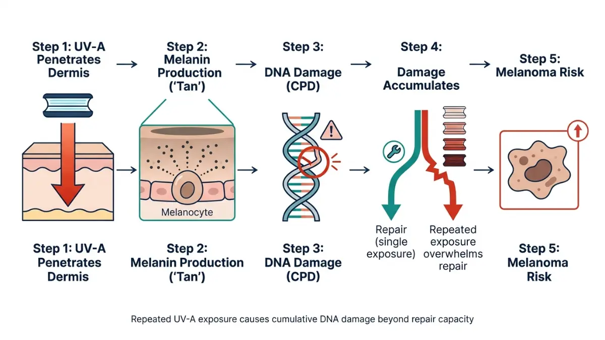
Cumulative DNA Damage: The Invisible Damage Scorecard
There is no mirror that shows UV damage at the cellular level. Your skin can look bronze and healthy while harboring thousands of unrepaired mutations.
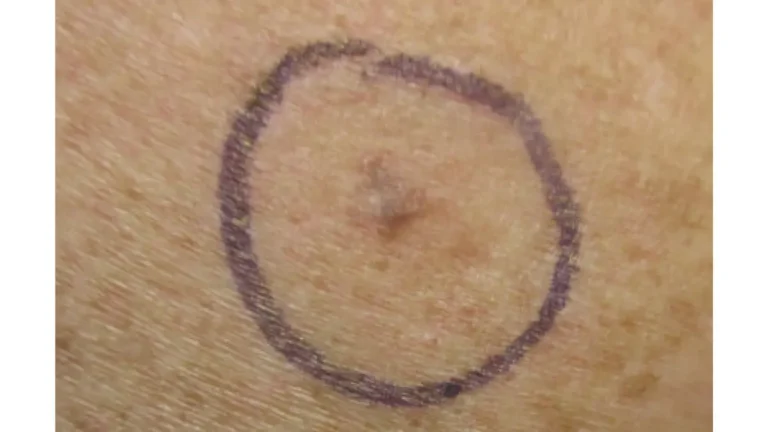
This is why UV-induced melanomas — especially amelanotic melanoma, a pigment-free variant — are so frequently missed in early self-exams. Understanding the full picture of what causes melanoma at the molecular level is the foundation of meaningful risk prevention.
What This Means For You: The tan you achieve in a tanning bed is your body’s emergency response to DNA damage — not a sign of health or vitality. Every session adds silently to a cumulative damage total your dermatologist can assess but no cosmetic mirror can reveal.
Tanning Beds vs. Natural Sun — The Honest Risk Comparison
Side-by-Side Breakdown
| Factor | Natural Sun (Midday Summer) | Tanning Bed |
|---|---|---|
| UV-A dose | Baseline | 10–15× higher |
| UV-B dose | Present | Variable/reduced |
| Equivalent sun session | 60–90 minutes | 10–20 minutes |
| Marketed as “controlled”? | No | Yes — misleadingly |
| Annual cumulative dose (regular user) | High | Very High |
| IARC classification | UV broadly carcinogenic | Group 1 specifically |
According to the American Cancer Society’s risk factor research, both natural and artificial UV exposure elevate melanoma risk — but tanning beds stack an additional concentrated UV-A dose on top of whatever natural exposure a user already accumulates throughout their day.
Why “Just 5 Minutes” Is Not a Safe Starting Point
The “gentle starter session” promoted by tanning salons has no scientific basis. Research supported by NIH-funded indoor tanning studies shows that even 1 to 4 lifetime tanning bed sessions is associated with a statistically measurable increase in melanoma risk.
No evidence establishes a safe exposure threshold for tanning bed UV-A on melanocyte DNA.
The Vitamin D Argument: What 2025 Research Actually Shows
Many tanning bed users cite Vitamin D synthesis as justification. Current science has decisively countered this:
- Oral Vitamin D3 supplementation (1,000–2,000 IU/day) achieves equivalent or superior serum 25(OH)D levels with zero UV exposure
- Updated NIH guidelines (2024) do not recommend tanning beds as a Vitamin D source under any clinical circumstances
- All major dermatology bodies — AAD, ACS, Skin Cancer Foundation — explicitly advise against UV tanning as a Vitamin D strategy
For a comprehensive guide from detection through treatment, our melanoma symptoms, stages, and treatment pillar article covers the full clinical spectrum.
What This Means For You: Tanning beds offer zero health benefit that cannot be achieved more safely via supplementation or moderate outdoor activity. The Vitamin D justification has been scientifically retired.
Who Faces the Highest Melanoma Risk From Tanning Beds? A 2026 Stratification Guide
Age: Why First Use Under 35 Is a Clinical Red Flag
| Age at First Use | Melanoma Risk Increase | Primary Data Source |
|---|---|---|
| Under 20 | +87–100% | JNCI 2022 |
| 20–25 | +75–87% | WHO/IARC |
| 25–35 | +59–75% | NCI SEER Database |
| 35–50 | +30–45% | CDC Surveillance 2024 |
| Over 50 | +15–25% | AAD Clinical Review |
Younger skin contains more rapidly dividing melanocytes, which are disproportionately vulnerable to CPD accumulation during peak cell replication cycles.
Skin Type, Hair Color, and Genetic Predisposition
Highest-risk profile:
- Fitzpatrick Type I–II: Very fair skin, red or light blonde hair, blue or green eyes
- 10+ atypical moles (dysplastic nevi): Pre-existing precursor lesion burden
- First-degree relatives with melanoma: Documented familial clustering
If melanoma has appeared in your family, our detailed breakdown of whether melanoma is hereditary covers CDKN2A, BRCA2, and MC1R gene variants that interact directly with UV exposure.
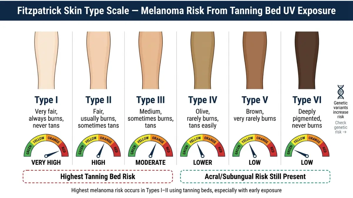
Gender Patterns: Why Young Women Are Disproportionately Affected
Melanoma in women aged 15–29 is now the most common cancer diagnosis in that age bracket in the US, according to CDC skin cancer surveillance data.
Young women account for the largest share of tanning salon customers. The epidemiological link between this demographic and rising early-onset melanoma rates is well-established across 25+ years of data.
Countries That Have Banned Tanning Beds in 2026
| Country/Region | Status |
|---|---|
| Brazil | Full commercial ban |
| Australia | Full ban — all states |
| United Kingdom | Under-18 ban; adult regulations tightening |
| Germany, France, Belgium | Various commercial restrictions |
| USA | No federal ban; FDA Class II device; under-18 banned in 42 states |
The Skin Cancer Foundation’s tanning risk page notes that the US regulatory framework remains significantly weaker than most peer nations — placing greater individual responsibility on American consumers.
With AI-assisted dermoscopy now changing early-detection outcomes, our melanoma 2026 AI screening survival guide covers how these new tools work in clinical practice.
What This Means For You: If you are a woman who used tanning beds before age 30, you fall squarely in the highest-risk demographic tracked by US cancer surveillance. Proactive screening is not a precaution — it is clinically indicated.
Used Tanning Beds Before? Your Exact Action Plan for 2026
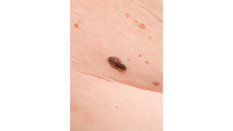
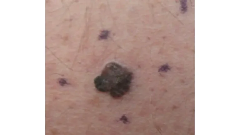
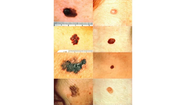
The ABCDE Rule — With Tanning-Bed-Specific Red Flags
| Letter | What to Check | Tanning Bed-Specific Note |
|---|---|---|
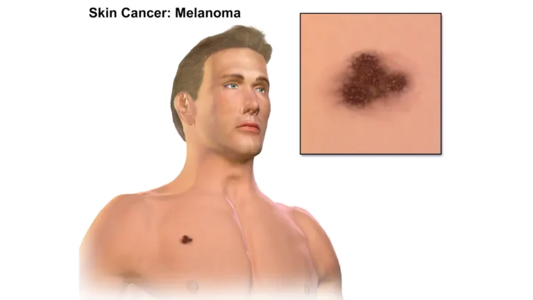
| A – Asymmetry | Halves don’t match | UV melanomas often asymmetric early |
| B – Border | Ragged, notched, blurred edges | Can appear as faint irregular smudging |
| C – Color | Multiple shades, brown to black to red | Watch for pink-red variants (amelanotic) |
| D – Diameter | Larger than 6mm | Some UV-induced melanomas present smaller |
| E – Evolving | Any change in size, shape, or color | Most critical sign — act within 2 weeks |
For complete guidance on every variant, our melanoma warning signs and symptoms guide and the ABCDE mole assessment guide cover diagnostic nuances that most general health sites miss.
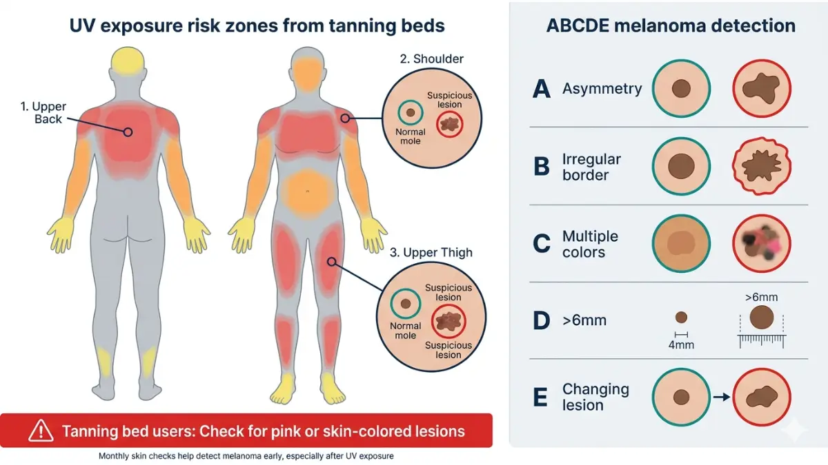
Screening Frequency by Risk Level
| Risk Level | Screening Frequency |
|---|---|
| Low (1–5 lifetime sessions, Type III+) | Annual full-body skin exam |
| Moderate (6–15 sessions or family history) | Every 6 months |
| High (16+ sessions, Type I–II, atypical moles) | Every 3–4 months with dermoscopy |
| Very High (prior biopsy finding, gene mutation) | Every 3 months + mole mapping |
7-Step Monthly Skin Self-Examination Checklist
Do this monthly in good lighting using a full-length mirror and a hand mirror:
- ✅ Face and scalp — part hair section by section, examine entire scalp
- ✅ Neck, chest, and upper back — high UV exposure zone, frequently missed
- ✅ Arms, underarms, and between fingers — include elbow creases
- ✅ Torso front and back — use hand mirror for the full back
- ✅ Hips, groin, and lower abdomen — tanning beds expose these areas directly
- ✅ Legs, behind the knees, and between toes
- ✅ Soles of feet and nail beds — melanoma beneath nails is rare but aggressive
If you identify any ABCDE warning signs, use our Symptom Checker for a structured preliminary assessment, then book a dermatologist appointment within two weeks — not months.
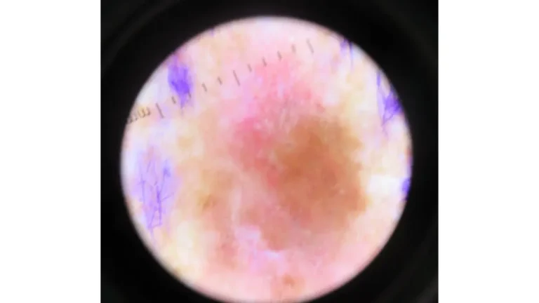
According to the Melanoma Research Foundation, professional dermoscopy — a magnified digital skin surface examination — is the clinical gold standard for evaluating any suspicious lesion identified during self-examination.
What This Means For You: Stage I melanoma carries a 98%+ five-year survival rate. Stage IV drops below 30%. The difference between those two outcomes is almost entirely determined by when the lesion is first found. Monthly self-exams take 10 minutes. That time investment has a documented life-or-death return.
Safer Alternatives — What Dermatologists Actually Recommend in 2026
Self-Tanners and DHA: Are They Actually Safe?
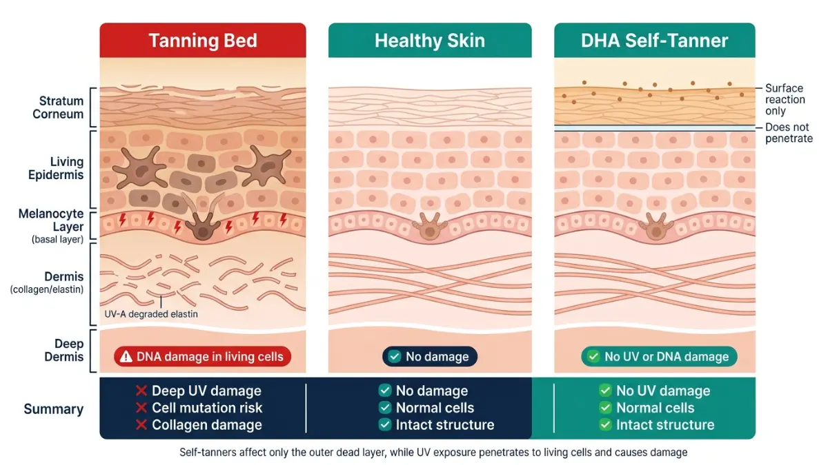
Dihydroxyacetone (DHA), the active ingredient in self-tanning creams and sprays, is FDA-approved and works exclusively on the outermost layer of dead skin cells. Key facts:
- Zero UV radiation — no melanocyte stimulation of any kind
- No DNA penetration — DHA does not reach living skin cells
- No documented melanoma risk across multiple independent dermatology reviews
- Color develops in 2–4 hours and lasts approximately 5–7 days as dead cells shed naturally
Spray Tan vs. Tanning Bed: Full Risk Comparison
| Factor | Tanning Bed | DHA Self-Tanner / Spray Tan |
|---|---|---|
| UV exposure | High (UV-A dominant) | None |
| DNA damage | Yes — cumulative | None |
| Melanoma risk | High | None documented |
| Vitamin D production | Yes (unsafe method) | No |
| FDA status | Class II device with restrictions | Approved ingredient |
| Recommended by dermatologists | No | Yes, universally |
The FDA’s sunless tanning consumer guide confirms DHA as safe for topical skin use, with one practical precaution: avoid inhaling aerosolized spray formulations directly.
Can Tanning Bed Skin Damage Be Reversed?
This is one of the most commonly searched questions by former tanning bed users. The answer is nuanced but honest:
What can improve with treatment:
- Surface photodamage, uneven pigmentation, and fine texture changes
- Some photoaged collagen — responds to professional microneedling or fractional laser
- Immune surveillance capacity — improves measurably with consistent sun avoidance
What cannot be fully reversed:
- Deep melanocyte DNA mutations already embedded before treatment begins
- Chronic UV-A-induced dermal elastin degradation
- Pre-existing atypical moles triggered by UV stimulation
Evidence-based daily recovery protocol:
- Broad-spectrum SPF 30–50 daily — the single most impactful intervention
- Topical tretinoin (0.025–0.05%) — clinically proven to reduce photodamage and improve cellular turnover
- Niacinamide 5% — supports skin barrier integrity and DNA repair enzyme activity
- Antioxidant serum (Vitamin C) — neutralizes ongoing free radical damage from residual sun exposure
- Annual professional skin checks — minimum; more frequent if your risk profile is elevated
For a broader guide to identifying all forms of skin cancer early, our 12 warning signs of skin cancer guide covers basal cell, squamous cell, and melanoma presentations side by side.
The AAD’s official skin self-examination resource provides board-certified dermatologist-verified visual guides for every skin type and body area.
What This Means For You: You cannot undo every UV session — but every protective decision you make from today forward actively reduces your ongoing risk. Start SPF daily. Book your skin check this month. Replace all future tanning bed use with DHA-based alternatives. The choices you make in 2026 directly determine your melanoma risk curve over the next two decades.
⚠️ Medical Disclaimer: This article is for educational purposes only and does not constitute medical advice. All information is based on peer-reviewed research and clinical guidelines current as of April 2026. Consult a board-certified dermatologist or licensed healthcare provider for personal evaluation and treatment decisions.
Frequently Asked Questions
1. Do tanning beds cause melanoma?
Yes. Tanning beds are IARC Group 1 carcinogens — the same classification as tobacco. They increase melanoma risk by up to 75% compared to never using them, based on WHO data.
2. How many tanning bed sessions does it take to cause skin cancer?
There is no proven safe number. Research shows even 1–4 lifetime sessions produce a statistically significant melanoma risk increase. Risk compounds cumulatively with each additional session.
3. Are tanning beds more dangerous than natural sun exposure?
In key ways, yes. Tanning beds deliver UV-A radiation 10–15× stronger than midday summer sun in a fraction of the time, creating a concentrated carcinogenic dose that exceeds most natural daily sun exposure.
4. Can one tanning bed session increase melanoma risk?
Yes. A single session causes measurable DNA damage in melanocytes. While the absolute risk from one session is low, it is not zero — and it adds permanently to cumulative lifetime UV damage.
5. What age group faces the highest risk from tanning beds?
Users under 35 — especially under 25 — face the greatest risk increase (75–87%). Young women aged 15–29 are the single highest-risk demographic in the United States.
6. Are tanning beds banned anywhere in 2026?
Yes. Brazil and Australia have full commercial bans. The UK bans under-18 use. In the US, 42 states restrict under-18 use, but no federal commercial ban is in effect.
7. What type of melanoma do tanning beds most often cause?
Superficial spreading melanoma is most common. However, tanning beds also significantly raise the risk of amelanotic melanoma — a pigment-free variant that is frequently missed because it lacks the classic dark color change.
8. How long after using tanning beds can melanoma develop?
Melanoma can appear 5 to 30 years after the causative UV exposure due to the slow accumulation of DNA mutations. Tanning bed sessions in your 20s can directly contribute to diagnoses in your 40s or 50s.
9. Can tanning bed skin damage be reversed?
Surface photodamage can partially improve with retinoids, SPF, and professional treatments. Deep melanocyte DNA mutations cannot be fully reversed — which makes ongoing prevention and early detection critical.
10. Is spray tanning safer than a tanning bed for melanoma risk?
Yes — significantly safer. DHA-based self-tanners involve zero UV radiation, cause no DNA damage, and carry no documented melanoma risk. Dermatologists universally recommend them as the safe alternative to indoor tanning.
11. What should I do if I’ve used tanning beds for years?
Schedule a full-body skin exam with a board-certified dermatologist as soon as possible. Begin monthly ABCDE self-exams. Apply broad-spectrum SPF 30–50 daily without exception. If melanoma runs in your family, request genetic risk screening at your next appointment.
Medical disclaimer
The content on MyMedicineAdvisor is provided for general informational and educational purposes only and is not a substitute for professional medical advice, diagnosis, or treatment. Health information on this website should not be used to diagnose, treat, cure, or prevent any condition without guidance from a qualified healthcare professional. Always seek the advice of your doctor, physician, or another licensed healthcare provider with any questions you may have regarding a medical condition, symptoms, medications, or treatment decisions.