On This Page – Quick Medical Summary
Breast cancer: what you need to know right now
A breast cancer diagnosis changes everything about the way you move through your day.
It changes what you hear when your phone rings, what you think about when the room goes quiet, and what you type into Google at two in the morning when you cannot sleep and you need real answers from someone who actually knows what they are talking about.
This guide was written by a team of board-certified oncologists and reviewed by three physician specialists — because you deserve clinical accuracy, not vague reassurance, and because the decisions you make in the next weeks and months matter enormously for your outcome.
Breast cancer is the second most common cancer diagnosed in women in the United States. It develops when cells in the breast tissue begin dividing without control, forming a malignant tumor that can, without treatment, invade surrounding tissue and spread to lymph nodes and distant organs.
The word “cancer” carries weight that no physician — and no article — can fully neutralize. But there is something clinically true and genuinely important to say at the beginning of this guide: breast cancer is one of the most treatable cancers in medicine. The overwhelming majority of women diagnosed at the earliest stages go on to live full lives after treatment. Even for more advanced diagnoses, the landscape of available treatment has changed fundamentally in the last decade, and it continues to change.
Understanding your specific situation — your stage, your tumor subtype, your receptor status, and your treatment options — is the most powerful thing you can do right now.
That is what this article is for.
🩺 Physician Note: “Every patient I see who has just received a breast cancer diagnosis asks the same first question: ‘What does my stage mean — is this going to kill me?’ My answer is always the same: stage matters, but it is not the whole story. Receptor status, tumor biology, and your overall health are equally important factors in predicting your outcome and building a treatment plan. This guide will help you understand all of them.” — Dr. Nathaniel J. Hargrove, MD, Medical Oncology
The American Cancer Society’s 2026 breast cancer incidence and mortality figures confirm that breast cancer remains the most commonly diagnosed cancer among US women (excluding non-melanoma skin cancers). Early detection, improved systemic therapy, and personalized treatment based on tumor biology have driven meaningful improvements in survival outcomes over the past two decades.
This article is organized to meet you exactly where you are. Whether you are newly diagnosed, supporting a family member through treatment, researching for prevention, or deep into a treatment protocol that raises new questions every week — the sections below are written for your specific moment.
ℹ️ Medical Disclaimer: The diagnostic criteria, staging frameworks, treatment descriptions, medication information, surgical procedure details, genetic testing guidance, screening recommendations, insurance coverage information, and survival data in this article reflect current 2026 clinical guidelines and are provided for educational purposes only. This article does not constitute a physician-patient relationship and does not replace a consultation with a board-certified medical professional.
Individual diagnostic conclusions, treatment decisions, chemotherapy regimen selection, targeted therapy eligibility, surgical approach, radiation indication, hormone therapy duration, genetic testing eligibility, and prognosis depend on factors including tumor pathology, staging workup, receptor status, genomic testing results, comorbidities, menopausal status, genetic profile, and specialist assessment. Consult a board-certified oncologist, surgical oncologist, radiation oncologist, or genetic counselor — as appropriate to your specific situation — before acting on any clinical information in this article.
What is breast cancer and how does it develop?
Understanding what breast cancer actually is — at the cellular and structural level — makes every subsequent conversation about staging, treatment, and prognosis more meaningful and less frightening.
How normal breast cells become cancerous
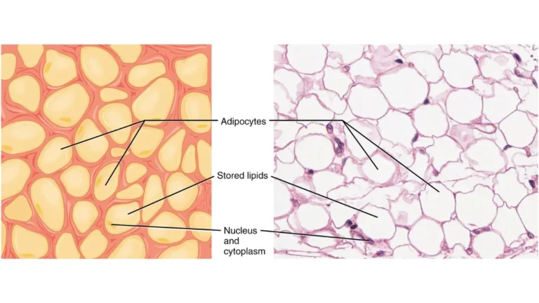
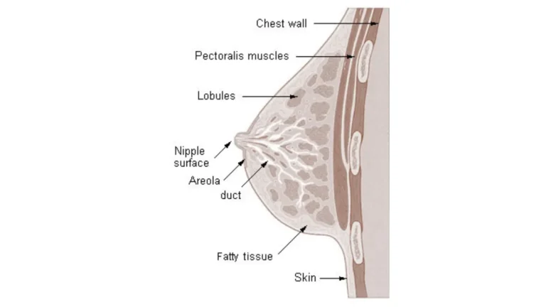
The breast is a complex glandular structure composed of mammary ducts, lobules, stromal tissue, fat, and blood vessels. Under normal conditions, cells in these structures divide in an orderly, regulated way — replacing old cells at a controlled rate determined by genetic programming.
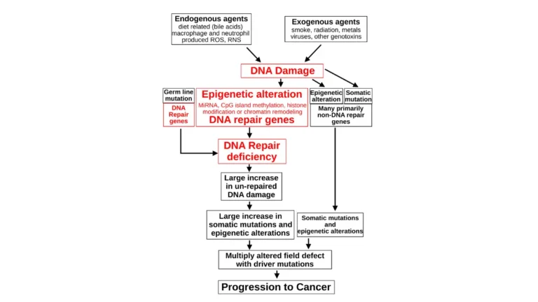
Cancer begins when that genetic regulation breaks down. Specific mutations — in tumor suppressor genes, proto-oncogenes, or DNA repair genes — cause a single cell to begin dividing without the normal stop signals that govern healthy tissue growth.
That cell’s descendants inherit the same unchecked growth capacity. Over time, they accumulate as a tumor mass — a growing cluster of abnormal cells that do not perform the function of normal breast tissue and that do not die when they should.
🔬 How It Works: Healthy cells receive constant molecular signals that tell them when to divide and when to stop. Two of the most important brake systems are tumor suppressor proteins encoded by genes called TP53 and RB1. When mutations disable these brakes — through inherited DNA variants, environmental DNA damage, or errors in DNA replication — cell division accelerates beyond the body’s ability to correct it. The result, over months to years, is a detectable tumor.
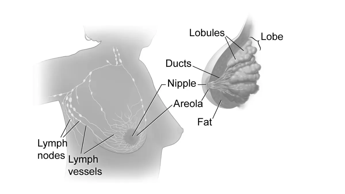
Where breast cancer typically starts: ducts vs. lobules
The two most common starting points for breast cancer are the ductal cells (which line the milk-carrying ducts) and the lobular cells (which form the milk-producing lobules at the end of each duct system).
Invasive ductal carcinoma (IDC) — which begins in the duct lining and grows through the duct wall into surrounding breast tissue — accounts for the large majority of breast cancer diagnoses in the United States. Invasive lobular carcinoma (ILC) is the second most common type, beginning in the lobules. IDC and ILC share many treatment principles but differ in how they appear on mammography, how they spread, and in some cases how they respond to systemic therapy.
The distinction matters clinically because ILC is more frequently bilateral (present in both breasts simultaneously), more often presents as a diffuse thickening rather than a discrete lump, and has a higher rate of detection on MRI than mammography alone — influencing the choice of imaging for staging and surgical planning.
How breast cancer spreads to lymph nodes and beyond
Metastasis — the spread of cancer beyond the primary tumor site — follows a predictable anatomical pathway in most cases, though not all cases follow this pathway in sequence.
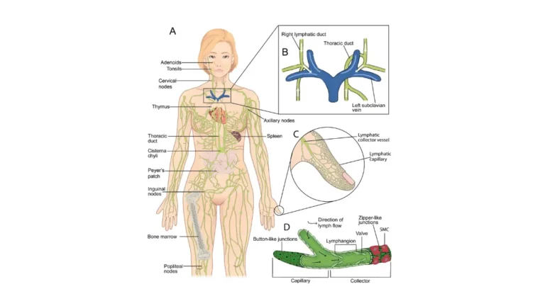
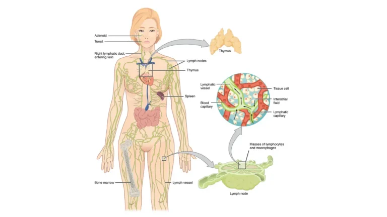
Local invasion occurs first: tumor cells penetrate the basement membrane of the duct or lobule and grow into surrounding breast tissue. As the tumor develops blood vessels (a process called angiogenesis), cancer cells gain access to the lymphovascular system. From there, the most common initial destination is the axillary lymph nodes — the cluster of nodes in the armpit on the same side as the tumor.
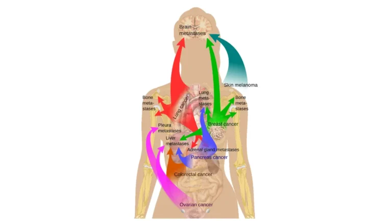
Lymph node involvement is one of the two most critical factors in staging (alongside tumor size) because it indicates that cancer cells have moved beyond the primary site. When lymph nodes are positive for cancer, the risk of distant metastasis — spread to the lungs, liver, bone, or brain — increases.
This anatomical pathway is why lymph node evaluation is a mandatory component of breast cancer staging and why surgical lymph node removal (either sampling or full dissection) plays an important role in both staging and local control.
The role of hormones in driving breast cancer growth
Approximately 70–80% of breast cancers express receptors for estrogen (ER) and/or progesterone (PR) on their cell surface. These receptors are molecular binding sites — when circulating estrogen attaches to them, it activates a signaling cascade inside the tumor cell that drives cell division.
🔬 How It Works: Estrogen binds to the estrogen receptor protein on the surface or inside the tumor cell. This binding activates the receptor, which then travels to the cell’s nucleus and acts as a transcription factor — turning on genes that promote cell growth. Aromatase inhibitors work by blocking the enzyme (aromatase) that converts androgens to estrogen in fat tissue, thereby removing the fuel for this cycle. Tamoxifen works differently — it binds to the estrogen receptor itself, blocking estrogen from attaching and triggering growth. Both approaches are targeting the same biological driver from different angles.
Understanding this hormonal mechanism is essential for understanding why treatment protocols differ so dramatically between hormone receptor-positive (HR+) breast cancer and hormone receptor-negative subtypes like triple-negative breast cancer — where this entire hormonal pathway is absent and treatment must rely on other approaches.
Breast cancer symptoms: what to look for
Most breast cancers are detected during routine mammography — before any symptoms develop. But approximately 20–30% of breast cancers are first identified by the patient or a clinician through physical examination, which means knowing what to look for remains genuinely clinically important.
The most common first sign: what a breast lump really feels like
The most frequently reported first sign of breast cancer is a new breast lump or thickening — but the clinical reality of what that lump feels and behaves like is more nuanced than most health articles convey.
Breast cancer lumps are typically:
- Hard or firm, with irregular edges rather than smooth borders
- Fixed in position — they do not slide freely under the skin as a benign cyst often does
- Usually painless, though not always — pain does not rule out cancer, and its absence does not rule it in
- Most commonly located in the upper outer quadrant of the breast (nearest the armpit), though they can appear anywhere in the breast tissue or along the chest wall
- Often detected first as an area of asymmetric firmness rather than a discrete, well-defined mass
Most breast lumps are not cancerous. Fibroadenomas (smooth, mobile, rubbery nodules — most common in women under 35) and fibrocystic changes (cyclical tenderness and lumpiness that worsens before menstruation) account for the large majority of breast lumps evaluated in clinical practice.
But a new lump that was not previously present, that does not resolve after a menstrual cycle, or that has any of the characteristics described above warrants prompt evaluation — not watchful waiting.
✅ Patient Action: If you have discovered a breast lump, schedule an evaluation with your OB/GYN or primary care physician within one to two weeks. At that appointment, specifically request a diagnostic mammogram — not just a clinical breast exam — because clinical exam alone has limited sensitivity for distinguishing benign from malignant masses. If you are under 30 and mammography is not indicated by age criteria, request a breast ultrasound as the appropriate first-line imaging modality.
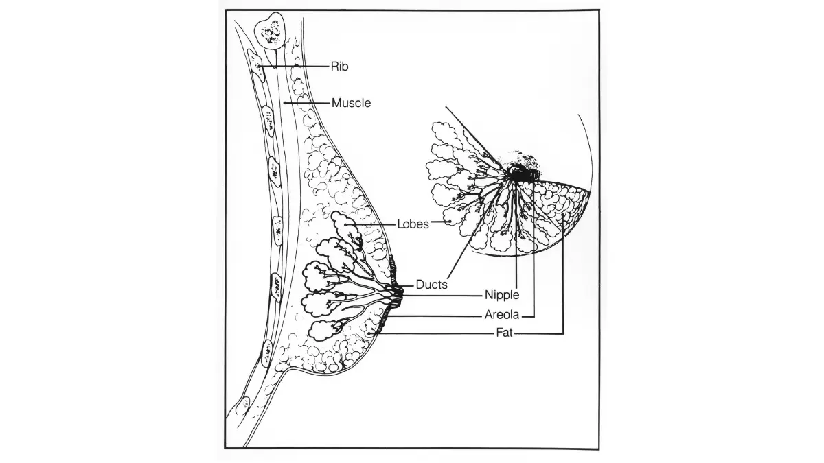
Symptoms that are not a lump: what many patients miss
Breast cancer symptoms extend well beyond the lump that most awareness campaigns have taught people to expect. A significant proportion of breast cancers are first detected not through a lump but through one of the following physical changes:
- Nipple discharge — particularly if it is spontaneous (without squeezing), unilateral (one breast only), clear or bloody in character, and associated with a mass
- Nipple retraction or inversion — a nipple that turns inward when it was previously everted, or that points in a different direction than usual
- Skin dimpling — a puckering or indentation in the breast skin, resembling the surface of an orange (peau d’orange), caused by cancer cells tethering the skin to deeper tissue through Cooper’s ligaments
- Breast asymmetry — one breast becoming noticeably larger, differently shaped, or positioned differently than the other, without an obvious mechanical cause
- Axillary lump — a firm lymph node in the armpit, sometimes the first detectable sign of breast cancer that has already spread to the regional lymph nodes before the primary tumor is palpable
- Persistent breast pain in one localized area — not cyclical, not bilateral, not relieved by ibuprofen or position changes
- Redness, warmth, or swelling that does not resolve within 7–10 days of appropriate antibiotic treatment (see the inflammatory breast cancer subsection below)
If you have noticed any of these changes, use our clinical symptom screening tool to check your symptoms for a structured evaluation before your appointment.
Inflammatory breast cancer: symptoms that look nothing like cancer
⚠️ Clinical Warning: Inflammatory breast cancer (IBC) is the most frequently delayed diagnosis in breast oncology — not because it is subtle, but because its symptoms mimic a breast infection so convincingly that patients, and sometimes clinicians, pursue a full antibiotic course before cancer is considered. Any breast redness, warmth, swelling, or skin thickening in a woman over 30 that does not resolve completely after 7–10 days of antibiotic treatment must be evaluated by diagnostic imaging and biopsy without further delay.
Inflammatory breast cancer is a rare but clinically distinct form of breast cancer that does not typically present with a discrete lump. Instead, IBC presents as:
- Rapid-onset redness (erythema) covering one-third or more of the breast skin
- Warmth to the touch, often mistaken for a breast infection (mastitis) or abscess
- Breast swelling and heaviness
- Skin texture changes — pitting, thickening, or the classic peau d’orange (orange-peel) appearance
- Nipple flattening or inversion that occurs quickly rather than gradually
- Occasionally a palpable mass, but frequently no discrete lump at all
IBC is always staged at Stage 3B or higher at diagnosis, regardless of whether a discrete tumor is identifiable, because the clinical presentation itself indicates dermal lymphatic invasion — a defining feature of the disease. This staging reality makes early recognition and biopsy urgency genuinely life-affecting.
How breast cancer is diagnosed: from mammogram to biopsy
Breast cancer diagnosis follows a defined clinical sequence when imaging or physical examination identifies a suspicious finding:
- Clinical breast examination — assessment of breast tissue, nipple, and regional lymph nodes by a trained clinician
- Diagnostic mammography — targeted imaging of the area of concern with additional views; not the same as a routine screening mammogram
- Breast ultrasound — used to characterize a mass (solid vs. fluid-filled) and to guide biopsy; often performed immediately after diagnostic mammography when a suspicious mass is identified
- Breast MRI — used in specific circumstances: evaluating the extent of disease before surgery, assessing the opposite breast, evaluating implant integrity, or characterizing findings that are indeterminate on mammography and ultrasound
- Core needle biopsy — the definitive diagnostic step; a needle removes multiple small cores of tissue from the suspicious area under ultrasound or stereotactic guidance; this is the test that establishes whether cancer is present and provides the tissue for all downstream receptor and genetic testing
- Pathology and receptor testing — the biopsy specimen is evaluated for: cancer cell type and grade, estrogen receptor (ER) status, progesterone receptor (PR) status, and HER2 protein expression (by immunohistochemistry and, when equivocal, by FISH testing)
📊 Clinical Data Point: Per NCCN Clinical Practice Guidelines in Oncology (Breast Cancer, 2026 edition), core needle biopsy — not excisional biopsy — is the recommended diagnostic procedure for evaluating a suspicious breast mass. Fine needle aspiration is insufficient for full receptor testing and is not the standard of care for initial diagnosis.
What your pathology report means: the key terms explained
The pathology report that follows a positive biopsy is the foundational document for every treatment decision that follows. Understanding its key terms gives patients the ability to participate meaningfully in treatment planning.
Invasive vs. in situ: Invasive cancer means tumor cells have grown beyond the basement membrane of the duct or lobule into surrounding tissue. In situ means cancer cells are confined within the duct (DCIS) or lobule (LCIS) and have not invaded beyond.
Tumor grade (1 through 3): Grade reflects how different the cancer cells look compared to normal cells under a microscope. Grade 1 (well-differentiated) — cells still resemble normal breast cells and divide slowly. Grade 2 (moderately differentiated) — intermediate characteristics. Grade 3 (poorly differentiated) — cells look very abnormal and divide rapidly. Higher grade generally correlates with more aggressive behavior.
Ki-67 proliferation index: A percentage reflecting how many tumor cells are actively dividing. A high Ki-67 (typically above 20–30%) indicates a rapidly growing tumor and influences decisions about chemotherapy and treatment sequencing.
ER/PR receptor status and HER2 status: These three receptor results — positive or negative for each — determine the entire treatment architecture. They are discussed in detail in the Breast cancer types: ER+, HER2+, and triple-negative section below.
Breast cancer stages: what Stage 0 through 4 means
Your stage is not your prognosis. It is a clinical classification system that describes the extent of the disease at the time of diagnosis — and it is the framework on which your treatment plan is built.
Breast cancer is classified into five stages based on three factors, known collectively as the TNM staging system: T (tumor size and local extension), N (lymph node involvement), and M (presence or absence of distant metastasis). The AJCC (American Joint Committee on Cancer) eighth edition staging manual governs the current classification framework, as referenced in NCCN 2026 Clinical Practice Guidelines.
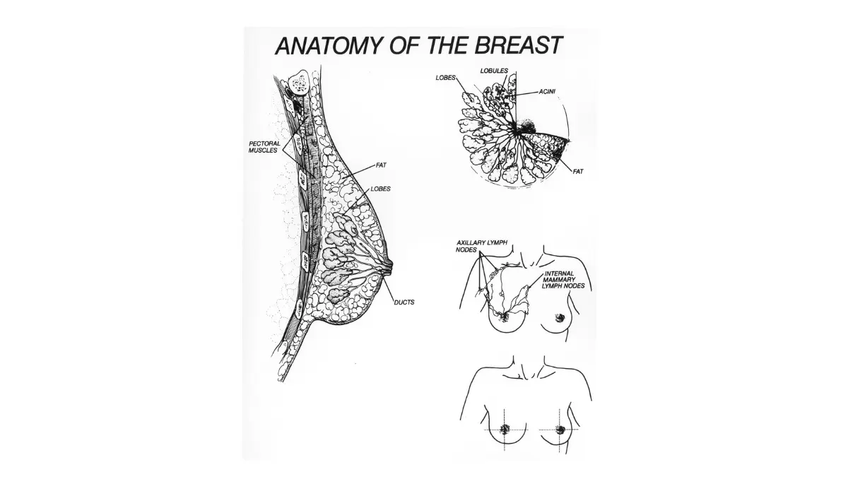
Breast cancer is classified into five stages (Stage 0 through Stage 4) based on tumor size, lymph node status, and presence of distant metastasis:
- Stage 0 (DCIS) — Non-invasive cancer confined within the milk ducts; has not grown through the duct wall into surrounding tissue
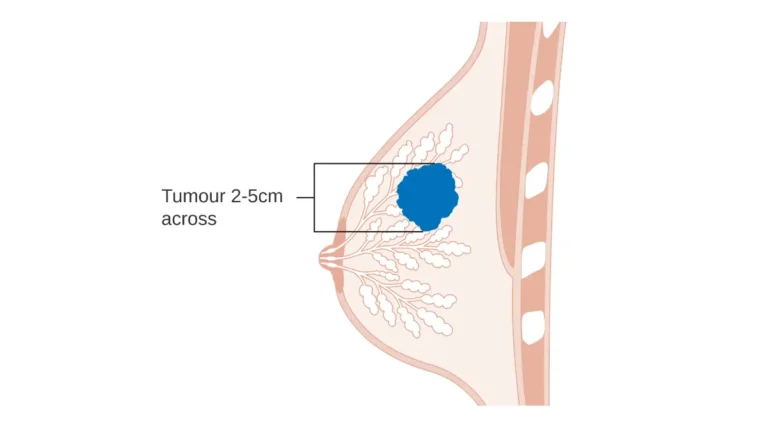
- Stage 1 — Small invasive tumor (2 cm or smaller) with no lymph node involvement or very limited micrometastasis to one lymph node
- Stage 2 — Larger tumor (up to 5 cm) with involvement of up to three axillary lymph nodes, or a smaller tumor with regional node involvement
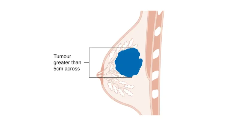
- Stage 3 — Locally advanced disease — larger tumors, more lymph node involvement, or tumor extension to chest wall or skin — without distant metastasis
- Stage 4 — Metastatic breast cancer — cancer has spread to distant organs including the lungs, liver, bone, or brain
For a complete reference including substage classifications (1A, 1B, 2A, 2B, 3A, 3B, 3C) and the pathological criteria that determine surgical eligibility, see our detailed breakdown of every breast cancer stage.
Stage 0 (DCIS): breast cancer that has not spread
Ductal carcinoma in situ (DCIS) is the presence of abnormal cells inside the milk ducts of the breast that have not yet grown through the duct wall into surrounding tissue. DCIS is not invasive cancer, but it is considered a precursor — without treatment, a proportion of DCIS lesions progress to invasive breast cancer over time.
DCIS is most often detected on screening mammography as a cluster of calcifications (tiny calcium deposits) that appear before any palpable mass has formed. It is not detectable by physical examination in most cases.
Treatment for DCIS typically involves lumpectomy with radiation, or mastectomy for more extensive disease. Hormone therapy (tamoxifen or an aromatase inhibitor) is often added for ER-positive DCIS to reduce the risk of future invasive cancer in either breast.
🩺 Physician Note: “A DCIS diagnosis sometimes feels almost unreal to patients — they had no symptoms, they had a routine mammogram, and suddenly they’re being told they have cancer. I always take time to distinguish DCIS from invasive breast cancer because the clinical implications are genuinely different. DCIS is treated with the goal of preventing invasive cancer — and the treatment options are real and effective. A DCIS diagnosis, caught on routine screening, is one of the best outcomes in breast oncology.” — Dr. Nathaniel J. Hargrove, MD
Stage 1: early-stage breast cancer with favorable outcomes
Stage 1 breast cancer describes a small invasive tumor — typically 2 cm or smaller — with no lymph node involvement (Stage 1A) or with only tiny microscopic cancer deposits in a sentinel lymph node that do not alter the overall staging or treatment significantly (Stage 1B).
Stage 1 disease is generally treated with lumpectomy and radiation (or mastectomy if the patient prefers or if tumor location precludes clear lumpectomy margins), with systemic therapy added based on receptor status and genomic testing results. For many women with Stage 1, ER+, HER2-negative disease, an Oncotype DX genomic test provides the key piece of information that determines whether chemotherapy is beneficial.
The prognosis at Stage 1 is among the most favorable of any solid tumor diagnosis in oncology.
Stage 2: locally confined cancer with curative treatment options
Stage 2 breast cancer includes tumors ranging from 2 to 5 cm with varying degrees of lymph node involvement, or smaller tumors with more significant regional lymph node involvement.
Stage 2A includes tumors smaller than 2 cm with one to three positive axillary nodes, or tumors 2 to 5 cm with no nodal involvement. Stage 2B includes tumors 2 to 5 cm with one to three positive axillary nodes, or tumors larger than 5 cm with no nodal involvement.
🩺 Physician Note: “In my clinical practice, Stage 2 breast cancer patients who ask about Oncotype DX genomic testing before committing to chemotherapy consistently make better-informed treatment decisions. For Stage 2, ER-positive, HER2-negative patients, a low recurrence score on Oncotype DX means that chemotherapy does not improve survival — and that information can spare months of treatment with significant side effects. I recommend every patient in this category ask their oncologist about this test before the treatment plan is finalized.” — Dr. Nathaniel J. Hargrove, MD
Is Stage 2 breast cancer curable?
Stage 2 breast cancer is treated with curative intent in the overwhelming majority of patients. The treatment plan — which typically includes surgery (lumpectomy or mastectomy), systemic therapy tailored to receptor status, radiation in most lumpectomy cases, and hormone therapy for ER+ disease — is designed to eliminate all cancer cells and prevent recurrence.
The concept of “cure” in breast cancer is generally expressed as disease-free survival — the absence of detectable cancer at defined time points after treatment ends. For Stage 2 patients who receive the complete recommended treatment course, long-term disease-free outcomes are the expected result, not the exception.
✅ Patient Action: Before your first oncology appointment, request a complete copy of your pathology report and confirm that your HER2 FISH test result is included alongside the IHC result. If your IHC result was 2+ (equivocal), the FISH test is the definitive determinant of HER2 status — and HER2 status determines whether targeted therapies like trastuzumab are part of your protocol.
Stage 3: locally advanced breast cancer — what this means
Stage 3 breast cancer — also called locally advanced breast cancer — includes a range of presentations that share one defining characteristic: significant local or regional disease burden without confirmed distant metastasis.
Stage 3 is subdivided into three categories. Stage 3A includes tumors with extensive axillary lymph node involvement or involvement of internal mammary lymph nodes. Stage 3B includes tumors that have grown into the chest wall or the skin of the breast — including inflammatory breast cancer, which is always classified as Stage 3B or higher. Stage 3C describes any tumor size with extensive nodal involvement including infraclavicular, supraclavicular, or combined axillary and internal mammary node involvement.
Treatment for Stage 3 disease typically begins with neoadjuvant chemotherapy (systemic therapy before surgery), which has two goals: shrinking the tumor to improve surgical options, and testing the tumor’s responsiveness to treatment — with pathologic complete response (pCR) after neoadjuvant therapy being the strongest predictor of long-term disease-free survival available.
Stage 4: metastatic breast cancer — treatment and what to expect
Stage 4 — metastatic breast cancer — is the diagnosis that most frightens patients, and it is the diagnosis that requires the most careful clinical contextualization. Stage 4 breast cancer has spread beyond the breast and regional lymph nodes to distant organs: most commonly the bones, lungs, liver, and brain, though any organ can be affected.
Stage 4 breast cancer is not curable with current treatment. It is, however, treatable — and the distinction matters enormously for patients and families navigating this diagnosis.
Metastatic breast cancer is increasingly managed as a chronic condition, not a terminal event. Modern targeted therapies — CDK4/6 inhibitors for ER+ disease, HER2-directed antibody-drug conjugates for HER2-positive disease, PARP inhibitors for BRCA-mutated disease, and immunotherapy for eligible TNBC patients — have extended survival duration significantly from historical benchmarks. Many patients with Stage 4 disease live for years on active treatment while maintaining quality of life.
🩺 Physician Note: “When I tell a patient she has Stage 4 breast cancer, I am very careful about the conversation that follows. I tell her that this diagnosis means we are no longer talking about cure in the traditional sense — we are talking about long-term disease management. That is a real and meaningful distinction. And then I tell her that the treatment landscape for metastatic breast cancer has changed so dramatically in the last decade that the trajectory her mother faced in 2010, or her grandmother faced in 2000, is not her trajectory. We have more options than we have ever had.” — Dr. Nathaniel J. Hargrove, MD
For patients and families navigating a metastatic diagnosis, a complete guide to treatment options and survival outlook for Stage 4 breast cancer covers palliative treatment, systemic therapy options, and clinical trial enrollment in full detail.
The difference between Stage 3 and Stage 4 breast cancer
The clinical distinction between Stage 3 and Stage 4 is whether cancer has spread to distant organs. Stage 3, regardless of how locally extensive, remains regional — confined to the breast, nearby lymph nodes, and local structures (chest wall, breast skin). Stage 4 has crossed into systemic disease.
This distinction has profound treatment implications. Stage 3 is treated with curative intent — the full treatment plan is designed to eliminate all disease. Stage 4 treatment is designed to control disease, extend survival, and preserve quality of life, with treatment continuing indefinitely rather than concluding after a defined protocol.
The staging workup — CT scan of chest, abdomen, and pelvis, plus a bone scan or PET-CT — is the test that definitively distinguishes Stage 3 from Stage 4, and it is a mandatory component of the initial evaluation for any Stage 2 or higher diagnosis.
How doctors determine your stage: the TNM system explained
The TNM staging system assigns three values to every breast cancer diagnosis:
T (Tumor) describes the primary tumor:
- T1: tumor 2 cm or smaller
- T2: tumor 2.1 to 5 cm
- T3: tumor larger than 5 cm
- T4: tumor of any size that extends into the chest wall or breast skin (including IBC)
N (Nodes) describes regional lymph node involvement:
- N0: no regional lymph node involvement
- N1: 1–3 axillary lymph nodes involved (or internal mammary node micrometastasis)
- N2: 4–9 axillary lymph nodes involved, or internal mammary lymph node involvement
- N3: 10 or more axillary nodes, or infraclavicular or supraclavicular node involvement
M (Metastasis) describes distant spread:
- M0: no distant metastasis identified
- M1: confirmed distant metastasis present
These three values combine to determine the overall AJCC stage, which then drives treatment planning per NCCN 2026 guidelines.
Breast cancer types: ER+, HER2+, and triple-negative
Two patients can be diagnosed with Stage 2 breast cancer and receive completely different treatment plans. This is not an inconsistency in oncology — it is precision medicine. The treatment differences are driven by tumor subtype, which is determined by three receptor markers tested on every breast cancer biopsy.
Understanding your receptor status is as important as understanding your stage. In some cases, it is more important.
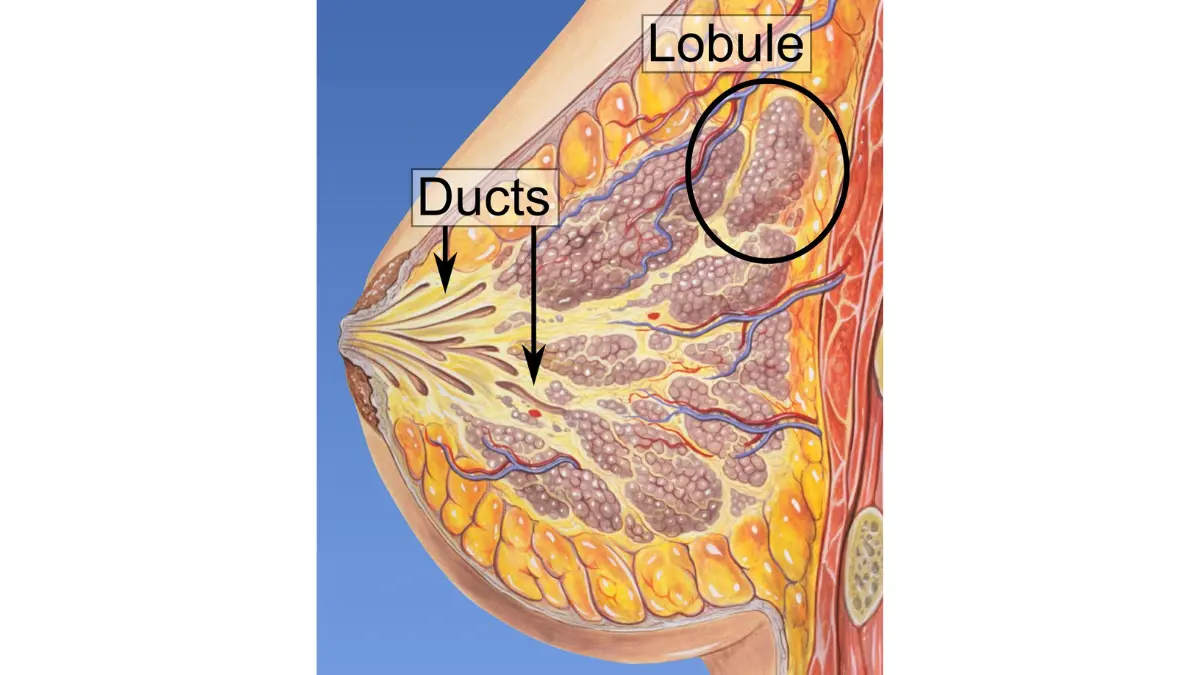
Estrogen receptor positive (ER+): the most common subtype
Estrogen receptor-positive (ER+) breast cancer is the most common molecular subtype in the United States, representing approximately 70–80% of breast cancer diagnoses.
In ER+ tumors, cancer cells have estrogen receptor proteins on their surface that act as growth accelerators when circulating estrogen binds to them. This hormone-driven growth mechanism creates a vulnerability that physicians exploit directly: treatments that block or remove estrogen from the tumor environment — tamoxifen, aromatase inhibitors, and ovarian suppression — can slow or stop tumor growth.
🔬 How It Works: In ER-positive breast cancer cells, the estrogen receptor protein sits either on the cell membrane or inside the cell cytoplasm. When estrogen (or the synthetic progestin in some hormone medications) binds to this receptor, it activates the receptor-protein complex, which travels to the cell’s nucleus and acts as a transcription factor — directly switching on genes that drive cell division. Blocking this pathway — either by blocking the receptor itself (tamoxifen) or by reducing the amount of estrogen available to bind it (aromatase inhibitors) — deprives the tumor of its primary growth signal.
ER+ breast cancer is generally associated with a relatively lower proliferation rate than ER-negative subtypes, though grade and Ki-67 index vary widely within the ER+ category. Importantly, ER+ tumors have the capacity for late recurrence — reappearing five, ten, or even fifteen years after the initial treatment ends — which is why extended hormone therapy (five to ten years) is a critical component of the treatment plan.
HER2-positive breast cancer: targeted therapy changes outcomes
HER2-positive (HER2+) breast cancer occurs when tumor cells overproduce a protein called HER2 (human epidermal growth factor receptor 2) — driving faster-than-normal cell division. Approximately 15–20% of breast cancers are HER2-positive, and HER2-targeted therapies have significantly improved survival outcomes for this subtype compared to historical benchmarks.
🔬 How It Works: HER2 is a receptor tyrosine kinase — a protein on the cell surface that, when activated, triggers downstream signaling pathways (including PI3K/AKT and MAPK/ERK) that drive aggressive cell proliferation. In HER2-positive tumors, the HER2 gene is amplified, meaning the cell produces far more HER2 receptor protein than normal — creating a state of chronic, unchecked growth signaling. Trastuzumab (Herceptin) works by binding to the extracellular domain of the HER2 receptor, blocking downstream signaling and simultaneously recruiting immune cells to attack the cancer cell (a process called antibody-dependent cellular cytotoxicity, or ADCC).
For a full explanation of treatment options and outcomes for this subtype, see HER2-positive breast cancer and targeted therapy.
HER2-positive tumors were historically associated with a more aggressive clinical course. Modern dual HER2 blockade (trastuzumab plus pertuzumab) and antibody-drug conjugates (particularly trastuzumab deruxtecan, or T-DXd) have dramatically altered this trajectory.
Triple-negative breast cancer: challenges and new treatment options
Triple-negative breast cancer (TNBC) tests negative for estrogen receptors, progesterone receptors, and HER2 protein overexpression — meaning it does not respond to hormone therapy or HER2-targeted drugs and is treated primarily with chemotherapy and, in eligible patients, immunotherapy.
TNBC represents approximately 10–15% of breast cancer diagnoses and is disproportionately represented in younger women and women of African ancestry. It is associated with higher proliferation rates and a more aggressive initial course compared to ER+ subtypes, but it also responds frequently and dramatically to chemotherapy — and pembrolizumab (an immune checkpoint inhibitor) has improved pathologic complete response rates in eligible Stage 2 and Stage 3 TNBC patients treated with neoadjuvant chemotherapy plus pembrolizumab.
For a complete guide to treatment options and prognosis for this subtype, see triple-negative breast cancer treatment options.
⚠️ Clinical Warning: Triple-negative breast cancer is sometimes described colloquially as “the worst subtype” — a characterization that is both clinically inaccurate and harmful to patients who receive this diagnosis. TNBC patients who achieve a pathologic complete response after neoadjuvant chemotherapy have excellent long-term disease-free survival outcomes. Pembrolizumab has added a meaningful new treatment dimension for eligible patients. A TNBC diagnosis is serious — but it is not a sentence.
Inflammatory breast cancer: a rare but aggressive presentation
Inflammatory breast cancer (IBC) is classified as its own subtype not because of a specific receptor profile but because of its distinctive clinical presentation and staging. IBC presents with rapid-onset breast skin changes — redness, warmth, peau d’orange texture, swelling — that result from dermal lymphatic invasion by tumor emboli.
IBC is always staged at Stage 3B or higher at diagnosis, regardless of tumor size, because the skin involvement itself meets the T4d staging criterion. IBC is always treated with neoadjuvant chemotherapy first — surgery is not the initial treatment step.
What your receptor status means for your treatment plan
The combination of your stage and your receptor status determines which of four primary treatment pathways applies to you:
| Subtype | First-Line Systemic Therapy | Surgical Approach | Hormone Therapy | Key Eligibility |
|---|---|---|---|---|
| ER+/HER2- | Chemo (if high-risk or high Oncotype DX score), then hormone therapy | Lumpectomy or mastectomy | Yes — 5–10 years | Oncotype DX guides chemo decision for Stage 1–2 |
| ER+/HER2+ | Neoadjuvant chemo + dual HER2 blockade (trastuzumab + pertuzumab) | Lumpectomy or mastectomy | Yes + HER2-targeted therapy | Trastuzumab for 12 months adjuvant |
| TNBC | Neoadjuvant chemo ± pembrolizumab | Lumpectomy or mastectomy | No | Pembrolizumab for eligible Stage 2–3 |
| ER-/HER2+ | Neoadjuvant chemo + dual HER2 blockade | Lumpectomy or mastectomy | No | T-DXd for residual disease post-neoadjuvant |
Per NCCN Clinical Practice Guidelines in Oncology (Breast Cancer), 2026 edition. Individual treatment plans depend on patient-specific factors assessed by a board-certified oncologist.
If you are uncertain about the treatment implications of your receptor status, patients are increasingly seeking a second board-certified oncologist opinion through telehealth platforms before committing to a treatment plan — a step that has meaningfully altered treatment protocols in a clinically significant proportion of cases.
Breast cancer survival rates by stage in 2026
The survival rate question is the first thing most patients want answered — and it deserves the most careful, most honest, most clinically grounded answer in this entire article.
Before any statistics, there is something you need to understand that most health websites never explain: a survival rate is a population statistic, not a personal prediction.
The 5-year relative survival rate for a given breast cancer stage describes what happened to a large group of patients with that stage diagnosis over a defined period. It does not — and cannot — tell you, sitting in your specific circumstances with your specific tumor biology on a specific day in 2026, what your outcome will be.
🩺 Physician Note: “When I present survival statistics to a newly diagnosed patient, I begin every conversation the same way: I tell her that these numbers describe thousands of people across different treatment eras, different tumor biologies, and different health backgrounds. They are genuinely useful for understanding the landscape of the disease — but they describe a population, not a person. The two most important predictors of your individual outcome are your specific tumor biology and whether you complete the full recommended treatment course. Those are factors we can act on.” — Dr. Nathaniel J. Hargrove, MD
What “5-year relative survival rate” actually means
A 5-year relative survival rate is the percentage of patients with a given diagnosis who are alive five years after diagnosis compared to people in the general population who do not have cancer, matched for age and sex.
“Relative” survival accounts for deaths from all causes in the comparison group, which means the survival rate for breast cancer specifically — rather than overall mortality from any cause — is what is being measured.
This measurement has two important limitations that patients should understand:
- It reflects patients diagnosed years ago under treatment protocols that may have since been updated. Survival statistics published in 2026 reflect patients diagnosed in approximately 2018–2020 — before several current therapies reached standard of care status.
- It does not disaggregate by receptor subtype, genomic risk score, comorbidity burden, or treatment adherence — all of which meaningfully influence individual outcomes.
Survival rates for Stage 0 and Stage 1 breast cancer
📊 Clinical Data Point: Per the NCI SEER program, 5-year relative survival data for breast cancer by AJCC stage is reported annually. The 2026 SEER Cancer Statistics Review data will be published at NCI’s 2026 breast cancer survival statistics by stage — specific percentages should be inserted at publication from the current NCI SEER release. The narrative context below is based on physician-level interpretation of established survival trends confirmed in NCCN 2026 guidelines.
For Stage 0 (DCIS): The vast majority of women diagnosed with DCIS who receive standard treatment go on to live full lifespans without ever developing invasive breast cancer. DCIS is not invasive cancer, and its treatment is specifically designed to prevent it from becoming invasive.
For Stage 1 invasive breast cancer: Among women diagnosed at Stage 1, survival outcomes are among the most favorable of any solid tumor diagnosis. The overwhelming majority of Stage 1 patients are alive and disease-free at five years, with outcomes reflecting the fact that the cancer was detected before lymph node involvement or distant spread occurred.
Survival rates for Stage 2 and Stage 3 breast cancer
For Stage 2 breast cancer: Five-year outcomes remain highly favorable, with the large majority of patients achieving disease-free status after completing the full recommended treatment protocol. The specific proportion varies by subtype — HER2+ patients who achieve pathologic complete response after neoadjuvant therapy have outcomes comparable to Stage 1; TNBC patients who achieve pCR also have excellent long-term outcomes.
For Stage 3 breast cancer: Outcomes at Stage 3 are meaningfully influenced by two factors: whether pathologic complete response is achieved with neoadjuvant treatment, and adherence to the complete treatment plan. The survival differential between Stage 3 patients who achieve pCR and those who do not is one of the most clinically significant prognostic distinctions in breast oncology.
🔬 How It Works: Pathologic complete response (pCR) — defined as no invasive cancer remaining in the breast tissue or lymph nodes after neoadjuvant chemotherapy — is the strongest surrogate endpoint for long-term disease-free survival in the neoadjuvant setting. Achieving pCR means the tumor’s biology was responsive to the selected chemotherapy regimen. Patients who achieve pCR after neoadjuvant therapy typically follow up with less intensive adjuvant therapy; those with residual disease may receive additional targeted agents (T-DXd for HER2+ patients, capecitabine for TNBC) to further reduce recurrence risk.
Stage 4 breast cancer survival: what the data shows in 2026
Stage 4 breast cancer survival data requires the most careful contextualization of any survival statistic in oncology.
The historical median survival figures for metastatic breast cancer that many patients find when searching online reflect treatment eras when CDK4/6 inhibitors did not exist, when T-DXd had not been approved, when PARP inhibitors were not standard of care for BRCA-mutated patients, and when pembrolizumab was not yet part of the TNBC treatment algorithm. Those historical figures do not describe the trajectory of a patient diagnosed and treated in 2026.
Current systemic therapy for Stage 4 ER+ breast cancer — CDK4/6 inhibitor plus aromatase inhibitor as first-line treatment — has produced progression-free survival durations that were unimaginable under earlier treatment standards. Many Stage 4 patients live for multiple years on this regimen before requiring a second-line agent.
For a detailed, subtype-specific guide to what metastatic breast cancer management looks like in 2026, see our complete resource on treatment options and survival outlook for Stage 4 breast cancer.
⚠️ Clinical Warning: Do not apply historical median survival figures for metastatic breast cancer to your current situation without discussing them with your oncologist. The median survival data widely available online reflects patient cohorts treated before the current standard-of-care regimens were approved. Ask your oncologist specifically what survival data is available for patients on the regimen you are being offered.
How receptor status affects survival beyond stage
Receptor status modifies survival outcomes within each stage in ways that population-level staging statistics cannot capture.
Two patients with identical Stage 2B diagnoses — one ER+/HER2- with a low Oncotype DX recurrence score, one TNBC — are facing different disease biology, different treatment requirements, and different prognostic trajectories. Grouping them under the same stage-level survival statistic obscures more than it reveals.
The take-home clinical principle: your survival outlook depends on your stage AND your receptor subtype AND your response to treatment. The staging statistic gives you a starting point. Your oncologist’s assessment of your complete pathology gives you the specific picture.
Why your individual survival outlook differs from population statistics
The population of patients described by any 5-year survival statistic includes patients who:
- Did and did not complete their full treatment course
- Were treated with older and newer regimens
- Had different receptor subtypes grouped together
- Had varying comorbidities, ages, and performance status
- Did and did not have access to academic cancer centers or clinical trials
Your individual outcome is shaped by none of those averages. It is shaped by your specific tumor biology, the completeness of your staging workup, the expertise of your treatment team, the accuracy of your receptor testing, your treatment adherence, and the ongoing evolution of therapy options — several of which may be different in two years from what they are today.
✅ Patient Action: If a survival statistic you’ve read has raised questions about your specific prognosis, bring that statistic to your oncologist at your next appointment and ask specifically: “Does this population’s receptor subtype profile and treatment era match my situation? And what does my specific tumor biology tell you about my individual trajectory?” These are the questions that move the conversation from population statistics to personalized prognosis.
How breast cancer is treated: a complete overview
Breast cancer treatment is not a single protocol. It is a personalized architecture built from your stage, your receptor status, your tumor’s genomic behavior, your menopausal status, and your overall health — assembled specifically for you by an oncology team working from the NCCN 2026 Clinical Practice Guidelines in Oncology.
How your treatment plan is built: stage plus receptor subtype
The treatment planning conversation begins with two documents: your pathology report (which gives stage, grade, receptor status, and HER2 result) and your staging imaging results (CT, PET-CT, or bone scan, which confirm or rule out distant spread).
From those two data sources, your medical oncologist identifies your NCCN risk category, which determines the recommended treatment intensity and the sequencing of the three primary treatment components: systemic therapy (chemotherapy, targeted therapy, or hormone therapy), local therapy (surgery and/or radiation), and maintenance therapy (extended hormone therapy or targeted therapy continuation).
📊 Clinical Data Point: Per NCCN Clinical Practice Guidelines in Oncology (Breast Cancer), 2026 edition, all patients with newly diagnosed invasive breast cancer should undergo complete receptor testing (ER, PR, HER2 by IHC, and FISH for equivocal HER2 IHC results) and a staging workup appropriate to their clinical stage before a final treatment plan is determined. For Stage 1 disease, staging imaging is not routinely required in asymptomatic patients; for Stage 2 and above, CT and bone scan or PET-CT is recommended.
Neoadjuvant vs. adjuvant therapy: what the sequence means
Two terms that appear constantly in breast cancer treatment discussions cause persistent confusion for patients:
Neoadjuvant therapy — systemic treatment (chemotherapy, targeted therapy, or hormone therapy) given before surgery. The goal is to shrink the tumor, improve surgical options (converting a mastectomy candidate to a lumpectomy candidate in some cases), and test the tumor’s biological responsiveness. Achieving pathologic complete response (pCR) — no cancer remaining in the breast or lymph nodes at surgery — is the strongest positive prognostic indicator available.
Adjuvant therapy — systemic treatment given after surgery, with the goal of eliminating any microscopic cancer cells that may remain in the body despite surgery. Adjuvant chemotherapy, targeted therapy, and extended hormone therapy all operate in this framework.
The choice of neoadjuvant vs. adjuvant sequencing is determined by tumor size, nodal involvement, and subtype. For most Stage 3 patients, all HER2-positive patients with Stage 2 or 3 disease, and most TNBC patients with Stage 2 or 3 disease, neoadjuvant chemotherapy with or without targeted therapy is the preferred approach.
The four treatment pathways by receptor status
| Receptor Status | Neoadjuvant Systemic Therapy | Surgery | Adjuvant/Post-Surgical Therapy | Hormone Therapy |
|---|---|---|---|---|
| ER+/HER2- (Stage 1–2) | Chemo if Oncotype DX high-risk; skip if low-risk | Lumpectomy or mastectomy | Complete Oncotype DX; extend hormone therapy 5–10 years | Yes — aromatase inhibitor or tamoxifen |
| ER+/HER2- (Stage 3) | Neoadjuvant chemo, consider neoadjuvant hormone therapy | Lumpectomy or mastectomy; radiation usually required | Adjuvant CDK4/6 inhibitor for high-risk residual disease | Yes — 5–10 years |
| ER+/HER2+ or ER-/HER2+ | Neoadjuvant chemo + trastuzumab + pertuzumab | Surgery; reconstruction if mastectomy | T-DXd for residual disease; trastuzumab 12 months total | Yes if ER+; No if ER- |
| TNBC | Neoadjuvant chemo ± pembrolizumab | Surgery | Capecitabine for residual disease; pembrolizumab continuation | No |
Per NCCN 2026. All treatment decisions require individual oncologist assessment.
How long breast cancer treatment typically takes
Treatment duration varies significantly by subtype and stage:
- Stage 1, ER+/HER2- with low Oncotype DX score: Surgery + radiation (approximately 3–6 weeks of radiation) + hormone therapy beginning after radiation (continuing 5–10 years)
- Stage 2–3, neoadjuvant chemotherapy pathway: 16–24 weeks of neoadjuvant chemotherapy → surgery → radiation → adjuvant targeted therapy continuation → hormone therapy for 5–10 years
- Metastatic Stage 4: Continuous treatment — regimen changes as disease responds, progresses, or side effects develop
The five-to-ten-year duration of hormone therapy for ER+ disease is one of the most under-discussed aspects of breast cancer treatment, and one of the most clinically important.
Clinical trials: when to ask about enrollment
Clinical trial enrollment is not a “last resort.” It is a treatment pathway that should be considered at the beginning of treatment planning — not after first-line therapy has failed.
Active breast cancer clinical trials are investigating: new combination regimens for TNBC, antibody-drug conjugates for HER2-low expression, next-generation CDK4/6 inhibitors, and novel targeted agents for BRCA-mutated and non-BRCA hereditary breast cancers. Many trials offer access to agents that are years away from standard-of-care approval.
Search active breast cancer clinical trials at ClinicalTrials.gov to identify currently enrolling studies at institutions near you, filtered by stage and receptor subtype.
Getting a second opinion before treatment begins
A second oncology opinion before beginning treatment is clinically appropriate, not disloyal.
Studies have consistently demonstrated that second opinions at academic cancer centers alter treatment recommendations in a meaningful proportion of cases — through more precise staging workup, additional receptor testing, identification of clinical trial eligibility, or recognition of a less aggressive approach than initially proposed.
The timeline anxiety of a new breast cancer diagnosis is real — but for most diagnoses, taking one to two weeks to obtain a second opinion does not compromise outcomes. Ask your oncologist specifically: “Is there any reason my diagnosis is so urgent that I cannot take a week to seek a second opinion at an NCI-designated cancer center?”
✅ Patient Action: Before signing a consent form for any breast cancer treatment, ask your oncologist three specific questions: (1) Am I eligible for a clinical trial that offers this treatment plus an investigational agent? (2) If I am Stage 2, ER-positive, and HER2-negative, has my Oncotype DX recurrence score been ordered? (3) Would a second opinion at a designated NCI cancer center change my treatment plan? These three questions have altered treatment decisions for a meaningful proportion of patients in my practice. — Dr. Nathaniel J. Hargrove, MD
Breast cancer surgery: lumpectomy vs. mastectomy
A lumpectomy removes only the tumor and a margin of surrounding tissue while preserving the breast. A mastectomy removes the entire breast. For patients who are eligible for both, 2026 clinical evidence confirms that survival outcomes are equivalent — but radiation therapy is required after lumpectomy to achieve that equivalence.
This is one of the most important clinical facts in breast cancer surgery — and it is one that many patients arrive at the surgical consultation having already gotten wrong.
What is a lumpectomy and when is it recommended?
Lumpectomy (also called breast-conserving surgery or partial mastectomy) removes the tumor and a rim of normal tissue around it, called the surgical margin. The goal is to achieve clear margins — no cancer cells at the edge of the removed tissue — while preserving as much of the breast as possible.
Lumpectomy is appropriate when:
- The tumor is small enough relative to the breast size that adequate margins can be achieved while preserving a cosmetically acceptable breast
- The tumor is not multicentric (multiple tumors in different quadrants of the same breast)
- The patient does not carry a BRCA1 or BRCA2 mutation that substantially increases the risk of a new cancer in the preserved breast tissue
- The patient is willing to complete the required course of radiation therapy after surgery
Radiation after lumpectomy is not optional — it is a required component that reduces local recurrence risk to levels equivalent to mastectomy. Patients who decline radiation after lumpectomy have meaningfully higher local recurrence rates than either radiation-treated lumpectomy patients or mastectomy patients.
What is a mastectomy and when is it necessary?
Mastectomy — removal of the entire breast — is recommended or preferred when:
- The tumor is large relative to breast size and clear lumpectomy margins cannot be achieved with acceptable cosmetic outcome
- Multiple tumors are present in different breast quadrants (multicentric disease)
- Radiation therapy is contraindicated (prior radiation to the chest wall, certain connective tissue disorders)
- The patient carries a pathogenic BRCA1 or BRCA2 mutation, and prophylactic removal of the affected breast substantially reduces future cancer risk — in which case contralateral (opposite breast) mastectomy is often also discussed
- The patient has a personal preference for mastectomy after being informed that survival outcomes are equivalent for eligible candidates
🩺 Physician Note: “The surgical decision conversation is one of the most emotionally complex I have in oncology. Patients often arrive having decided they want ‘the whole thing taken out,’ believing a more radical surgery produces better survival odds. In appropriately selected patients, that assumption is clinically incorrect. A lumpectomy with radiation and a mastectomy produce equivalent survival outcomes — and choosing to preserve the breast is a medically legitimate decision that does not compromise cure. What I ask every patient to consider is: which of these two options gives you the best quality of life going forward, given that both give you the same survival odds?” — Dr. Nathaniel J. Hargrove, MD
Survival outcomes: do lumpectomy and mastectomy produce the same results?
For patients who meet eligibility criteria for lumpectomy, decades of randomized clinical trial data — referenced in NCCN 2026 guidelines — confirm that lumpectomy plus radiation and mastectomy produce equivalent long-term survival outcomes.
Local recurrence rates (cancer returning in the breast or chest wall) are similarly equivalent when lumpectomy is performed with adequate margins and followed by the required radiation. For most patients, the choice between lumpectomy and mastectomy is not a survival decision — it is a quality-of-life and personal preference decision made within a framework of clinical eligibility.
The situation changes for BRCA1/2 carriers, who face a substantially higher lifetime risk of a new primary cancer in the remaining breast tissue — a risk that bilateral mastectomy significantly reduces and that lumpectomy does not.
For a complete guide to surgical decision-making, eligibility criteria, reconstruction options, and recovery expectations, see our detailed resource on comparing lumpectomy and mastectomy outcomes.
Breast reconstruction: what are the options after mastectomy?
Breast reconstruction restores the breast shape after mastectomy. It can be performed immediately (at the time of mastectomy) or delayed (weeks to months later, after chemotherapy or radiation is complete).
The two primary approaches are:
- Implant-based reconstruction: A tissue expander is placed at the time of mastectomy, gradually expanded over several months, then replaced with a permanent silicone or saline implant. This is the most common approach and generally involves shorter initial surgery and recovery.
- Autologous (flap) reconstruction: Uses the patient’s own tissue — from the abdomen (TRAM or DIEP flap), back (latissimus dorsi flap), or other sites — to reconstruct the breast. Produces more natural results but involves more complex surgery and longer recovery.
📊 Clinical Data Point: The Women’s Health and Cancer Rights Act (WHCRA) is a federal law requiring group health plans that cover mastectomy to also cover all stages of breast reconstruction, surgery on the other breast to produce a symmetrical appearance, prostheses, and physical complications of mastectomy including lymphedema. This is not a benefit option — it is a federal legal requirement. Insurance plans cannot deny coverage for reconstruction following a medically necessary mastectomy.
What is a sentinel lymph node biopsy and why does it matter?
Sentinel lymph node biopsy (SLNB) is the surgical procedure used to evaluate whether cancer has spread to the axillary lymph nodes without removing all of the nodes — which carries significant risks including chronic arm swelling (lymphedema).
The sentinel lymph node is the first lymph node (or nodes) in the axillary chain to receive drainage from the primary tumor site. If cancer has spread from the tumor to the lymph nodes, it is most likely in the sentinel node first. By injecting a radioactive tracer or blue dye near the tumor before surgery, the surgeon can identify and remove only the sentinel node(s) for pathological analysis.
If the sentinel node is negative (no cancer), the remaining axillary nodes are statistically very unlikely to contain cancer, and full axillary node dissection — with its higher lymphedema risk — is avoided. If the sentinel node is positive, further axillary management (additional nodes or targeted radiation) is determined by the extent of involvement per NCCN 2026 guidelines.
✅ Patient Action: Before finalizing your surgical plan, ask your surgeon these two specific questions: (1) Based on my tumor size, location, and imaging results, am I a candidate for lumpectomy with radiation rather than mastectomy? (2) If I carry a BRCA mutation or have a strong family history, does that change the recommendation for prophylactic surgery on the other breast? These two questions frame the complete surgical decision conversation.
Chemotherapy for breast cancer: what to expect
Breast cancer chemotherapy is one of the most feared words in oncology — and one of the most misunderstood. Chemotherapy is not always required. When it is required, its side effects are real and deserve honest preparation — not minimization. And the decision about whether you need it has been transformed in the last decade by genomic testing that can now identify patients for whom chemotherapy provides no benefit.
Who needs chemotherapy and who may not?
Chemotherapy is not recommended for every breast cancer patient. The decision depends on stage, subtype, and in some cases genomic testing results:
- Stage 1, ER+/HER2-, low Oncotype DX score: Chemotherapy does not improve survival — hormone therapy alone is the recommended approach per NCCN 2026 guidelines
- Stage 2, ER+/HER2-, low to intermediate Oncotype DX score: Chemotherapy benefit depends on the specific score and the patient’s age and menopausal status — the decision is made in consultation with the oncologist based on the Oncotype DX result
- Stage 2–3, HER2+: Neoadjuvant chemotherapy plus HER2-targeted therapy is the standard approach regardless of hormone receptor status
- Stage 2–3, TNBC: Chemotherapy (typically with pembrolizumab for eligible patients) is always part of the treatment plan
- Any stage with positive lymph nodes: Chemotherapy is generally recommended unless Oncotype DX testing shows low risk in the ER+/HER2- subset
The most common chemotherapy regimens for breast cancer
Breast cancer chemotherapy is delivered in cycles — a treatment period followed by a recovery period — typically over 16 to 24 weeks depending on the regimen:
- AC-T (dose-dense): Doxorubicin (Adriamycin) plus cyclophosphamide for four cycles, followed by paclitaxel (Taxol) for four cycles — one of the most commonly used regimens for high-risk early breast cancer
- TC: Docetaxel (Taxotere) plus cyclophosphamide for six cycles — used for lower-risk patients who still require chemotherapy
- Carboplatin + paclitaxel: Often used for TNBC patients, particularly in combination with pembrolizumab in the neoadjuvant setting
- FEC-T or FEC-D: Fluorouracil, epirubicin, and cyclophosphamide followed by docetaxel — used in some Stage 2–3 protocols
Neoadjuvant chemotherapy: why chemo comes before surgery
When chemotherapy is administered before surgery — called neoadjuvant chemotherapy — the approach serves two distinct clinical goals.
First: tumor shrinkage. A tumor that is too large for lumpectomy may shrink enough after chemotherapy that breast-conserving surgery becomes possible. Second and more critically: pathologic complete response (pCR) — the absence of any residual invasive cancer in the breast or lymph nodes at the time of surgery — is the strongest prognostic marker available in the neoadjuvant setting. Achieving pCR after neoadjuvant chemotherapy correlates strongly with favorable long-term disease-free survival, particularly in HER2-positive and triple-negative subtypes.
For detailed information about the full chemotherapy experience — infusion schedules, anti-nausea medication protocols, and cycle-by-cycle expectations — see our complete resource on what to expect during breast cancer chemotherapy.
What does chemotherapy actually feel like? Managing side effects
🩺 Physician Note: “What clinical trial reports classify as ‘Grade 2 nausea’ and what my patients describe as ‘being completely unable to get out of bed for four days after an infusion’ are the same clinical event — and I think honest, specific preparation for what chemotherapy actually feels like is an ethical obligation. The patients who do best are the ones who know exactly what to expect and have a management plan in place before the first infusion, not the ones who are surprised by how difficult those first 72 hours can be.” — Dr. Nathaniel J. Hargrove, MD
The specific side effect profile depends on the chemotherapy regimen, but the most clinically significant effects across breast cancer regimens include:
- Hair loss (alopecia): Occurs with most breast cancer chemotherapy regimens, typically beginning 2–3 weeks after the first infusion; scalp cooling (cold cap therapy) can reduce hair loss in some regimens
- Nausea and vomiting: Most intense in the first 24–72 hours after infusion; managed with 5-HT3 receptor antagonists (ondansetron), NK-1 receptor antagonists (aprepitant), and dexamethasone — the current antiemetic protocol is highly effective for most patients
- Neutropenia: Low white blood cell count, typically at its lowest point (nadir) 10–14 days after infusion; G-CSF (filgrastim or pegfilgrastim) injections reduce infection risk during this window
- Peripheral neuropathy: Numbness, tingling, or pain in the hands and feet — cumulative with taxane regimens (paclitaxel, docetaxel); may persist after chemotherapy ends
- Chemotherapy-related cognitive effects (“chemo brain”): Difficulty with memory, concentration, and word-finding during and after treatment; typically improves but may persist for months
- Cardiotoxicity: Doxorubicin (Adriamycin) carries dose-dependent cardiac risk — cumulative cardiotoxicity is monitored with periodic echocardiograms; baseline cardiac evaluation is required before doxorubicin-containing regimens
Oncotype DX: how a genomic test is changing chemotherapy decisions
Oncotype DX is an FDA-cleared genomic assay that analyzes the activity of 21 genes within a breast cancer tumor sample to generate a Recurrence Score — a number between 0 and 100 that predicts the likelihood of cancer recurrence within 10 years and, critically, the likelihood of chemotherapy benefit.
📊 Clinical Data Point: Per NCCN Clinical Practice Guidelines in Oncology (Breast Cancer), 2026 edition, Oncotype DX is recommended for patients with Stage 1–2, ER-positive, HER2-negative, node-negative or limited node-positive breast cancer to guide decisions about adjuvant chemotherapy. Patients with a low Oncotype DX recurrence score have been shown in clinical trials to have equivalent survival outcomes with hormone therapy alone compared to chemotherapy plus hormone therapy — meaning chemotherapy provides no additional benefit at this score range.
Oncotype DX has eliminated chemotherapy for a substantial proportion of patients who would previously have received it based on stage alone. For patients in the low-score range, the conversation with the oncologist shifts from “should you have chemotherapy” to “your tumor biology does not warrant the toxicity risk of chemotherapy — hormone therapy alone is the recommended approach.”
Is chemotherapy always needed for breast cancer?
Chemotherapy is not required for every breast cancer patient. For women with early-stage, hormone receptor-positive, HER2-negative disease, the Oncotype DX recurrence score can identify patients whose tumors are biologically unlikely to benefit from chemotherapy, allowing them to safely avoid it per NCCN 2026 clinical guidelines. Patients with HER2-positive or triple-negative breast cancer will generally require chemotherapy regardless of stage, as both subtypes are chemotherapy-responsive and the addition of chemotherapy to HER2-targeted therapy and immunotherapy, respectively, produces meaningful survival improvements.
✅ Patient Action: If you are Stage 2, ER-positive, and HER2-negative, ask your oncologist before your treatment plan is finalized: “Has my Oncotype DX recurrence score been ordered? And what does my specific score mean for whether chemotherapy will benefit me?” A score in the low range, per NCCN 2026 guidelines, indicates that chemotherapy does not improve survival in this setting — and that information can spare you months of treatment with significant cumulative side effects.
Targeted therapy and immunotherapy for breast cancer
The last decade has produced more FDA-approved targeted therapies for breast cancer than the previous five decades combined. These agents — designed to attack specific molecular vulnerabilities in tumor cells rather than damaging all rapidly dividing cells indiscriminately — have fundamentally altered the treatment landscape for HER2-positive, ER-positive, and BRCA-mutated breast cancer.
Understanding which targeted therapies are relevant to your specific receptor status and diagnosis is one of the most high-value conversations you can have with your oncologist.
HER2-targeted therapy: trastuzumab, pertuzumab, and T-DXd
Trastuzumab (Herceptin) — a monoclonal antibody that binds the extracellular domain of the HER2 receptor and blocks downstream growth signaling — was the first HER2-targeted therapy approved by the FDA and remains a cornerstone of HER2-positive breast cancer treatment.
Pertuzumab (Perjeta) — a second HER2-targeted antibody that binds a different domain of the HER2 receptor and prevents HER2 from dimerizing (pairing) with other HER family receptors — is combined with trastuzumab in what is called dual HER2 blockade. Per FDA’s current list of approved breast cancer therapies, dual HER2 blockade (trastuzumab + pertuzumab) in combination with chemotherapy is standard of care for neoadjuvant treatment of Stage 2 and Stage 3 HER2-positive breast cancer.
Trastuzumab deruxtecan (T-DXd, Enhertu) — an antibody-drug conjugate (ADC) that links an anti-HER2 antibody to a topoisomerase I inhibitor payload — has transformed the treatment of HER2-positive metastatic breast cancer and is now also approved for patients with residual HER2-positive disease after neoadjuvant chemotherapy plus HER2-targeted therapy.
🔬 How It Works: T-DXd works through a dual mechanism that makes it uniquely powerful. The HER2-antibody component binds to the HER2 receptor on the tumor cell surface, where the entire complex is internalized into the cell. Once inside, the payload — a topoisomerase I inhibitor (DXd) — is released, causing DNA double-strand breaks and tumor cell death. The additional dimension of T-DXd is its “bystander effect”: the released DXd payload diffuses through the cell membrane and attacks neighboring tumor cells that may express lower levels of HER2 — expanding the drug’s reach beyond high-HER2-expressing cells alone.
CDK4/6 inhibitors: palbociclib, ribociclib, and abemaciclib for ER+ disease
CDK4/6 inhibitors — palbociclib (Ibrance), ribociclib (Kisqali), and abemaciclib (Verzenio) — target cyclin-dependent kinases 4 and 6, enzymes that regulate the transition from the G1 growth phase to the S (DNA synthesis) phase of the cell cycle.
🔬 How It Works: In normal cells, CDK4 and CDK6 partner with cyclin D to phosphorylate (activate) the retinoblastoma protein (Rb), releasing the cell to proceed through the cell cycle into DNA replication. In ER-positive breast cancer cells — where estrogen drives cyclin D production — this pathway is chronically activated. CDK4/6 inhibitors block this kinase activation, keeping the Rb protein in its growth-suppressive state and effectively pausing tumor cell division in the G1 phase. The result is cytostatic rather than cytotoxic — the drug halts cell division without directly killing cells, which explains its more favorable toxicity profile compared to standard chemotherapy.
CDK4/6 inhibitors are used in combination with aromatase inhibitors or fulvestrant for ER-positive, HER2-negative metastatic breast cancer as first- and second-line therapy. Abemaciclib is also FDA-approved as adjuvant therapy for high-risk early-stage ER-positive, HER2-negative breast cancer with lymph node involvement.
PARP inhibitors: olaparib and talazoparib for BRCA-positive patients
PARP inhibitors — olaparib (Lynparza) and talazoparib (Talzenna) — are FDA-approved for patients with germline BRCA1 or BRCA2 pathogenic variants and HER2-negative metastatic breast cancer.
🔬 How It Works: BRCA1 and BRCA2 are DNA repair proteins — they fix double-strand DNA breaks through a high-fidelity repair pathway called homologous recombination. When BRCA1 or BRCA2 is mutated and non-functional, cancer cells rely on a backup repair mechanism involving the PARP enzyme. PARP inhibitors block this backup pathway, leaving BRCA-mutated tumor cells unable to repair DNA damage from normal replication — a concept called synthetic lethality. The result is selective cancer cell death, because normal cells can still use homologous recombination for DNA repair while the BRCA-mutated cancer cells cannot.
PARP inhibitors are FDA-approved specifically for patients with confirmed germline (inherited) BRCA1/2 pathogenic variants — not somatic (tumor-only) BRCA mutations and not all BRCA “mutations” detected on commercial panels. Eligibility requires germline genetic testing through a certified laboratory.
Immunotherapy for breast cancer: pembrolizumab and when it is used
Pembrolizumab (Keytruda) is an FDA-approved immune checkpoint inhibitor used in two distinct breast cancer settings:
- High-risk early-stage triple-negative breast cancer: Pembrolizumab combined with neoadjuvant chemotherapy, then continued as monotherapy for up to one year after surgery, is FDA-approved for patients with Stage 2 or Stage 3 TNBC regardless of PD-L1 expression status
- Metastatic triple-negative breast cancer: Pembrolizumab combined with chemotherapy is FDA-approved for patients with PD-L1 expressing metastatic TNBC (as defined by specific PD-L1 assay and scoring criteria)
🔬 How It Works: Pembrolizumab is a PD-1 inhibitor. PD-1 (programmed death-1) is a checkpoint receptor on T cells that, when bound by the PD-L1 protein expressed on tumor cells, signals the T cell to stand down — allowing the tumor to evade immune destruction. Pembrolizumab blocks the PD-1/PD-L1 interaction, releasing the brake on the T cell immune response. The result is reactivation of the patient’s own immune system against tumor cells — particularly effective in TNBC, which tends to be more immunogenic than ER-positive subtypes.
How to know if you qualify for targeted therapy
Eligibility for targeted therapy depends entirely on molecular testing results:
- HER2-targeted therapy: Requires confirmed HER2-positive status by IHC 3+ or FISH-positive result
- CDK4/6 inhibitors: Require ER-positive, HER2-negative status; specific indications vary by agent and disease setting (early-stage vs. metastatic)
- PARP inhibitors: Require confirmed germline BRCA1/2 pathogenic variant by certified germline genetic testing; somatic BRCA testing alone is insufficient
- Pembrolizumab (early TNBC): Requires TNBC subtype; no PD-L1 requirement for the early-stage neoadjuvant indication
- Pembrolizumab (metastatic TNBC): Requires TNBC subtype and PD-L1 positivity assessed by the FDA-approved 22C3 assay using a combined positive score (CPS) threshold
✅ Patient Action: If your breast cancer is HER2-positive, ask your oncologist: “Is my treatment plan using trastuzumab plus pertuzumab as dual HER2 blockade, and if I have residual disease after neoadjuvant therapy, am I a candidate for T-DXd?” If you have a germline BRCA mutation, ask: “Am I eligible for a PARP inhibitor as part of my treatment plan?” These are the two targeted therapy questions that most commonly open treatment options that patients were not previously aware of.
Hormone therapy for ER-positive breast cancer
For the approximately 70–80% of breast cancer patients whose tumors are hormone receptor-positive, hormone therapy (also called endocrine therapy) is the longest-running component of the treatment plan — continuing for five to ten years after surgery and chemotherapy are complete.
It is also the component of treatment most frequently discontinued prematurely — and early discontinuation is one of the most common preventable causes of breast cancer recurrence.
How tamoxifen and aromatase inhibitors work
Tamoxifen and aromatase inhibitors (AIs) — letrozole (Femara), anastrozole (Arimidex), and exemestane (Aromasin) — are the two primary hormone therapy categories. Both reduce the estrogen-driven growth signal to ER+ tumor cells, but through completely different mechanisms.
🔬 How It Works: Tamoxifen is a selective estrogen receptor modulator (SERM). It binds to the estrogen receptor on breast cancer cells and blocks estrogen from attaching — occupying the binding site without triggering the growth-promoting activation that estrogen would cause. Tamoxifen is effective in both premenopausal and postmenopausal women because it directly blocks the receptor regardless of how much estrogen is circulating.
Aromatase inhibitors work differently — they block the aromatase enzyme in fat tissue and adrenal glands, which converts androgens into estrogen. By blocking this conversion, AIs reduce circulating estrogen levels by greater than 90% in postmenopausal women. AIs are NOT effective in premenopausal women who have functional ovaries producing estrogen through a pathway that AIs cannot block — unless ovarian suppression is added.
Who receives hormone therapy and for how long?
All patients with ER-positive and/or PR-positive breast cancer are candidates for hormone therapy. The specific agent and duration depend on:
- Menopausal status: Premenopausal patients begin with tamoxifen (with or without ovarian suppression); postmenopausal patients typically receive an aromatase inhibitor
- Stage and recurrence risk: Higher-risk patients (positive lymph nodes, high Oncotype DX score, Grade 3 tumors) are generally recommended for extended therapy — 7.5 to 10 years rather than the standard 5 years
- Tolerability: Side effects of hormone therapy are real and affect quality of life — management options exist for most side effects, and switching between agents is often possible
Per NCCN 2026 Clinical Practice Guidelines, the standard recommendation for most ER-positive breast cancer patients is a minimum of five years of hormone therapy, with extended therapy to seven to ten years recommended for patients with lymph node involvement or other high-risk features.
Managing the side effects of hormone therapy
The side effects of hormone therapy are among the most common reasons for premature discontinuation — and they are manageable in most cases with appropriate clinical support:
- Hot flashes: The most common side effect of both tamoxifen and AIs; managed with low-dose venlafaxine, gabapentin, or clonidine for patients who cannot use hormonal therapies
- Joint pain and stiffness (arthralgias): The leading cause of AI discontinuation; managed by switching between AI agents (different AI agents have different side effect profiles for individual patients), adding vitamin D and calcium, and supervised physical activity
- Bone density loss: AIs suppress estrogen sufficiently to accelerate bone loss; baseline DEXA scan before or shortly after starting an AI is recommended, with bisphosphonate therapy considered for patients with low bone density
- Vaginal dryness and sexual dysfunction: Affects a significant proportion of patients on AIs; managed with non-hormonal vaginal moisturizers, localized low-dose vaginal estrogen (in selected patients where the risk-benefit has been assessed by the oncologist), and pelvic floor physical therapy
Why stopping hormone therapy early increases recurrence risk
⚠️ Clinical Warning: Early discontinuation of hormone therapy is one of the most common and most preventable causes of breast cancer recurrence in ER-positive patients. Clinical data consistently demonstrates that the recurrence risk reduction from hormone therapy accumulates over the full treatment duration — stopping at year 2 or 3 because side effects are difficult does not preserve the benefit already gained; it eliminates the protection of years 4 through 10.
🩺 Physician Note: “I tell every patient starting hormone therapy the same thing: if you are experiencing side effects that are affecting your quality of life significantly, call my office before you stop the medication. We have options — switching from one AI to another, adding a bone-protective agent, referring to gynecology for vaginal dryness management, or in some cases discussing the risk-benefit of a brief treatment hold. What I never want is for a patient to quietly stop their hormone therapy because they didn’t want to ‘bother’ me with a side effect they thought they had to tolerate. Stopping early is the highest-risk independent decision you can make in this phase of treatment.” — Dr. Nathaniel J. Hargrove, MD
✅ Patient Action: If you are experiencing joint pain, hot flashes, or vaginal dryness severe enough that you are considering stopping your hormone therapy, contact your oncologist’s office before stopping. Specifically ask about switching between tamoxifen and an aromatase inhibitor, adding non-hormonal symptom management options, or adjusting your treatment approach. The side effects of hormone therapy are real — but managing them is far preferable to the alternative of recurrence.
Radiation therapy for breast cancer: who needs it and why
Radiation therapy is standard after lumpectomy in nearly all patients, because it significantly reduces the risk of local recurrence and produces survival outcomes equivalent to mastectomy. After mastectomy, radiation is recommended when lymph nodes were involved, when the tumor was Stage 3 or larger, or when surgical margins were close or positive — per NCCN 2026 Clinical Practice Guidelines.
When is radiation therapy used in breast cancer treatment?
Radiation is indicated in the following clinical situations:
- After lumpectomy: Standard for virtually all patients who undergo breast-conserving surgery; significantly reduces local recurrence in the preserved breast
- After mastectomy with lymph node involvement: Post-mastectomy radiation therapy (PMRT) is recommended for patients with four or more positive axillary lymph nodes; it is also discussed for patients with one to three positive nodes and other high-risk features per NCCN 2026
- After mastectomy with close or positive surgical margins: Radiation to the chest wall reduces local recurrence when surgical margins are inadequate
- After neoadjuvant chemotherapy with residual nodal disease: Even when mastectomy is performed, radiation to the chest wall and regional nodes is typically recommended when cancer cells remain in the lymph nodes after neoadjuvant treatment
- Inflammatory breast cancer: PMRT is always part of the trimodality treatment plan for IBC regardless of surgical response
📊 Clinical Data Point: Per NCCN Clinical Practice Guidelines in Oncology (Breast Cancer), 2026 edition, post-lumpectomy radiation therapy is standard of care for invasive breast cancer treated with breast-conserving surgery. Hypofractionated radiation (larger doses given over fewer treatment sessions — typically 15–16 treatments over 3 weeks rather than 25–28 treatments over 5–6 weeks) produces equivalent outcomes to standard fractionation for whole-breast irradiation and is now preferred by most radiation oncology practices because of convenience and equivalent toxicity profile.
External beam radiation vs. brachytherapy: what is the difference?
External beam radiation therapy (EBRT) is the standard approach for breast cancer — radiation is delivered from a machine outside the body, targeting the breast tissue or chest wall from multiple angles to minimize dose to the heart and lungs.
Brachytherapy (accelerated partial breast irradiation, or APBI) delivers radiation from a device placed inside the breast at the lumpectomy cavity, treating only the area immediately around the tumor site rather than the entire breast. It is delivered over 5 days (twice daily for one week) rather than the several weeks required for whole-breast EBRT. APBI is appropriate for a selected low-risk patient population — typically older patients with small, ER-positive, node-negative, lower-grade tumors.
What does radiation therapy feel like — and how long does it take?
Radiation therapy sessions are brief — typically 10 to 20 minutes from arrival to completion, with the actual radiation delivery lasting only seconds to minutes. Treatment is painless during delivery. The cumulative side effects develop over the treatment course:
- Skin changes (radiation dermatitis): Redness, irritation, and occasionally moist peeling develop in the treated skin; typically peaks in the last week of treatment and resolves within 2–4 weeks after completion; gentle non-fragrance moisturizers reduce discomfort
- Fatigue: Accumulates over the treatment course, particularly in the final 2 weeks; most patients can continue routine daily activities
- Breast swelling and tenderness: Typically resolves within weeks to months after treatment ends
Side effects of breast cancer radiation therapy
Long-term side effects of radiation depend on treatment field and technique:
- Lymphedema: When axillary nodes are included in the radiation field, arm lymphedema risk increases; specialized physical therapy is available for lymphedema management
- Cardiac exposure (left-sided breast treatment): Older radiation techniques carried elevated risk of cardiac toxicity for left-sided breast tumors; modern treatment planning with respiratory gating (treating only during specific phases of breathing that move the heart away from the radiation field) has substantially reduced this risk
- Secondary malignancy: Very rare long-term risk; radiation-induced malignancies in the treated field occur in a small proportion of patients and are substantially outweighed by the local recurrence reduction benefit
✅ Patient Action: If your tumor is on the left side and radiation has been recommended, ask your radiation oncologist specifically: “Does your radiation planning use deep inspiration breath-hold (DIBH) or other cardiac-sparing techniques? And what is my estimated mean heart dose with this plan?” These questions confirm your treatment center is using current standard techniques to protect cardiac function during left-sided breast radiation.
Breast cancer risk factors: what actually increases your risk?
Understanding breast cancer risk is not about identifying whose fault a diagnosis is. Breast cancer develops in women who do everything right — who maintain healthy weight, who exercise regularly, who limit alcohol, who breastfeed their children. Risk factors are population-level statistical associations. They describe probability in large groups. They do not determine individual destiny.
What risk factor awareness does accomplish: it helps identify who should be screened more intensively, who should consider genetic counseling, and which lifestyle modifications genuinely shift the population-level risk curve.
Non-modifiable risk factors: age, sex, and genetic predisposition
The non-modifiable risk factors for breast cancer — factors that cannot be changed — are the most powerful predictors of elevated individual risk:
- Age: Breast cancer risk increases with age throughout a woman’s lifetime; the large majority of breast cancer diagnoses occur in women over 50, though breast cancer in younger women occurs and is often more aggressive
- Female sex: Men can develop breast cancer, but female sex is the strongest single demographic risk factor; breast cancer in men accounts for less than 1% of all breast cancer diagnoses in the US
- Personal history: Women who have had breast cancer in one breast have elevated risk in the other breast
- Dense breast tissue: Having dense breast tissue both mildly increases breast cancer risk (approximately 1.5–2x compared to average-density breast tissue) and reduces mammography sensitivity, creating a dual clinical concern
- Prior radiation to the chest: Women who received chest radiation (for example, for Hodgkin lymphoma) before age 30 have substantially elevated breast cancer risk beginning approximately 8–10 years after radiation exposure
Modifiable risk factors: lifestyle choices that affect breast cancer risk
Several factors associated with breast cancer risk are modifiable — though it is essential to note that eliminating all modifiable risk factors does not eliminate breast cancer risk, and many women diagnosed with breast cancer have none of these risk factors:
- Alcohol consumption: Even moderate alcohol intake (one drink per day) is associated with a small but measurable increase in breast cancer risk; the association is dose-dependent and is one of the most consistent findings in breast cancer epidemiology
- Postmenopausal obesity: Excess body fat in postmenopausal women is a source of estrogen (through aromatase enzyme activity in fat tissue), which may drive hormone receptor-positive breast cancer risk
- Physical inactivity: Regular physical activity is associated with reduced breast cancer risk through multiple proposed mechanisms including hormonal, inflammatory, and immune pathways
- Combined hormone replacement therapy: Long-term use of combined estrogen-progestin HRT is associated with a modest increase in breast cancer risk; estrogen-only HRT (used only in women who have had a hysterectomy) carries a lower or absent risk increase; risk generally returns toward baseline after discontinuation
Family history: how much does it actually raise your risk?
A first-degree relative (mother, sister, or daughter) diagnosed with breast cancer approximately doubles a woman’s lifetime risk compared to the general population average. Two first-degree relatives diagnosed with breast cancer increases risk further.
However, “doubled risk” is a relative measure that requires context. If the population average lifetime risk is approximately 12–13%, doubling that risk raises the individual estimate to approximately 24–26% — a real increase that warrants earlier or more intensive screening, but not a certainty of diagnosis.
The most clinically actionable family history scenarios — those that warrant genetic counseling referral — include: a first-degree relative diagnosed before age 50, multiple relatives in the same bloodline with breast or ovarian cancer, a male relative with breast cancer, a relative with bilateral breast cancer, or any family member with a known BRCA1 or BRCA2 pathogenic variant.
Dense breast tissue and mammography: what you need to know
Dense breast tissue is a radiological classification — not something felt on physical examination — determined by the proportion of fibroglandular tissue to fat on mammography. Approximately 40–50% of US women have dense breasts by mammographic criteria.
Dense tissue has two clinical implications: it mildly increases breast cancer risk, and it reduces mammography sensitivity. Dense tissue appears white on mammography, as does a breast tumor — meaning small tumors can be obscured against a dense background in ways that would be more detectable against the dark, fatty background of non-dense breast tissue.
An increasing number of US states have passed breast density notification laws requiring mammography facilities to inform patients of their breast density result and discuss supplemental screening options. Supplemental ultrasound and, for very high-risk patients, MRI can identify cancers hidden in dense tissue that mammography misses.
BRCA testing for breast cancer: who should get tested?
BRCA1 and BRCA2 — two genes whose proteins are essential for high-fidelity DNA repair — are the most clinically important hereditary breast cancer genes identified. When either gene carries a pathogenic variant (a mutation that disables normal protein function), the cell loses a critical DNA repair mechanism, dramatically increasing the lifetime risk of breast cancer and other cancers (particularly ovarian cancer).
Genetic counseling and testing is not appropriate for every woman with a family history of breast cancer. It is specifically indicated for individuals who meet defined criteria — criteria established to identify those with the highest prior probability of carrying a clinically significant hereditary variant.
What do BRCA1 and BRCA2 mutations actually do?
🔬 How It Works: BRCA1 and BRCA2 proteins are central participants in homologous recombination repair — the cell’s most accurate mechanism for fixing double-strand DNA breaks, which occur naturally during DNA replication and in response to radiation or chemical damage. When a cell inherits a pathogenic BRCA variant from one parent, that copy is non-functional. If the remaining normal copy sustains a somatic mutation (as eventually happens in susceptible tissue), the cell loses all homologous recombination repair capacity. Without this backup, DNA damage accumulates rapidly, increasing the probability of oncogenic mutations — cancer-driving genetic events — in breast, ovarian, and other susceptible tissues.
BRCA1 and BRCA2 mutations carry different cancer risk profiles:
- BRCA1 pathogenic variants are more commonly associated with high-grade, triple-negative breast cancer and with a higher lifetime risk of ovarian cancer
- BRCA2 pathogenic variants are more commonly associated with ER-positive, HER2-negative breast cancer and are also associated with male breast cancer, prostate cancer, and pancreatic cancer
- Combined lifetime breast cancer risk for BRCA1/2 carriers ranges widely by specific variant and family history, but is substantially higher than the population average
Who qualifies for BRCA genetic testing per 2026 guidelines?
Per NCCN Genetic/Familial High-Risk Assessment guidelines (2026 edition), BRCA genetic testing is recommended for individuals who meet one or more of the following criteria:
- A personal history of breast cancer diagnosed at age 45 or younger
- A personal history of breast cancer diagnosed at age 46–50 with a first-degree relative with breast, ovarian, pancreatic, or prostate cancer at any age
- A personal history of triple-negative breast cancer diagnosed at age 60 or younger
- A personal history of two or more primary breast cancers (bilateral or sequential)
- A personal history of ovarian cancer
- A first-degree relative with a known pathogenic BRCA1 or BRCA2 variant
- A male first-degree relative with breast cancer at any age
- Two or more first- or second-degree relatives on the same side of the family with breast cancer (at least one diagnosed at age 50 or younger, or at any age if both were diagnosed under 50)
- Ashkenazi Jewish ancestry with any first- or second-degree relative with breast or ovarian cancer
- A personal or family history that includes both breast and ovarian cancer on the same side of the family
✅ Patient Action: If your personal or family history meets any of the criteria above, ask your oncologist or primary care physician for a referral to a board-certified genetic counselor — not just a genetic test order, but a formal genetic counseling session that includes pre-test education and a post-result interpretation appointment. Testing without counseling is a missed clinical opportunity for both the patient and the family members who might benefit from cascade testing.
What happens if you test positive for a BRCA mutation?
A pathogenic BRCA variant changes three clinical conversations: the treatment discussion for the current diagnosis, the surveillance and risk-reduction discussion for future cancers, and the cascade testing discussion for family members.
For a patient with newly diagnosed breast cancer who tests positive for a BRCA1 or BRCA2 variant:
- Surgical planning: Bilateral mastectomy is often discussed as an alternative to unilateral lumpectomy or mastectomy, because the lifetime risk of a new primary cancer in the contralateral breast is substantially elevated. This is a personal preference decision made within a framework of clinical risk-benefit discussion with the surgical oncologist.
- Systemic therapy: BRCA-mutated patients with HER2-negative metastatic breast cancer are eligible for PARP inhibitor therapy (olaparib or talazoparib)
- Surveillance: After completing primary treatment, BRCA carriers require more intensive surveillance — annual MRI plus annual mammography beginning at age 25–30, alternating every six months
Other hereditary breast cancer genes: PALB2, ATM, and CHEK2
BRCA1 and BRCA2 are not the only genes associated with elevated hereditary breast cancer risk. PALB2 (a BRCA2 binding partner involved in the same DNA repair pathway) carries breast cancer risk comparable to BRCA2 carriers. ATM and CHEK2 — genes involved in DNA damage signaling — are associated with moderate breast cancer risk elevation and are now commonly included on multi-gene hereditary cancer panel tests.
The clinical implications of pathogenic variants in these “moderate-risk” genes differ from BRCA1/2 in important ways — the risk levels are lower, the management recommendations are different, and the evidence base for specific interventions is less mature. A board-certified genetic counselor is specifically trained to interpret multi-gene panel results and translate them into individualized clinical recommendations.
Cascade testing: what BRCA-positive patients’ family members should know
A positive BRCA result in one family member opens a clinical opportunity — and an ethical responsibility — for their biological relatives.
First-degree relatives (parents, siblings, children) of a confirmed BRCA carrier have a 50% probability of having inherited the same variant. Testing for the specific, known familial variant is simpler, faster, and less expensive than full BRCA sequencing — typically covered by insurance when the variant is known in the family.
🩺 Physician Note: “When I refer a patient for genetic counseling, I always tell her that a BRCA-positive result is not a death sentence — it is the most powerful piece of information she can give her daughters, her sisters, and her mother. It opens doors to preventive options — enhanced surveillance, prophylactic surgery, chemoprevention — that can dramatically reduce the probability that they ever face the same diagnosis. A genetic result is not just about the person in my office. It is about the whole family.” — Dr. Nathaniel J. Hargrove, MD
To assess your personal breast cancer risk profile using our validated Genetic Risk Assessment Tool — which incorporates family history, age at first menstrual period, prior biopsy history, and density data into a structured risk summary — you can begin that process here and bring the results to your next clinical conversation.
For a complete guide to the genetic testing process, multi-gene panel options, and what different result categories mean clinically, see our detailed resource on who should consider BRCA genetic testing.
Does health insurance cover genetic testing for breast cancer?
Under the Affordable Care Act, genetic counseling and BRCA1/2 testing is covered at no cost-sharing (no copay, no deductible) for women who meet the USPSTF criteria for increased-risk screening — specifically, women with a personal or family history that increases their risk of carrying a pathogenic BRCA1/2 variant. This cost-sharing waiver applies to plans subject to ACA preventive services requirements.
Not all genetic testing is covered without cost-sharing — the ACA coverage applies specifically to testing that meets the USPSTF B-grade risk criteria for BRCA-related cancer. Multi-gene panel testing beyond BRCA1/2 may be subject to different coverage rules. Confirming coverage and prior authorization requirements with your insurance carrier before testing prevents unexpected out-of-pocket costs.
Breast cancer screening: mammography guidelines for 2026
Per 2026 ACS and NCCN screening guidelines, women at average risk should begin annual mammography at age 40. Women at high risk — including those with a BRCA1/2 mutation, a lifetime risk estimated above 20%, or a history of chest radiation before age 30 — should begin annual mammography and annual breast MRI at age 30, or 10 years before the youngest age at which a first-degree relative was diagnosed, whichever comes first.
When should you start mammograms? Current 2026 guidelines
Screening mammography recommendations in 2026 are organized by risk category:
Average-risk women (lifetime risk below 15%):
- Annual mammography beginning at age 40 per ACS 2026 recommendations
- Continue annually for as long as the patient is in good health and has a life expectancy of 10 or more years
Intermediate-risk women (lifetime risk 15–20%):
- Annual mammography beginning at age 40 with consideration of supplemental screening (ultrasound) depending on breast density and other risk factors
- Formal risk assessment using a validated tool (Tyrer-Cuzick, BOADICEA) to confirm risk category
High-risk women (lifetime risk above 20%, BRCA1/2 carriers, prior chest radiation):
- Annual mammography plus annual contrast-enhanced breast MRI beginning at age 25–30
- Alternating mammography and MRI every 6 months for BRCA carriers and other highest-risk groups per NCCN 2026
📊 Clinical Data Point: Per NCCN Clinical Practice Guidelines in Oncology (Breast Cancer Screening and Diagnosis), 2026 edition, high-risk screening with annual MRI in addition to annual mammography is recommended for: confirmed BRCA1/2 pathogenic variant carriers; first-degree relatives of confirmed BRCA carriers who have not yet been tested; women with a calculated lifetime risk above 20% using a validated risk assessment model; and women who received chest radiation between ages 10 and 30.
High-risk screening: when mammography is not enough
For women whose lifetime breast cancer risk exceeds 20%, mammography alone has insufficient sensitivity to serve as the sole screening modality. Annual breast MRI is added to the screening protocol because MRI is substantially more sensitive than mammography for detecting cancers in dense breast tissue — the tissue composition most common in younger, high-risk women.
Contrast-enhanced breast MRI requires an intravenous contrast agent (gadolinium), takes approximately 30–60 minutes, and is interpreted by a radiologist with breast imaging subspecialty training. The additional sensitivity comes at the cost of lower specificity — MRI finds more findings that require follow-up biopsy, and more of those biopsies return benign results. For women at genuinely high risk, this tradeoff is clinically appropriate.
What is breast density and how does it affect your screening plan?
Mammography facilities in the United States are required under federal law to inform patients whether their mammograms show dense breast tissue. The four BI-RADS density categories range from A (almost entirely fatty) to D (extremely dense), with categories C and D considered “dense.”
Dense breast tissue:
- Reduces mammography sensitivity — small cancers can be hidden against the bright-white appearance of dense tissue
- Is associated with a modest increase in breast cancer risk independent of screening sensitivity effects
- May prompt discussion of supplemental screening (ultrasound or MRI) depending on overall risk level
For women with dense breasts and average overall risk, supplemental ultrasound may detect some additional cancers that mammography missed, but at the cost of additional false-positive findings and follow-up procedures. The decision about supplemental screening should be individualized based on breast density category, overall lifetime risk estimate, and patient preference.
For a complete guide to mammography scheduling by age and risk category, screening modalities, and how to request supplemental imaging, see our detailed resource on current mammography screening guidelines for women.
Does insurance cover mammograms? ACA rules explained
Under the Affordable Care Act, screening mammography for women age 40 and older is covered as a preventive service at no cost-sharing — no copay, no deductible — in plans subject to ACA preventive services requirements. This applies to annual screening mammography for asymptomatic women at average risk.
Diagnostic mammography — ordered when a symptom, clinical finding, or screening mammogram result requires additional evaluation — is typically subject to standard cost-sharing (copay and/or deductible) under most plans. The distinction between screening and diagnostic mammography is a billing classification, not a clinical one, and it affects out-of-pocket costs.
Women who received notification of dense breast tissue and who are seeking supplemental screening ultrasound or MRI should confirm coverage terms with their insurance carrier before scheduling — coverage for supplemental screening varies by plan and state law.
After treatment: breast cancer survivorship and recurrence risk
Completing active breast cancer treatment — finishing the last chemotherapy infusion, completing the last radiation session, leaving the operating room — is a moment that patients often anticipate as a return to normal life. The reality of survivorship is more complex.
The end of active treatment is the beginning of a new clinical phase: long-term surveillance, management of treatment side effects, ongoing hormone therapy, and living with the uncertainty that every survivor carries about whether the cancer might return.
What is the surveillance schedule after breast cancer treatment ends?
The post-treatment surveillance schedule per NCCN 2026 Clinical Practice Guidelines includes:
- Clinical breast examination: Every 3–6 months for the first 3 years after treatment ends; every 6–12 months for years 4 and 5; annually thereafter
- Annual mammography: For patients who have had lumpectomy, annual mammography of the treated breast (and the other breast) is standard; after mastectomy, mammography of the remaining breast tissue (if any) is continued annually
- Bone density assessment: For patients on aromatase inhibitors, baseline DEXA scan is recommended at the start of AI therapy and periodically during treatment
- Pelvic examination: Annually for patients on tamoxifen, due to a small but real association between tamoxifen and uterine endometrial changes; report any vaginal bleeding promptly regardless of timing
⚠️ Clinical Warning: Routine full-body CT scanning or PET imaging is NOT recommended for asymptomatic breast cancer survivors after Stage 1–3 treatment per NCCN 2026 guidelines. These tests generate false-positive findings that require follow-up procedures, amplify anxiety substantially, and have not been shown to improve survival outcomes in the asymptomatic surveillance population. Surveillance imaging should be symptom-directed — performed when a specific symptom warrants evaluation, not as routine reassurance.
How to recognize the signs of breast cancer recurrence
Breast cancer recurrence can be either locoregional (in the original treatment area or nearby lymph nodes) or distant (in organs away from the breast, most commonly bone, liver, lungs, or brain).
Signs that warrant prompt evaluation — contact your oncologist rather than waiting for a scheduled appointment:
- A new lump or thickening in the treated breast, chest wall, or armpit on the treated side
- Swelling in the arm on the treated side (lymphedema) that appears suddenly or worsens significantly
- Persistent, localized bone pain that does not resolve with standard analgesics
- Unexplained and persistent shortness of breath or chronic cough
- New neurological symptoms: severe headache, visual changes, arm or leg weakness, or speech difficulty
- Unexplained significant weight loss combined with fatigue and loss of appetite
- Jaundice (yellowing of the skin or whites of the eyes) or upper abdominal pain
For a complete guide to understanding the risk, timeline, and management of cancer after treatment ends, see our full resource on recognizing the early signs of breast cancer recurrence.
Locoregional vs. distant recurrence: what is the difference?
Locoregional recurrence — cancer returning in the treated breast, the chest wall, or the regional lymph nodes — carries a different clinical trajectory than distant recurrence. Locoregional recurrence is often treatable with salvage surgery, radiation (if not previously given to that area), and systemic therapy, and many patients with isolated locoregional recurrence achieve durable control.
Distant recurrence — cancer appearing in organs outside the original treatment region — constitutes metastatic (Stage 4) disease and is managed accordingly. The treatment approach for distant recurrence depends on the receptor status of the recurrent disease (which should be re-biopsied, as receptor status can change between primary and metastatic disease).
Late effects of breast cancer treatment: what to expect years later
Treatment side effects do not always resolve when chemotherapy ends. Late and long-term effects of breast cancer treatment include:
- Peripheral neuropathy: Numbness or tingling in the hands and feet from taxane chemotherapy; may persist for months to years, with gradual improvement in most patients
- Chemotherapy-related cognitive effects: Difficulty with memory and concentration; typically improves over 1–2 years but may persist in some patients
- Cardiotoxicity from doxorubicin: Dilated cardiomyopathy can develop years after anthracycline chemotherapy; cardiac function surveillance (echocardiogram) is indicated in patients who received cumulative doxorubicin doses above established thresholds
- Lymphedema: Arm swelling from axillary node surgery and/or radiation; can develop months to years after treatment and is managed with specialized compression garments and manual lymphatic drainage therapy
- Radiation-related secondary malignancies: Very rare late effect; surveillance for radiation-related changes includes clinical examination of the treated field
Emotional recovery and mental health after breast cancer treatment
The psychological impact of breast cancer does not end when active treatment does. The transition from “patient in active treatment” to “cancer survivor” is a disorienting shift that many patients are not prepared for.
During active treatment, the medical structure of appointments, infusion schedules, and surgical procedures provides a framework of action. When treatment ends, that structure disappears — replaced by the open-ended uncertainty of surveillance and the ongoing reality of hormone therapy. Many survivors describe this transition as paradoxically more anxiety-provoking than treatment itself.
🩺 Physician Note: “I tell every patient who is finishing treatment the same thing: the transition from active treatment to survivorship is one of the most emotionally challenging moments in the breast cancer journey — not because your situation has gotten worse, but because the structure of ‘being treated’ has ended and the uncertainty of ‘waiting to know’ has begun. The surveillance schedule is your new protocol. Following it consistently, reporting new symptoms promptly, and maintaining honest communication with your oncology team is the most important thing you can do in this phase.” — Dr. Nathaniel J. Hargrove, MD
Breast cancer and insurance: coverage, costs, and your rights
Under the Affordable Care Act, health insurance plans are required to cover screening mammography for women age 40 and older at no out-of-pocket cost. Breast cancer treatment coverage — including surgery, chemotherapy, radiation, and FDA-approved targeted therapies — varies by plan, but federal law prohibits lifetime and annual dollar limits on essential health benefits, which includes cancer treatment.
What does insurance cover for breast cancer treatment under the ACA?
Essential health benefits — a category defined under the ACA — includes cancer treatment services. Insurance plans sold on the ACA marketplace, and most employer-sponsored plans subject to ACA rules, are prohibited from imposing annual or lifetime dollar limits on these benefits.
Specific covered services include:
- Diagnostic imaging (mammography, MRI, ultrasound, CT, PET) when medically indicated
- Biopsy and pathology services
- Surgery (lumpectomy or mastectomy) and anesthesia
- Chemotherapy and infusion services
- Radiation therapy
- FDA-approved targeted therapies and immunotherapy
- Genetic testing and genetic counseling (covered without cost-sharing for patients meeting USPSTF criteria)
- Post-mastectomy breast reconstruction (required by federal law — see below)
The Women’s Health and Cancer Rights Act: mastectomy reconstruction rights
The Women’s Health and Cancer Rights Act (WHCRA) is a federal law that has been in effect since 1998 and applies to group health plans that cover mastectomy. Under WHCRA, if a plan covers mastectomy, it must also cover:
- All stages of breast reconstruction on the affected breast
- Surgery on the other breast to produce a symmetrical appearance
- Prostheses and physical complications of mastectomy, including lymphedema
This is not optional coverage — it is a federal legal mandate. Insurance plans cannot deny reconstruction coverage, impose different deductibles than other surgical benefits, or require preauthorization for reconstruction beyond what is required for other surgical procedures.
📊 Clinical Data Point: The Women’s Health and Cancer Rights Act (WHCRA) is enforced by the US Department of Labor (for employer-sponsored plans) and state insurance commissioners (for individual market plans). If your insurance carrier has denied coverage for post-mastectomy reconstruction, you have the right to file a formal appeal and may also file a complaint with your state insurance commissioner or the US Department of Labor.
Financial assistance programs for breast cancer patients
The financial burden of breast cancer treatment — including out-of-pocket costs for targeted therapies, transportation to treatment, and lost work income — is one of the most consistently underaddressed challenges in oncology care.
Resources available to breast cancer patients include:
- Pharmaceutical manufacturer patient assistance programs: Every major manufacturer of targeted cancer therapies (including CDK4/6 inhibitors, HER2-targeted agents, and PARP inhibitors) operates a patient assistance program for eligible uninsured or underinsured patients; ask your oncology social worker about specific programs for your medications
- American Cancer Society financial assistance resources: The ACS maintains a network of assistance programs including transportation to treatment, lodging near treatment centers, and connection to insurance navigation services
- Oncology social work services: NCI-designated cancer centers and most major cancer programs employ oncology social workers specifically trained to identify financial assistance resources; requesting a social work consultation is a clinical service, not an admission of financial difficulty
Understanding prior authorization for targeted therapies
Prior authorization is required by most insurance carriers before targeted therapies — including CDK4/6 inhibitors, HER2-directed agents, and PARP inhibitors — are covered. Prior authorization is the process by which the insurer reviews clinical documentation to confirm the therapy is medically indicated for the patient’s specific diagnosis.
When prior authorization for a recommended therapy is denied:
- Your oncology team’s office handles the initial prior authorization and, if denied, can submit a peer-to-peer review request — a direct physician-to-physician conversation between your oncologist and the insurer’s medical reviewer
- You have the right to a formal appeal of any coverage denial; your oncologist’s office typically assists with the clinical documentation required for an appeal
- Your state insurance commissioner’s office can receive complaints about unjustified denials of medically necessary care
Breast cancer FAQ: your most important questions answered
1. Can breast cancer be cured?
Breast cancer diagnosed at Stage 0, 1, or 2 is treated with curative intent — the full treatment plan is designed to eliminate all detectable disease and prevent recurrence. Most patients who complete the full recommended treatment course for early-stage breast cancer achieve durable long-term remission. Stage 4 (metastatic) breast cancer is not curable with current treatments but is increasingly managed as a chronic disease with systemic therapies that extend survival meaningfully. Consult a board-certified oncologist for prognosis specific to your stage and receptor subtype.
2. What are the first signs of breast cancer?
The most commonly reported first sign is a new, firm, fixed breast lump — though breast cancer also presents as skin dimpling (peau d’orange), nipple retraction or discharge, axillary lymph node enlargement, or unexplained breast asymmetry. Inflammatory breast cancer presents as breast redness, warmth, and swelling rather than a discrete lump. Most breast cancers are asymptomatic at early stages and are detected on routine screening mammography before symptoms develop. Consult your OB/GYN or primary care physician promptly if you notice any new breast change.
3. Is Stage 2 breast cancer curable?
Stage 2 breast cancer is treated with curative intent in the overwhelming majority of patients. Surgery (lumpectomy or mastectomy), chemotherapy when indicated, radiation for lumpectomy patients, and hormone therapy for ER-positive disease are all components of a treatment plan designed to eliminate all cancer and prevent return. Genomic testing (Oncotype DX) helps determine whether chemotherapy adds benefit for Stage 2, ER-positive, HER2-negative patients. Ask your medical oncologist specifically whether your Oncotype DX recurrence score has been ordered before your treatment plan is finalized.
4. What does HER2-positive breast cancer mean?
HER2-positive breast cancer occurs when tumor cells overproduce a protein called HER2 (human epidermal growth factor receptor 2), which drives accelerated tumor growth. Approximately 15–20% of breast cancers are HER2-positive. HER2-targeted therapies — including trastuzumab, pertuzumab, and the antibody-drug conjugate trastuzumab deruxtecan (T-DXd) — directly attack this molecular vulnerability. HER2-positive breast cancer, once among the more aggressive subtypes, now has substantially improved outcomes due to these targeted agents. Consult a board-certified medical oncologist to review your HER2-targeted therapy eligibility.
5. What is triple-negative breast cancer?
Triple-negative breast cancer (TNBC) tests negative for estrogen receptors, progesterone receptors, and HER2 overexpression — meaning hormone therapy and HER2-targeted drugs do not work against it. TNBC is treated with chemotherapy and, for eligible patients with Stage 2 or 3 disease, immunotherapy (pembrolizumab). Despite its more aggressive initial behavior, TNBC responds frequently and dramatically to chemotherapy. Patients who achieve pathologic complete response after neoadjuvant treatment have excellent long-term outcomes. Consult a board-certified oncologist about pembrolizumab eligibility in your specific TNBC treatment plan.
6. Is chemotherapy always required for breast cancer?
Chemotherapy is not required for every breast cancer patient. For women with early-stage, hormone receptor-positive, HER2-negative disease, the Oncotype DX genomic assay generates a recurrence score that identifies patients whose tumors are biologically unlikely to benefit from chemotherapy — allowing safe omission per NCCN 2026 guidelines. HER2-positive patients and TNBC patients generally require chemotherapy regardless of stage. Stage 3 patients across all subtypes typically require chemotherapy as part of neoadjuvant treatment. Ask your oncologist whether your Oncotype DX result has been reviewed before any chemotherapy decision is made.
7. What is DCIS (Stage 0 breast cancer)?
Ductal carcinoma in situ (DCIS) is the presence of abnormal cells confined within the breast milk ducts that have not grown through the duct wall into surrounding tissue. DCIS is a non-invasive precancer — not yet invasive breast cancer — but without treatment, a proportion of cases progress to invasive disease over time. DCIS is most commonly detected by screening mammography as a cluster of calcifications before any symptoms develop. Treatment typically involves lumpectomy with radiation, or mastectomy for more extensive DCIS, often followed by hormone therapy for ER-positive DCIS. Consult a surgical oncologist and medical oncologist to determine the treatment approach for your specific DCIS characteristics.
8. What is the difference between Stage 3 and Stage 4 breast cancer?
Stage 3 breast cancer is locally advanced — extensive lymph node involvement or tumor growth into the chest wall or skin — but has not spread to distant organs. Stage 4 breast cancer has spread to distant organs including the bone, lungs, liver, or brain. Both stages are serious, but Stage 3 is treated with curative intent using neoadjuvant chemotherapy, surgery, and radiation; Stage 4 is treated to control disease, extend survival, and preserve quality of life on an ongoing basis. The staging workup — CT scan and bone scan or PET-CT — is the definitive test that distinguishes these two categories.
9. Does breast cancer run in families?
Approximately 5–10% of breast cancer diagnoses are attributed to inherited pathogenic gene variants — most commonly in BRCA1 or BRCA2. Having a first-degree relative (mother, sister, or daughter) with breast cancer approximately doubles an individual’s lifetime risk, though most women with a family history of breast cancer do not carry a BRCA mutation and do not develop the disease. Genetic counseling is indicated when the family history includes early-onset diagnoses, multiple affected relatives, or both breast and ovarian cancer in the same family line. Consult a board-certified genetic counselor if your family history raises concern.
10. What is a sentinel lymph node biopsy?
Sentinel lymph node biopsy (SLNB) is a surgical procedure that identifies and removes only the first one to three lymph nodes in the armpit that drain from the primary tumor site. If these sentinel nodes are free of cancer, the remaining axillary nodes are very unlikely to contain cancer — and full axillary lymph node dissection (which carries risk of chronic arm lymphedema) can be avoided. SLNB has replaced full axillary dissection as the standard surgical staging procedure for early breast cancer per NCCN 2026 guidelines. Ask your surgical oncologist whether your surgical plan includes SLNB and what the protocol is if the sentinel node is positive.
11. What is Oncotype DX and how is it used?
Oncotype DX is an FDA-cleared genomic assay that analyzes 21 tumor genes to generate a Recurrence Score between 0 and 100, predicting both the likelihood of distant recurrence within 10 years and the likely benefit of chemotherapy. For patients with Stage 1–2, ER-positive, HER2-negative breast cancer, a low Oncotype DX recurrence score identifies patients for whom chemotherapy does not improve survival — hormone therapy alone is the appropriate treatment. Oncotype DX has eliminated chemotherapy for a meaningful proportion of patients who would previously have received it based on stage criteria alone. Ask your oncologist whether you qualify for Oncotype DX testing before any chemotherapy recommendation is finalized.
12. What are the most common side effects of breast cancer chemotherapy?
The most clinically significant side effects of breast cancer chemotherapy depend on the specific regimen but commonly include: hair loss (begins 2–3 weeks after the first infusion); nausea and vomiting (most intense in the first 72 hours, managed with antiemetics); neutropenia (low white blood cell count — most severe 10–14 days after infusion, often managed with G-CSF injections); peripheral neuropathy (numbness and tingling in hands and feet, particularly with taxane regimens); fatigue; and chemotherapy-related cognitive effects.
Cardiotoxicity is a late risk with doxorubicin-containing regimens. Consult your medical oncologist about the specific expected side effects of your protocol and the management plan for each.
13. How often does breast cancer come back after treatment?
Recurrence risk varies substantially by stage, receptor subtype, and treatment response. ER-positive breast cancer has a pattern of late recurrence — it can return years to decades after initial treatment, which is why extended hormone therapy and annual mammography surveillance continue for many years. TNBC has a higher risk of recurrence within the first three to five years; patients who achieve pathologic complete response have substantially lower recurrence risk. Stage 3 patients have higher recurrence risk than Stage 1 or 2. Consult your oncologist for a personalized assessment of your recurrence risk based on your specific pathology, staging, and treatment response.
14. What is neoadjuvant chemotherapy and when is it used?
Neoadjuvant chemotherapy is chemotherapy administered before surgery rather than after. It is used for most patients with Stage 2–3 HER2-positive breast cancer, Stage 2–3 TNBC, and Stage 3 locally advanced disease regardless of subtype. The goals are tumor shrinkage (to improve surgical options or convert a mastectomy candidate to a lumpectomy candidate) and testing the tumor’s biological responsiveness to treatment. Achieving pathologic complete response — no cancer remaining at surgery — is the strongest available predictor of long-term disease-free survival in the neoadjuvant setting. Ask your oncologist why neoadjuvant versus adjuvant sequencing has been recommended for your specific situation.
15. Can men get breast cancer?
Men can develop breast cancer, though it is uncommon — accounting for less than 1% of all breast cancer diagnoses in the United States. Male breast cancer most often presents as a breast lump beneath the nipple and is most commonly ER-positive.
BRCA2 pathogenic variants are significantly associated with male breast cancer risk, and male breast cancer in a family history is itself a criterion for BRCA genetic testing referral. Treatment for male breast cancer follows the same principles as female breast cancer — surgery, chemotherapy when indicated, HER2-targeted therapy if applicable, and hormone therapy (typically tamoxifen) for ER-positive disease. Men who notice a new breast lump should seek medical evaluation without delay.
Your next steps after reading this guide
You have just read one of the most detailed, physician-authored clinical resources on breast cancer available to any patient or caregiver in the United States today.
The most important action before your next appointment
Before your first or next oncology appointment, do three specific things:
First, obtain a complete copy of your pathology report and confirm that it includes your ER, PR, HER2 IHC, HER2 FISH (if IHC was 2+), Ki-67, and tumor grade. These are the foundational data points that determine your treatment pathway.
Second, confirm that your staging workup is complete for your stage — if you are Stage 2 or higher and have not had CT imaging and bone scan or PET-CT, ask why staging imaging has not been ordered.
Third, use the physician-authored checklist below to prepare for your appointment — because the questions you ask your oncologist in the first two appointments are the ones that shape the entire treatment plan.
About this content
How this article was put together: researched from recognised health sources, drafted with the help of AI tools, and edited by hand, with sources linked throughout.

Sameer Patel is the founder and editor of My Medicine Advisor. He is not a doctor or medical professional — before starting this site he worked in banking,…
Medical disclaimer
The content on MyMedicineAdvisor is provided for general informational and educational purposes only and is not a substitute for professional medical advice, diagnosis, or treatment. Health information on this website should not be used to diagnose, treat, cure, or prevent any condition without guidance from a qualified healthcare professional. Always seek the advice of your doctor, physician, or another licensed healthcare provider with any questions you may have regarding a medical condition, symptoms, medications, or treatment decisions.