
On This Page – Quick Medical Summary
What breast cancer radiation actually means for you
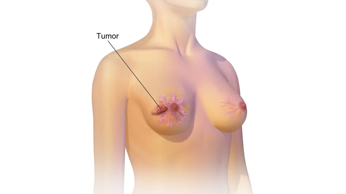
Breast cancer radiation is one of the most feared words in an oncology appointment — and one of the most misunderstood treatments in all of medicine. If you are reading this immediately after receiving a radiation recommendation, this guide was written for exactly that moment.
Before radiation begins, understanding your breast cancer stage and treatment plan clarifies exactly why your oncologist made this recommendation — and what the treatment is designed to achieve.
Why your oncologist recommended radiation after surgery
Radiation therapy is recommended after breast cancer surgery because even a technically complete surgical removal leaves behind microscopic cancer cells that no imaging can detect. For most patients who have had a lumpectomy or mastectomy, radiation is the step that converts a surgical result into a durable, long-term outcome.
The recommendation follows specific evidence-based criteria: your tumor stage, size, lymph node findings, and surgical margin status all factor into the decision.
How to use this article for your specific situation
This article is organized to match where you are in your treatment journey — not as a textbook chapter to read start to finish. Each section answers one clinical question that matters at one specific moment.
Go to the section that answers the question keeping you awake right now. That is the right place to start.
ℹ️ Medical Disclaimer: The radiation therapy protocols, dose parameters, fractionation schedules, side effect descriptions, long-term risk data, and at-home management guidance discussed in this article reflect current clinical guidelines and are provided for educational purposes only. Individual radiation therapy decisions — including schedule selection, technique choices such as deep inspiration breath hold (DIBH), boost dose planning, and side effect management — depend on factors specific to each patient, including tumor characteristics, surgical findings, lymph node status, cardiac anatomy, and comorbid conditions.
This article does not constitute a diagnosis, a treatment recommendation, or medical advice for any individual patient. Consult a board-certified radiation oncologist and your full oncology team before acting on any clinical information in this article. Coverage and insurance questions related to radiation therapy should be verified directly with your insurer and treating facility. If you experience a medical emergency, call 911 or go to the nearest emergency room immediately.
How radiation therapy targets breast cancer cells
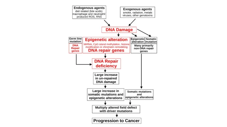
Radiation therapy kills breast cancer cells by delivering precisely aimed ionizing energy that causes irreparable double-strand DNA breaks — molecular damage that rapidly dividing cancer cells cannot recover from between treatment sessions. This targeted destruction covers not just the visible tumor bed but a margin of surrounding tissue where microscopic cancer cells may reside undetected by imaging.

🔬 How It Works: Cancer cells divide rapidly and have compromised DNA repair mechanisms compared to healthy tissue. Each daily session of radiation causes double-strand DNA breaks that accumulate in tumor cells. Normal breast tissue repairs itself between sessions. Cancer cells — unable to repair efficiently — accumulate lethal damage until replication is no longer possible. This biological asymmetry between cancer cells and normal tissue is the mechanistic foundation of fractionated radiation therapy.
The science behind radiation and cancer cell destruction
The primary delivery method for breast cancer is external beam radiation therapy — a linear accelerator delivers calibrated X-ray beams from outside the body to a precisely mapped target volume. The beams are aimed, not scattered, which is why the idea that radiation “goes everywhere in your body” is a clinical misconception.
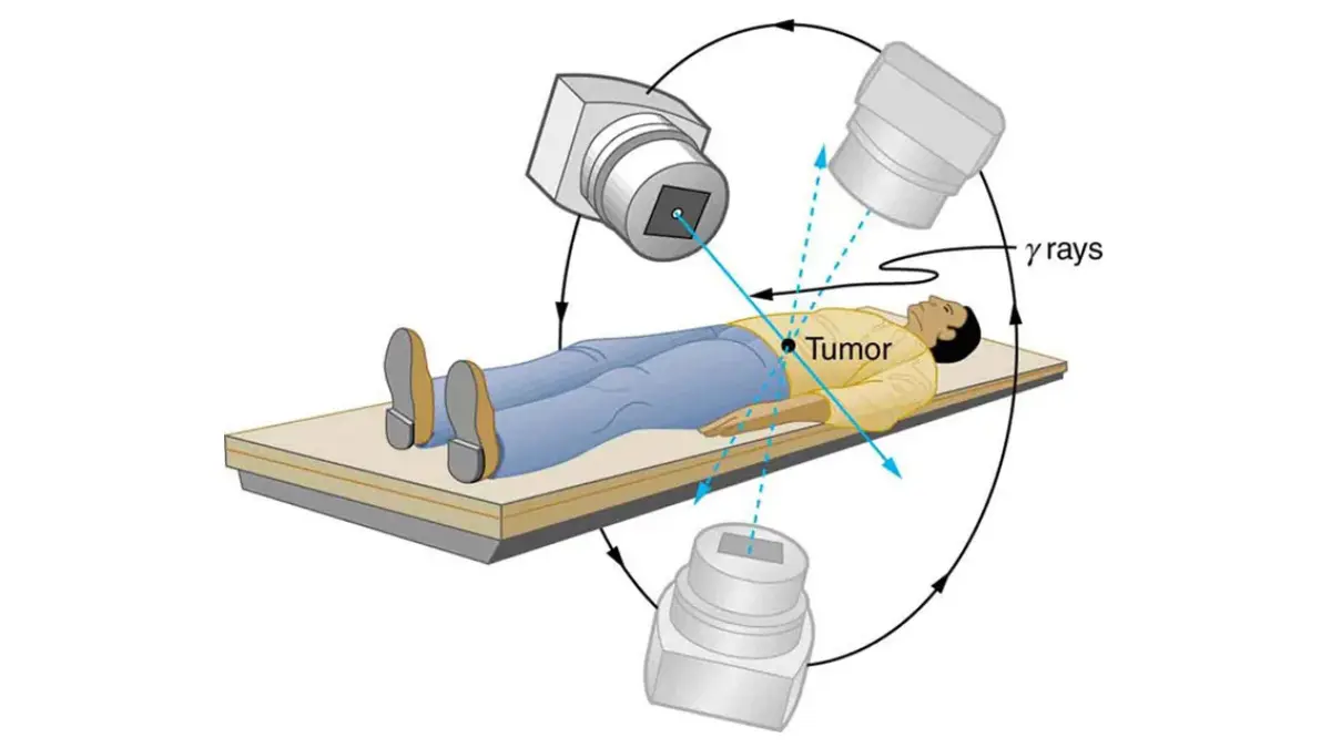
Modern intensity-modulated radiation therapy (IMRT) shapes the radiation dose to conform to the exact contour of the treatment target, reducing exposure to surrounding structures including the heart, lungs, and skin beyond the treatment field.
📊 Clinical Data Point: External beam radiation using IMRT and other modern delivery systems is endorsed as a standard of care option in the NCCN Clinical Practice Guidelines in Oncology: Breast Cancer, 2026 edition for eligible patients following lumpectomy or mastectomy with high-risk features. Source: NCCN, 2026. The NCI breast cancer radiation treatment overview provides a full clinical summary of radiation indications by stage.
Why radiation is given in daily sessions instead of one large dose
Delivering radiation in small daily amounts — a clinical principle called fractionation — is not a scheduling convenience. It is the mechanism that makes radiation safe for the tissue surrounding your tumor.
Each session deposits a measured dose in Gray (Gy). Healthy tissue repairs that damage overnight. Cancer cells, lacking efficient repair pathways, do not. Fractionation is what separates a precision cancer treatment from indiscriminate tissue damage — it is the core logic your radiation oncologist uses when designing your entire plan.
Short-term side effects of breast cancer radiation
The most common side effects of breast cancer radiation include skin changes, fatigue, and breast swelling — most of which begin in weeks 2–3 of treatment and resolve within 4–6 weeks after the final session. Knowing the difference between a side effect that is expected and manageable at home and one that requires an immediate call to your oncology team is the information that prevents both unnecessary fear and dangerous delays.
Skin changes: what’s normal and what needs medical attention
Radiation dermatitis follows a predictable clinical arc for most patients. Grade 1 — mild pink erythema, warmth, and tenderness in the treatment area — appears around week 2 in most patients and is expected. It is manageable at home with fragrance-free moisturizer and does not require a clinical visit on its own.
Grade 2 — dry desquamation with peeling skin and moderate discomfort — affects a smaller proportion of patients in weeks 3–4 and should be reported to your radiation oncology team at your next scheduled visit, not managed silently at home. Grade 3 — moist desquamation, blistering, or open sores — occurs in a minority of patients and requires immediate contact with your oncology team.
⚠️ Clinical Warning: Contact your radiation oncology team immediately — not your primary care physician — if you develop blistering or open sores in the treatment area, a fever above 100.4°F, or sudden shortness of breath during your radiation course. These symptoms may indicate a Grade 3 skin reaction or early radiation pneumonitis requiring urgent clinical assessment. Do not wait for your next scheduled appointment.
📊 Clinical Data Point: Radiation dermatitis severity is graded on a standardized 1–3 scale that your oncology team uses to assess skin response and determine whether to continue, modify, or pause treatment. Grading criteria and recommended management protocols are detailed in the ASTRO patient guide to breast cancer radiation. Source: ASTRO, 2026.
Radiation fatigue: why it happens and when it peaks
Radiation fatigue is not the acute crash associated with chemotherapy. It is a cumulative heaviness that builds gradually over the weeks of treatment — driven by ongoing cellular repair, mild systemic inflammation, and the logistical toll of daily appointments across weeks of treatment.
For most patients on a standard 5-week course, fatigue peaks in weeks 4–5 and begins improving within 2–4 weeks of completing treatment. A minority of patients experience fatigue lasting 3–6 months, particularly those who received chemotherapy before radiation — discuss cumulative toxicity management with your radiation oncologist before your first session if this applies to you.
Use our Symptom Checker to track your radiation side effects between appointments so your team has an accurate clinical picture at each visit rather than a summary recalled from memory.
Breast swelling, tenderness, and other common reactions
Breast swelling and heaviness in the treated area are common in the first weeks of radiation and typically resolve within weeks to months after treatment ends. Some patients notice temporary changes in breast firmness or contour during treatment — this is a normal inflammatory tissue response, not evidence of disease progression.
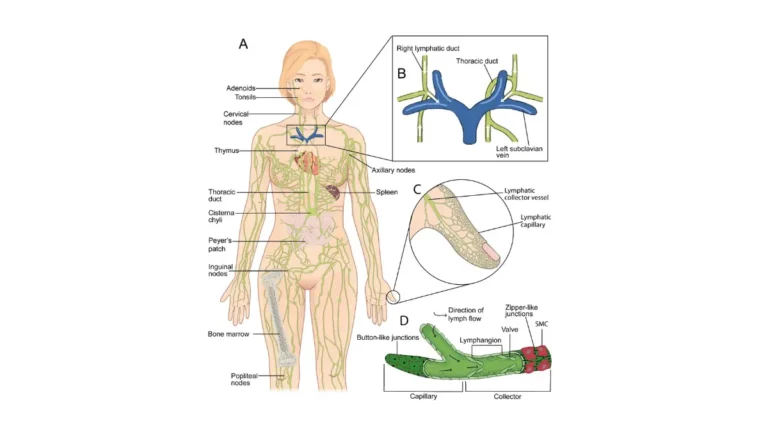
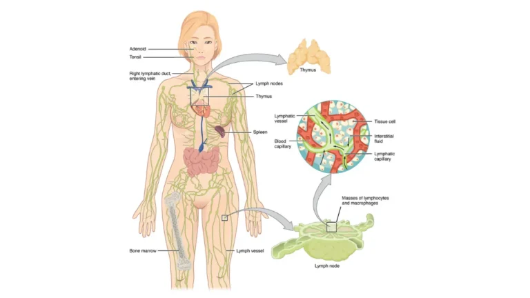
Lymphedema — fluid accumulation caused by lymphatic disruption — is a longer-term concern more specifically relevant to patients who also received lymph node removal or regional nodal irradiation. If your treatment plan includes nodal irradiation, ask your team about lymphedema prevention before treatment begins.
✅ Patient Action: Before your first radiation session, ask your radiation oncologist: “What Grade skin reaction should I realistically expect given my skin type, treatment field size, and whether I’m receiving a boost dose — and at exactly what point should I call your office rather than wait for my next appointment?” A specific threshold replaces generalized anxiety with a clear action plan.
Breast cancer radiation schedules: what your options are
Hypofractionated radiation therapy delivers a higher dose per session over fewer total appointments — typically 40 Gy in 15 fractions over 3 weeks — compared with the standard whole breast irradiation protocol of approximately 50 Gy in 25 fractions over 5 weeks, per 2026 NCCN guidelines. Your radiation oncologist selects a schedule based on your tumor characteristics, surgical findings, and cardiac anatomy — but knowing your options before that conversation puts you in a substantially better position.
Standard vs. hypofractionated radiation: what the research shows
The three primary breast cancer radiation schedules differ in total dose, number of sessions, treatment duration, and patient eligibility.
| Protocol | Dose | Sessions | Duration | Best For Patient Profile | Cardiac Risk Note |
|---|---|---|---|---|---|
| Standard WBI | ~50 Gy | 25 | 5 weeks | Post-lumpectomy or post-mastectomy patients with high-risk features; patients ineligible for shorter schedules | Lower dose per session; longer total course duration |
| Hypofractionated WBI | ~40 Gy | 15 | 3 weeks | Early-stage, node-negative post-lumpectomy patients; 2026 NCCN preferred option for most eligible patients | Fewer sessions; modern DIBH reduces cardiac exposure effectively |
| APBI | ~38.5 Gy | 10 | ~1 week | Age ≥50, T1 tumor, node-negative, hormone-receptor positive; per 2026 ASTRO consensus criteria | Smallest treatment volume; lowest cardiac exposure of all three protocols |
Source: NCCN Clinical Practice Guidelines in Oncology: Breast Cancer, 2026. See NCCN breast cancer treatment guidelines for full eligibility criteria by protocol.
📊 Clinical Data Point: Per the NCCN Clinical Practice Guidelines in Oncology: Breast Cancer, 2026 edition, hypofractionated whole breast irradiation is the preferred schedule for most eligible post-lumpectomy patients — specifically those with early-stage, node-negative disease without prior breast or chest wall irradiation. Source: NCCN, 2026.
Who qualifies for accelerated partial breast irradiation (APBI)
Accelerated partial breast irradiation (APBI) delivers radiation to the tumor cavity only — not the entire breast — completing treatment in 10 sessions over approximately one week (38.5 Gy in 10 fractions). APBI is not an abbreviated version of standard treatment for everyone; it is a specific indication for a specific patient profile.
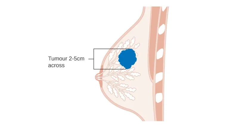
Per 2026 ASTRO consensus criteria, eligible patients are typically age 50 or older, with a tumor of 2 cm or smaller (T1), clear surgical margins, no lymph node involvement, and a hormone-receptor positive cancer subtype.
🩺 Physician Note: In my clinical experience, patients are often surprised to hear that a shorter schedule is not a downgraded option. For eligible patients, the evidence base for equivalent local disease control is solid. The question to bring to your radiation oncologist is not “Can I have the shorter schedule?” — it is “Do my specific pathology results and anatomical measurements meet the eligibility criteria?” If you cannot get a clear answer to that question during your consultation, a second radiation oncology opinion is entirely appropriate.
Your first radiation appointment: simulation and planning
Your first radiation appointment is a simulation session — no radiation is delivered on that day. Understanding each step in advance removes the anxiety that comes from walking into an unfamiliar clinical environment.
- CT simulation scan — a planning CT maps your breast tissue, chest wall, lymph nodes, heart, and lungs in precise anatomical detail.
- Permanent skin markings — small tattoo dots or temporary surface markers are placed so the linear accelerator reproduces the exact same beam geometry at every session.
- Treatment planning — a radiation physicist and your oncologist use the CT data to design your individualized dose plan; this takes 1–2 weeks after simulation.
- First treatment session — actual radiation delivery takes approximately 10–20 minutes per session; most of that time is positioning, not beam delivery.
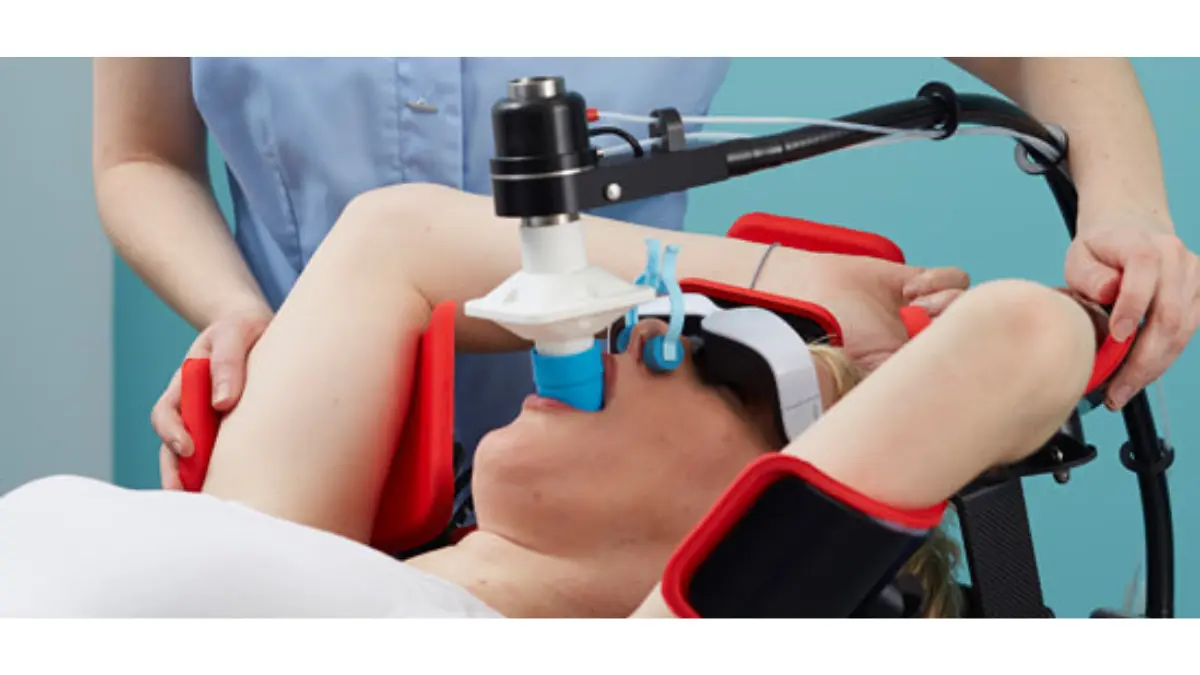
✅ Patient Action: Before your radiation simulation appointment, prepare three specific questions: “Will deep inspiration breath hold (DIBH) be used in my plan for cardiac protection? What is my planned boost dose and to which specific area? And what is the exact clinical reason my schedule was selected over the alternatives?” These three answers will give you a complete picture of your treatment before the first beam is delivered.
Long-term risks of breast cancer radiation therapy
Left-sided breast cancer radiation can increase cardiac exposure — but modern dose-sparing techniques, including deep inspiration breath hold, keep mean cardiac dose below 2 Gray for most patients. That is the threshold associated with clinically meaningful cardiac risk reduction per 2026 ASTRO guidelines. Understanding these risks in specific terms — not as a reason to decline treatment, but as a reason to ask the right questions before it begins — is the clinical literacy that protects you over the long term.
Heart risk: what left-sided breast radiation means for cardiac health
For patients with left-sided breast cancer, the proximity of the heart to the treatment field creates a dose-exposure risk that older radiation systems could not adequately solve. Modern technique has changed this materially.
🔬 How It Works: The deep inspiration breath hold (DIBH) technique asks the patient to inhale deeply and hold their breath during each beam delivery. This expands lung volume, physically increasing the distance between the chest wall target and the heart — reducing mean cardiac dose to below the ASTRO 2026 threshold of less than 2 Gray. Not every center offers DIBH; ask your radiation oncologist explicitly whether it is part of your treatment plan.
📊 Clinical Data Point: ASTRO 2026 clinical practice guidelines establish a mean cardiac dose of less than 2 Gray as the target threshold for left-sided breast cancer radiation patients treated with modern techniques. Source: ASTRO, 2026. The ACS guide to long-term breast cancer radiation effects provides a patient-facing overview of cardiac and pulmonary outcomes following treatment.
✅ Patient Action: If you are receiving radiation to the left breast, ask your radiation oncologist: “Will DIBH be used in my plan, and what will my mean cardiac dose be?” Then ask your cardiologist to establish a cardiac baseline before your first session. Annual cardiac monitoring for at least 10 years post-radiation is recommended for left-sided patients by 2026 cardio-oncology consensus guidelines — confirm this is part of your long-term follow-up plan before treatment ends.
Lung effects and radiation pneumonitis: who is most at risk
Radiation pneumonitis is an inflammatory response in lung tissue that receives incidental radiation during breast treatment — driven by a cytokine cascade that includes TGF-β and IL-6 signaling. It does not appear during treatment; onset typically occurs 6 weeks to 6 months after the final session.
Symptoms include dry cough, low-grade fever, and shortness of breath on exertion. These symptoms overlap with respiratory infections — any respiratory symptoms in this timeframe should prompt a call to your oncology team before you attribute them to a seasonal illness or cold.
Secondary cancer risk: putting the numbers in context
The absolute increase in secondary malignancy risk from modern breast radiation — primarily affecting the ipsilateral lung over decades — is real but small relative to the benefit of radiation in reducing local breast cancer recurrence. Your radiation oncologist can provide your individual risk-benefit calculation based on your specific treatment field and dose parameters.
For patients carrying a BRCA1 or BRCA2 mutation, secondary cancer risk after radiation requires a dedicated conversation with a genetic counselor. Assess your long-term cancer risk after treatment using our Genetic Risk Assessment Tool as a structured starting point. Review your breast cancer survival rates by stage to understand the full clinical context of the treatment decisions in front of you.
How to manage breast cancer radiation side effects at home
Caring for your skin during radiation therapy starts with a fragrance-free moisturizer applied twice daily to the treatment area — beginning on day one, not after irritation appears. Prevention consistently outperforms reaction. What follows is what oncology teams actually recommend, not generic skincare advice.
Skin care during radiation: what your team actually recommends
Radiation dermatitis management is clinical, not cosmetic. Follow these steps throughout your treatment course:
- Apply a fragrance-free, alcohol-free moisturizer (calendula-based formula or plain aloe vera gel) to the treatment area twice daily — morning and before bed.
- Avoid aluminum-containing antiperspirants on the treated side for the duration of your radiation course.
- Wear soft, loose cotton clothing that does not create friction over the treatment field.
- Protect the treatment area from direct sun exposure throughout and for several months after treatment.
- Contact your radiation oncology team immediately — not your primary care physician — if blistering, open sores, or weeping skin develops.
✅ Patient Action: If skin changes worsen despite consistent home care — specifically if you develop open sores, severe pain, or blistering — contact your radiation oncology nurse navigator immediately rather than waiting for your next scheduled appointment. You may need a brief treatment break and prescription topical management that cannot be substituted with over-the-counter products.
Managing fatigue: energy conservation and when to rest
Radiation fatigue is cumulative and physiological — it is your body doing the cellular repair work that radiation requires, not a sign that something is wrong. It is not weakness. It is biology.
Protect your highest-energy hours for what is most important — a work commitment, time with a family member, a meal you cook yourself. Rest during low-energy windows without requiring a reason. Most patients find fatigue manageable in weeks 1–2, with peak difficulty in weeks 3–5.
Nutrition, hydration, and what to avoid during treatment
Staying well-nourished and hydrated directly supports your body’s repair capacity throughout radiation. Calculate your daily hydration needs during radiation treatment using our Water Intake Calculator — dehydration amplifies fatigue and slows skin healing. Use our Protein Intake Calculator to ensure adequate daily protein, which directly supports tissue repair at the cellular level.
High-dose antioxidant supplements during active radiation — including megadose Vitamin C, Vitamin E, and beta-carotene — are clinically contested and should not be taken without discussing with your oncologist first. Ask specifically before starting any supplement during your radiation course.
A radiation oncologist’s perspective on treatment and risk
The evidence base for radiation therapy in breast cancer is not fragile — it is built on decades of Phase III clinical trial data showing that radiation after lumpectomy reduces 10-year local recurrence risk to a level that is clinically meaningful for the majority of patients with early-stage disease. Emerging 2026 trial data continues to confirm that hypofractionated schedules deliver equivalent local control to the standard 5-week course for most eligible patients.
What the evidence actually shows about radiation and recurrence
The clinical question patients rarely ask — but should — is not “Does radiation work?” It is “What happens to patients who do not complete it?”
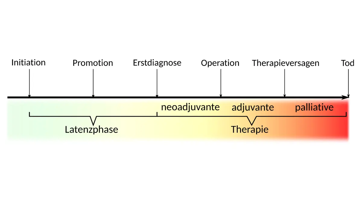
For early-stage, node-negative patients who decline radiation after lumpectomy, local recurrence rates at 10 years are substantially higher than for patients who complete a full course of radiation — a difference large enough that most board-certified radiation oncologists present this not as a persuasive argument but as the standard of care for eligible patients.
Patients interested in enrolling in current protocol research can review active breast cancer radiation clinical trials through the NCI clinical trials registry.
What I tell patients who are afraid of radiation side effects
As a gynecologic oncologist who works alongside radiation oncology teams throughout my patients’ treatment courses, I hear the same fear in almost every pre-radiation consultation. Patients want to know whether the treatment will change them permanently in ways that matter.
My answer is the same every time: modern radiation — IMRT, breath-hold protocols, precision cardiac-sparing planning — is not what the word “radiation” conjures in most patients’ minds. The permanent changes most patients experience are small. The permanent benefit of completing treatment is substantial.
Optimize your sleep schedule during radiation treatment using our Sleep Calculator — a consistent sleep schedule is one of the most evidence-supported daily tools for managing radiation fatigue across the weeks of treatment.
Breast cancer radiation: questions patients ask most
1. How many radiation sessions does breast cancer treatment require?
The number of sessions depends on your protocol. Standard whole breast irradiation runs 25 sessions over 5 weeks. Hypofractionated radiation — the 2026 NCCN preferred option for most eligible early-stage post-lumpectomy patients — runs 15 sessions over 3 weeks. Accelerated partial breast irradiation (APBI) for highly selected patients completes in 10 sessions over approximately one week. Consult a board-certified radiation oncologist to confirm which schedule applies to your specific case.
2. What does radiation therapy physically feel like for breast cancer?
The radiation beam is completely painless during delivery — you feel nothing while the machine is active. Most patients are genuinely surprised by the absence of any sensation. What develops over the treatment weeks is not pain from the beam itself but cumulative skin sensitivity, tenderness in the treated area, and mounting fatigue from the body’s tissue repair response. Reactions typically begin in weeks 2–3 and improve within 4–6 weeks of completing your breast cancer radiation course.
3. How long does fatigue last after breast cancer radiation?
Radiation fatigue peaks in weeks 4–5 of a standard 5-week course and begins improving within 2–4 weeks of completing treatment for most patients. A minority experience fatigue lasting 3–6 months, more commonly in patients who received prior chemotherapy. If fatigue significantly affects daily function beyond 6 weeks post-treatment, discuss evaluation for treatment-related anemia or thyroid changes with your oncologist — these are correctable contributing factors. Consult a board-certified oncologist if prolonged fatigue develops.
4. Is hypofractionated radiation as effective as the standard schedule?
Phase III non-inferiority trial evidence supports equivalent local recurrence control between hypofractionated whole breast irradiation (40 Gy in 15 fractions) and standard WBI (50 Gy in 25 fractions) for eligible early-stage patients. The 2026 NCCN guidelines designate hypofractionation as the preferred protocol for most post-lumpectomy, node-negative patients — not a compromise, but an evidence-based equivalent with a shorter course. Consult a board-certified radiation oncologist to confirm your specific eligibility for hypofractionation.
5. What are the long-term side effects of breast cancer radiation?
The primary long-term side effects of breast cancer radiation include cardiac toxicity (most significant for left-sided patients), radiation pneumonitis, breast tissue fibrosis, lymphedema for patients who received nodal irradiation, and a small absolute increase in secondary malignancy risk over decades. Modern cardiac-sparing techniques substantially reduce cardiac risk for left-sided patients. Consult a board-certified radiation oncologist and cardiologist to establish long-term monitoring protocols before your treatment course ends.
6. Can breast cancer radiation cause heart damage?
Left-sided breast cancer radiation carries a cardiac exposure risk — but modern DIBH (deep inspiration breath hold) technique reduces mean cardiac dose below the ASTRO 2026 threshold of less than 2 Gray for most patients. This is substantially lower than cardiac risk associated with older radiation techniques. Annual cardiac monitoring for at least 10 years is recommended for left-sided patients by 2026 cardio-oncology consensus guidelines. Consult a board-certified radiation oncologist and cardiologist to confirm your cardiac monitoring plan.
7. How should I care for my skin during breast cancer radiation?
Apply a fragrance-free moisturizer twice daily beginning on day one — before radiation dermatitis appears, not after. Avoid aluminum-containing antiperspirants on the treated side. Wear soft, loose cotton clothing over the treatment field. Protect the area from sun exposure throughout treatment. Report blistering, open sores, or weeping skin to your oncology team immediately — Grade 3 skin reactions require prescription management and cannot be safely addressed at home without clinical oversight.
8. What happens at a first radiation therapy appointment for breast cancer?
Your first appointment is a simulation session — no radiation is delivered on this day. The team performs a CT scan to map the treatment target, places small permanent skin markings for reproducible beam alignment, and begins building your individualized dose plan. Planning takes 1–2 weeks; actual treatment sessions then begin, each running approximately 10–20 minutes. Consult your radiation oncology team regarding what to bring, what to wear, and how long the simulation appointment typically runs at your center.
9. Can I keep working during breast cancer radiation treatment?
Most patients in desk-based or low-physical-demand roles maintain employment throughout breast cancer radiation therapy. Fatigue typically does not significantly affect function in weeks 1–2 but may require accommodations in weeks 4–5. Physical labor, outdoor work over the treatment field, or sustained concentration during peak fatigue windows may need temporary modification. Whether to take leave depends on your job type, commute logistics, and individual fatigue response — not a universal rule. Consult your board-certified radiation oncologist about your specific work situation.
10. Does breast cancer radiation increase the risk of developing another cancer?
Modern breast cancer radiation carries a small absolute increase in secondary malignancy risk — primarily affecting the treated-side lung over decades. The benefit of radiation in reducing local breast cancer recurrence is substantially larger than this secondary risk for the majority of eligible patients. Your radiation oncologist can provide your individual risk-benefit calculation based on your specific treatment field and dose parameters. Consult a board-certified radiation oncologist before making treatment decisions based on secondary cancer risk concerns alone.
11. What is accelerated partial breast irradiation (APBI)?
APBI delivers radiation to the tumor cavity only — not the entire breast — in 10 sessions over approximately one week (38.5 Gy in 10 fractions). Per 2026 ASTRO consensus criteria, eligible patients are typically age 50 or older, with a tumor of 2 cm or smaller, clear margins, no lymph node involvement, and hormone-receptor positive disease. Consult a board-certified radiation oncologist to determine whether your specific pathology and anatomy meet APBI eligibility criteria.
Your next step after reading this guide
The most useful thing you can do with this information is not to memorize it — it is to use it to have a better conversation with your radiation oncologist.
Before your simulation appointment, prepare a written list of the questions that matter most to you: your fractionation schedule and why it was selected, your cardiac risk management plan if you have left-sided disease, and your home side effect protocol for the weeks ahead. Bring it, and do not leave that appointment without specific answers.
Breast cancer radiation is a clinical intervention with a strong evidence base and a physician team whose specific job is to guide you through it. Return to your complete breast cancer stages and treatment overview for the full clinical picture of where radiation fits within your entire care plan.
About this content
How this article was put together: researched from recognised health sources, drafted with the help of AI tools, and edited by hand, with sources linked throughout.

Sameer Patel is the founder and editor of My Medicine Advisor. He is not a doctor or medical professional — before starting this site he worked in banking,…
Medical disclaimer
The content on MyMedicineAdvisor is provided for general informational and educational purposes only and is not a substitute for professional medical advice, diagnosis, or treatment. Health information on this website should not be used to diagnose, treat, cure, or prevent any condition without guidance from a qualified healthcare professional. Always seek the advice of your doctor, physician, or another licensed healthcare provider with any questions you may have regarding a medical condition, symptoms, medications, or treatment decisions.