
On This Page – Quick Medical Summary
You just heard the words “DCIS” — here is what that diagnosis actually means
If you are holding a biopsy report that says DCIS breast cancer and you do not know whether to feel relieved or afraid — that uncertainty is exactly right. This article is written for three specific readers: the woman who just received a Stage 0 diagnosis and needs to understand what it means before her first surgical consultation; the caregiver researching on behalf of a family member who cannot yet process the pathology report alone; and the patient who has heard the phrase “non-invasive cancer” and is unsure how seriously to take it.
Not yet sure whether your symptoms need evaluation? Use our free breast symptom checker first. For the full clinical context of where Stage 0 fits within the broader framework, the breast cancer stages and survival guide covers the complete picture.
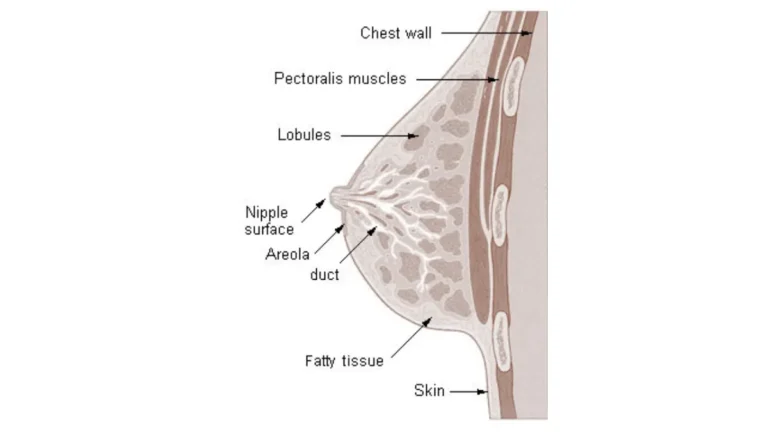
Ductal carcinoma in situ — DCIS — is a Stage 0 breast cancer in which abnormal cells have formed inside the milk ducts of the breast but have not broken through the duct wall into surrounding tissue. Every major oncology guideline classifies it as cancer. It is also one of the most treatable diagnoses in breast oncology when detected and managed correctly.
This article covers what your nuclear grade means clinically, how DCIS is found, what treatment options are available in 2026, and what the current survival data shows. Dr. Fairweather walks you through every question you are likely to face before your first specialist appointment.
ℹ️ Medical Disclaimer: The diagnostic criteria, treatment options, and medication information discussed in this article — including DCIS nuclear grading, lumpectomy and mastectomy indications, radiation therapy recommendations, tamoxifen and aromatase inhibitor prescribing, sentinel lymph node biopsy, Oncotype DX DCIS testing, and health insurance coverage considerations — reflect current 2026 clinical guidelines and are provided for educational purposes only. Individual diagnostic conclusions, treatment decisions, and medication choices depend on factors including patient history, comorbidities, pathology results, BRCA mutation status, and specialist assessment. Consult a board-certified breast surgeon or gynecologic oncologist before acting on any clinical information in this article.
What is DCIS breast cancer — and is it considered real cancer?
DCIS, or ductal carcinoma in situ, is classified as a malignancy by the National Cancer Institute, the American Cancer Society, and every major oncology guideline currently in use. The Latin phrase “in situ” means “in place” — and it describes the single biological feature that separates DCIS from all higher breast cancer stages.
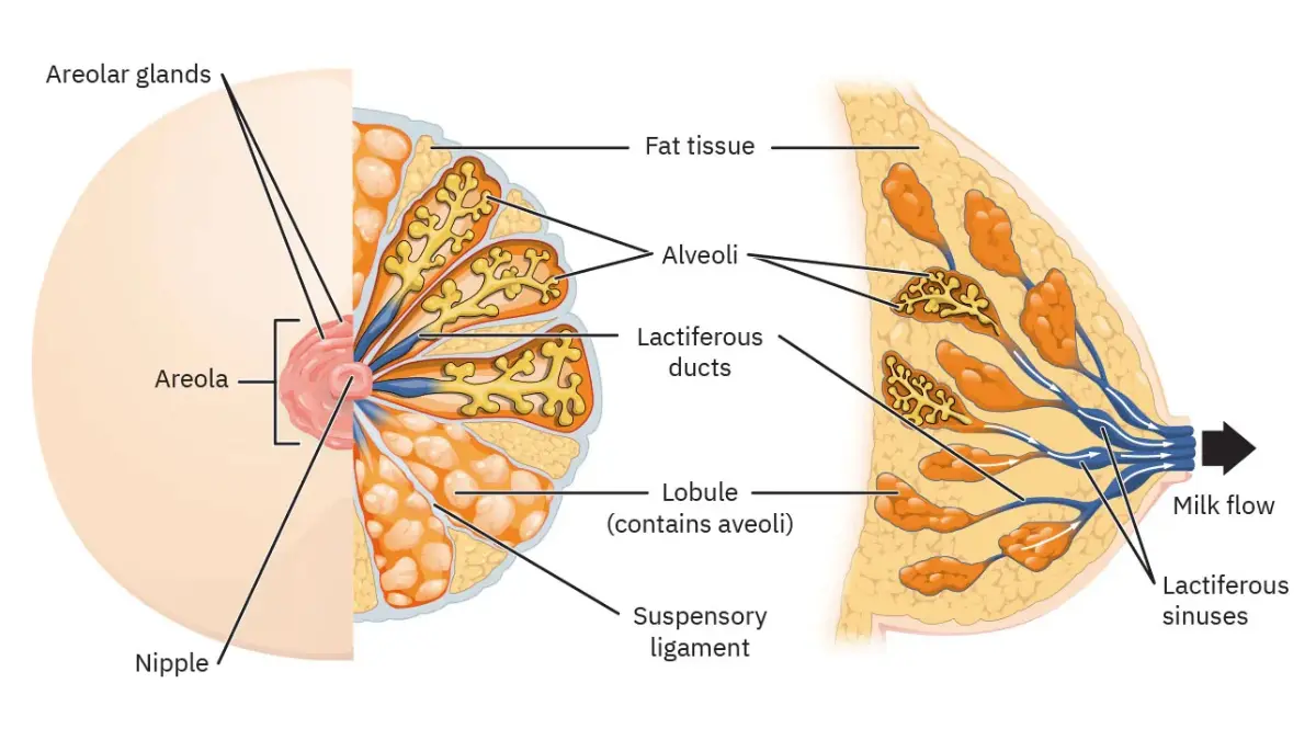
🔬 How It Works: Inside each breast duct runs a thin structural wall called the basement membrane. In DCIS, abnormal cells have multiplied inside the duct, but this wall remains intact. With the basement membrane unbreached, cancer cells have no pathway to the lymphatic system or bloodstream — which is why DCIS cannot spread to other organs in its current state.
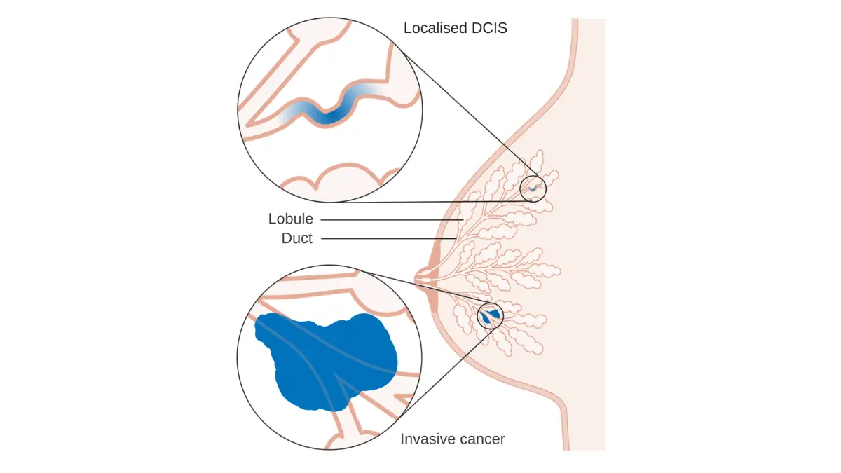
How DCIS is different from invasive breast cancer
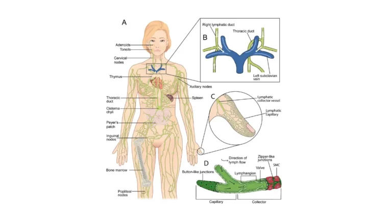
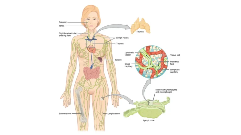
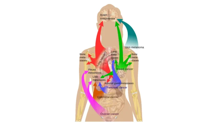
In invasive breast cancer, cancer cells have broken through the basement membrane into surrounding breast tissue, gaining access to lymph nodes and the bloodstream. In DCIS, that boundary has not been crossed. This is not a matter of degree — it is a fundamental difference in biology that defines the diagnosis, the prognosis, and the treatment approach simultaneously.
Most DCIS is estrogen receptor positive (ER-positive), meaning the cancer cells are stimulated by the hormone estrogen. Cases that are receptor-negative behave more similarly to hormone receptor-negative breast cancer, including triple-negative subtypes, and require different clinical management.
Why doctors call DCIS Stage 0
Stage 0 is the lowest position in the breast cancer staging system — below Stage I, II, III, and IV. The designation does not mean the diagnosis is insignificant. Clinical evidence consistently shows that a subset of untreated DCIS will progress to invasive cancer over time, which is precisely why active treatment is the standard recommendation for most patients.
📊 Clinical Data Point: The National Cancer Institute defines DCIS as a non-invasive carcinoma originating in the breast ducts with an intact basement membrane — the structural basis for its Stage 0 classification and its distinct treatment rationale. Source: NCI’s clinical overview of DCIS and breast cancer treatment. NCI, 2026.
✅ Patient Action: If you are uncertain whether your pathology report describes DCIS or an invasive component, ask your oncologist specifically: “Does my biopsy show any evidence of microinvasion — tumor cells that have crossed the duct wall by 1mm or less?” The answer changes your staging and your treatment discussion from the ground up.
DCIS grades explained: low, intermediate, and high — why your grade matters
The most clinically important finding on your pathology report — beyond the DCIS diagnosis itself — is your nuclear grade. Grade tells your surgeon how abnormal the cancer cells look under microscopy, how rapidly they are dividing, and which treatment intensity the evidence supports for your case.
DCIS is classified into three nuclear grades:
- Grade 1 — Low-grade: Cancer cell nuclei are small and uniform, closely resembling normal breast duct cells. Mitotic activity is low. Low-grade DCIS tends to grow slowly and carries the lowest ipsilateral recurrence risk after treatment.
- Grade 2 — Intermediate-grade: Nuclear size and shape fall between low and high grade, with moderate mitotic activity. Intermediate-grade DCIS requires treatment, though the specific approach is individualized based on size, margin status, and receptor profile.
- Grade 3 — High-grade: Cancer cell nuclei are large, irregular, and variable in size — a pattern called pleomorphism. Mitotic activity is high. High-grade DCIS carries the greatest risk of local recurrence and the highest likelihood of progressing to invasive cancer if left untreated.
Understanding personal breast cancer risk factors — including age, BRCA mutation status, and family history — alongside your nuclear grade gives you a more complete picture of your individual clinical profile before the treatment conversation begins.
What comedo necrosis means — and why it changes your treatment conversation
Comedo necrosis is a pathological finding that frequently accompanies high-grade DCIS. It refers to areas of central cell death — visible as dark debris inside the duct lumen on microscopy. Its presence signals biologically aggressive tumor behavior and consistently elevates recurrence risk beyond what grade alone predicts.
🩺 Physician Note: In my clinic, the combination of grade 3 nuclear grade and comedo necrosis on biopsy is the finding that most consistently changes the treatment conversation — specifically toward radiation after lumpectomy, and sometimes toward a mastectomy discussion. These two findings together indicate a higher recurrence risk than either finding alone. If both appear on your pathology report, ask your surgeon directly: “How does comedo necrosis change my margin requirement and my radiation recommendation?” — Dr. Carolyn D. Fairweather, MD, Gynecologic Oncology
✅ Patient Action: Ask your pathologist or breast oncologist specifically whether comedo necrosis was identified in your biopsy specimen — not just your nuclear grade. This single finding directly changes the recurrence risk calculation your surgeon uses when designing your treatment plan.
How DCIS is found: what happens from mammogram to diagnosis
DCIS produces no physical symptoms in approximately 80% of patients. There is no lump to feel. There is no pain. For the vast majority of women, a DCIS diagnosis begins not with a symptom — but with a routine annual mammogram.
Why DCIS almost never causes symptoms
Because DCIS cells are confined inside the breast ducts, they rarely form a palpable mass or cause the visible external changes — nipple discharge, skin dimpling, or breast shape changes — that characterize more advanced disease. The early signs of breast cancer most patients recognize simply do not apply to Stage 0 disease.
What a mammogram detects instead are microcalcifications — tiny calcium deposits, smaller than a grain of salt, that form inside the duct around abnormal cells. When these appear in clusters or linear branching patterns on the image, they are a recognized radiological signature of DCIS.
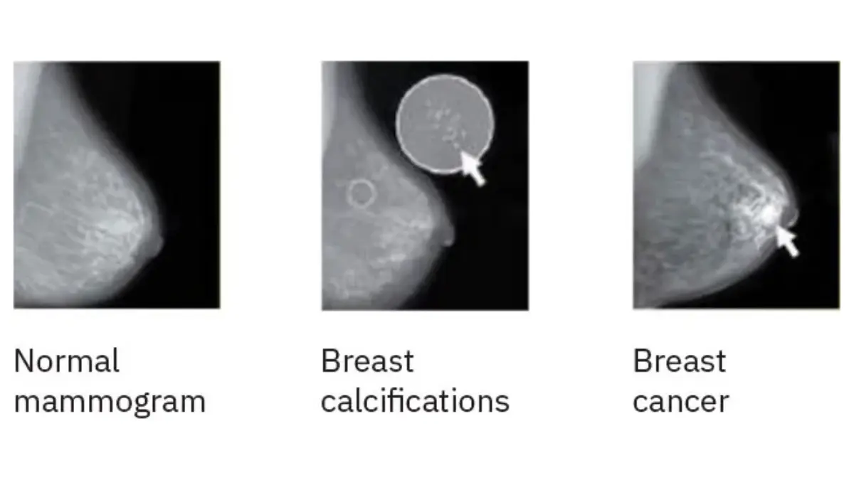
From abnormal result to biopsy: the diagnostic steps
If your mammogram shows suspicious microcalcifications, the diagnostic pathway follows this sequence:
- Callback for diagnostic mammogram or breast MRI: Additional imaging confirms the location and extent of the finding.
- Core needle biopsy under imaging guidance: A radiologist uses stereotactic or ultrasound guidance to extract tissue from the suspicious area.
- Pathology analysis: The tissue is examined for cell type, nuclear grade, hormone receptor status, and the presence or absence of comedo necrosis.
- Specialist referral: A confirmed DCIS diagnosis is referred to a breast surgeon for treatment planning.
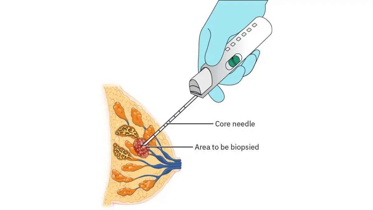
Because DCIS detection depends heavily on routine screening, patients with a confirmed diagnosis — particularly those with a family history of breast or ovarian cancer — should assess their hereditary breast cancer risk as part of the pre-treatment evaluation.
✅ Patient Action: Before agreeing to a core needle biopsy, ask your radiologist: “What specific finding on my mammogram is driving this recommendation, and which biopsy technique — stereotactic or ultrasound-guided — is most appropriate for my case?” Understanding the imaging rationale prepares you for every conversation that follows.
DCIS treatment options: how surgeons decide between lumpectomy, mastectomy, radiation, and hormone therapy
Treatment for DCIS breast cancer is not determined by diagnosis alone. Your surgeon’s recommendation will be shaped by four primary variables: nuclear grade, the size of the DCIS relative to your breast, surgical margin status, and your hormone receptor and BRCA mutation results.
The four established treatment options for DCIS are:
- Lumpectomy with radiation: The most common approach. The surgeon removes the DCIS-containing tissue with a surrounding rim of normal tissue. Per the NCCN Breast Cancer Treatment Guidelines current for 2026, an adequate surgical margin for DCIS lumpectomy is defined as 2mm or greater. Radiation follows to reduce ipsilateral recurrence risk in the remaining breast tissue.
- Mastectomy: Removal of the breast. Indicated when DCIS is multicentric (in two or more breast quadrants), when clear margins cannot be achieved after re-excision, when a BRCA1 or BRCA2 pathogenic variant is confirmed, or when the patient chooses mastectomy after fully informed discussion. What that surgery involves — and what surgeons often do not explain in advance — is covered in our complete guide to mastectomy.
- Hormone therapy: For ER-positive DCIS, FDA-approved tamoxifen for breast cancer risk reduction reduces the risk of recurrence and contralateral breast cancer. Aromatase inhibitors are the alternative for postmenopausal patients who cannot tolerate tamoxifen. This is not chemotherapy — it is a targeted hormonal therapy administered after surgery.
- Active surveillance: A highly selective option under clinical investigation, appropriate only for elderly patients with low-grade DCIS and significant medical comorbidities. Active surveillance is not a 2026 NCCN standard-of-care recommendation for most newly diagnosed patients.
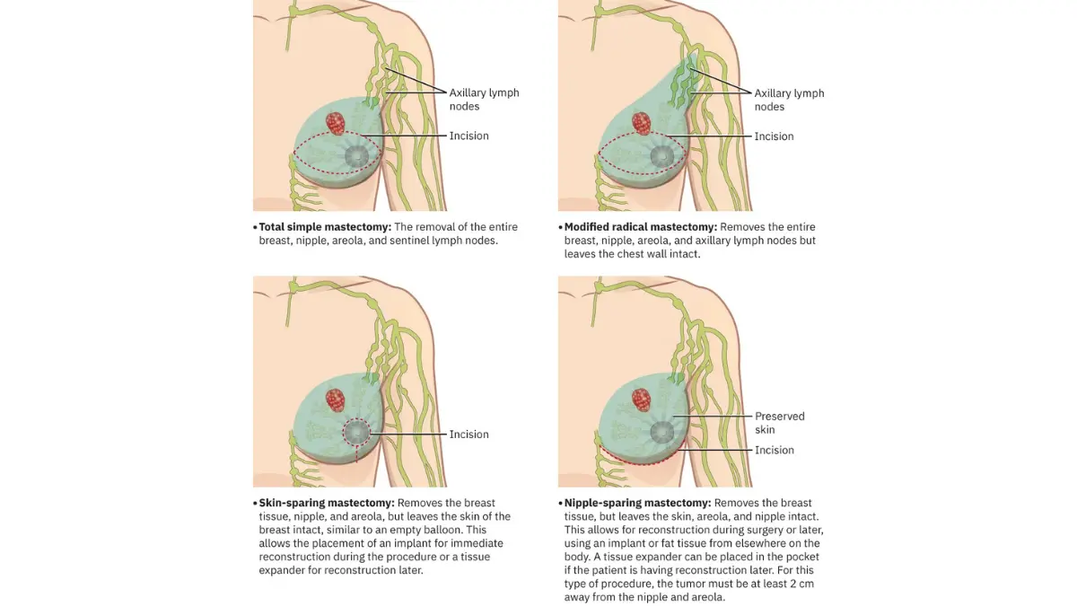
Lumpectomy vs. mastectomy: the four questions your surgeon weighs
The surgical decision is composite, not binary. Surgeons assess tumor size relative to breast volume, the ability to achieve a 2mm negative margin, confirmed BRCA mutation status, and the patient’s own informed preferences.
🩺 Physician Note: When a patient with high-grade DCIS asks me “Do I have to lose my breast?” — my answer begins with four specific questions. How large is the DCIS relative to your breast volume? Has your BRCA result been confirmed? Can we achieve a 2mm clear margin surgically? And what does your reconstruction timeline look like? Those four answers — not grade alone — determine the recommendation. If your surgeon has not addressed all four, ask them directly. — Dr. Carolyn D. Fairweather, MD, Gynecologic Oncology
A note on HER2 status and hormone therapy decisions
A subset of DCIS cases also show HER2 protein overexpression on immunohistochemistry — a biological finding that overlaps with HER2-positive breast cancer and that your oncologist will note in the treatment discussion. For ER-positive DCIS, tamoxifen is the primary pharmacologic risk-reduction tool post-surgery; it reduces future breast cancer events, not the DCIS that has already been surgically removed.
✅ Patient Action: Before consenting to any surgical procedure for DCIS, ask your breast surgeon two specific questions: “What is my Van Nuys Prognostic Index score, and does it support lumpectomy or mastectomy for my case?” and “Should BRCA genetic testing be completed before we finalize the surgical approach?” Both directly shape the recommendation you receive.
DCIS breast cancer survival rate and recurrence risk: what 2026 data shows
For most patients, the survival data for DCIS is the most reassuring statistic in oncology. For a small subset, it is also the most important number to interpret correctly — because DCIS left untreated does carry risk.
5-year and 10-year survival rates for Stage 0 breast cancer
📊 Clinical Data Point: According to NCI SEER data updated for 2026, the 5-year relative survival rate for localized breast cancer — the category that includes Stage 0 DCIS — exceeds 99% when treated. Ten-year survival for treated DCIS also remains high across all grade categories. Source: NCI SEER breast cancer survival statistics by stage.
The reason DCIS survival is near-complete is mechanistic, not coincidental. Because DCIS cells are biologically confined inside the breast duct with the basement membrane intact, they have no pathway to lymph nodes or distant organs. Mortality risk associated with DCIS arises only if the disease is left untreated and progresses to invasive cancer — which surgical and medical treatment is designed to prevent. For a complete comparison of survival rates across all breast cancer stages, see breast cancer survival rates by stage.
What raises your risk of DCIS coming back after treatment
Ipsilateral breast recurrence (IBR) — the return of cancer in the treated breast — is the primary clinical risk after DCIS surgery. Clinical evidence consistently identifies the following as recurrence risk elevators:
- High nuclear grade (grade 3) with comedo necrosis present
- Surgical margins that are close (less than 2mm) or positive after lumpectomy
- Younger patient age at diagnosis (under 45)
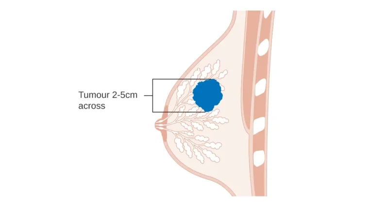
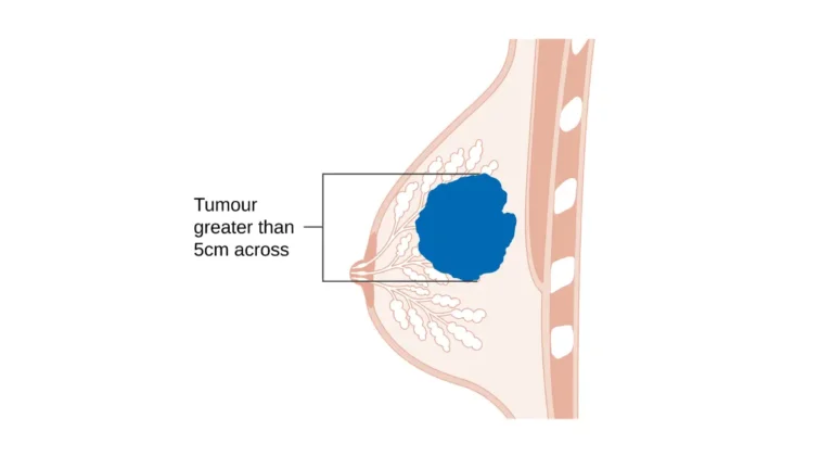
- Larger tumor size relative to breast volume
- Lumpectomy performed without subsequent radiation therapy
Approximately half of recurrences after DCIS treatment are DCIS again; the other half are invasive cancer. This is the clinical reality that makes margin status and radiation decisions consequential — not just technical.
⚠️ Clinical Warning: A local recurrence that is invasive — not DCIS — changes the entire treatment landscape, including systemic therapy considerations. Patients who decline radiation after lumpectomy should understand that this decision is associated with a measurably higher recurrence risk in most grade categories, per 2026 clinical evidence.
What I tell every new DCIS patient: a gynecologic oncologist’s perspective
The most common misconception I encounter in a new DCIS consultation is this: “Stage 0 means I can take my time.” That framing is clinically dangerous. DCIS has a window of manageability — and the decisions made in the first weeks after diagnosis significantly affect what that window looks like.
The three questions every new DCIS patient should ask their breast surgeon
Before leaving your first surgical consultation, ask these three questions specifically:
- “What is my Van Nuys Prognostic Index score, and what does it recommend for my case?” This validated scoring system integrates nuclear grade, tumor size, margin width, and patient age into a composite recurrence risk score that directly informs the lumpectomy vs. mastectomy decision.
- “Should I complete BRCA genetic testing before we finalize the surgical approach?” A confirmed BRCA1 or BRCA2 pathogenic variant changes the surgical recommendation for many patients — and reviewing how to read your BRCA results before your appointment will help you understand what that test means.
- “Am I a candidate for Oncotype DX DCIS testing to help decide whether I need radiation after lumpectomy?” This genomic test, developed specifically for DCIS, is emerging as a tool to individualize the radiation decision for low-grade and intermediate-grade patients — reducing overtreatment in some cases and identifying higher-risk patients in others.
Why a second opinion is always appropriate before DCIS surgery
A second opinion before any DCIS surgery is not a reflection of distrust toward your surgeon. It is a reflection of your right to a fully informed treatment decision — and any competent breast surgeon will support it. Patients who want a rapid second opinion from a board-certified breast surgeon can access telehealth consultation services within 48–72 hours.
Patients interested in emerging treatment protocols — including active surveillance validation studies and Oncotype DX DCIS outcome trials — can explore active clinical trials for DCIS breast cancer currently enrolling at centers across the United States.
✅ Patient Action: Before finalizing your DCIS treatment plan, ask your breast surgeon specifically: “Am I a candidate for Oncotype DX DCIS testing, and would the result change your recommendation about radiation after lumpectomy?” This single question can reshape the post-surgical treatment plan for eligible patients.
Frequently asked questions about DCIS breast cancer
Q1: What does DCIS stand for in breast cancer?
DCIS stands for ductal carcinoma in situ — a Stage 0 DCIS breast cancer in which abnormal cells have formed inside the milk ducts of the breast. “In situ” means the cancer cells remain confined within the duct wall and have not invaded surrounding breast tissue, lymph nodes, or other organs.
Q2: Is DCIS considered real cancer or pre-cancer?
DCIS breast cancer is classified as a malignancy by every major oncology organization, including the National Cancer Institute. The term “pre-cancer” understates the diagnosis. DCIS is biologically confined — its cells cannot spread in their current state — but clinical evidence consistently shows a meaningful proportion of untreated DCIS progresses to invasive cancer over time, which is why treatment is recommended for most patients.
Q3: What is the survival rate for DCIS breast cancer?
According to NCI SEER data updated for 2026, the 5-year relative survival rate for localized breast cancer — including Stage 0 DCIS — exceeds 99% when treated. Ten-year survival also remains high with standard care. DCIS itself cannot spread through the bloodstream or lymph nodes; mortality risk arises only if untreated DCIS progresses to invasive disease, which treatment prevents.
Q4: Does DCIS always require treatment?
The vast majority of DCIS breast cancer diagnoses are treated because untreated disease carries a documented risk of invasive progression. Active surveillance — watchful waiting without immediate surgery — is being studied in highly selected patients with low-grade DCIS and significant medical comorbidities, but it is not a standard 2026 NCCN recommendation for most patients. Consult a board-certified breast surgeon to determine whether active surveillance is appropriate for your specific pathology results.
Q5: What is the difference between DCIS and invasive breast cancer?
In DCIS breast cancer, abnormal cells are confined inside the breast duct — the basement membrane, the thin structural wall lining the duct, remains intact. In invasive breast cancer, cancer cells have broken through the basement membrane into surrounding tissue, gaining access to lymph nodes and the bloodstream. This structural boundary separates Stage 0 from every higher breast cancer stage and fundamentally changes the prognosis.
Q6: What grade is my DCIS and why does it matter?
DCIS breast cancer is graded 1 (low), 2 (intermediate), or 3 (high) based on how abnormal cancer cell nuclei appear under microscopy. Grade 3 carries the highest recurrence risk and the greatest likelihood of invasive progression without treatment. The presence of comedo necrosis alongside grade 3 elevates risk further. Ask your pathologist whether comedo necrosis appears in your specimen. Consult a board-certified breast oncologist to interpret how your grade affects your full treatment plan.
Q7: Can DCIS spread to the lymph nodes?
True DCIS breast cancer, with its basement membrane intact, does not spread to lymph nodes. However, sentinel lymph node biopsy is sometimes performed alongside mastectomy for high-grade DCIS, or when microinvasion — tumor cells extending just beyond the duct wall by 1mm or less — is suspected on biopsy. If your pathology report mentions microinvasion, ask your breast surgeon specifically whether lymph node evaluation is indicated in your case.
Q8: What is the best treatment for high-grade DCIS?
High-grade DCIS breast cancer is typically treated with lumpectomy and radiation — provided clear surgical margins of 2mm or greater can be achieved. When margins cannot be obtained, when disease is multicentric, or when a BRCA pathogenic variant is confirmed, mastectomy is recommended per 2026 NCCN guidelines. For ER-positive high-grade DCIS, tamoxifen is added after surgery. Consult a board-certified breast surgeon who can review your specific margin status and BRCA result before deciding.
Q9: Do I need chemotherapy for DCIS breast cancer?
Chemotherapy is not a standard treatment for DCIS breast cancer. The pharmacologic option for DCIS is hormone therapy — tamoxifen for premenopausal ER-positive patients, or an aromatase inhibitor for postmenopausal ER-positive patients. These are targeted hormone-blocking medications, not cytotoxic chemotherapy. Chemotherapy is reserved for invasive breast cancer with systemic spread risk. If chemotherapy is recommended for your DCIS without a confirmed invasive component, consult a board-certified oncologist for a second opinion.
Q10: What is my risk of DCIS coming back after treatment?
After lumpectomy with radiation, clinical evidence consistently shows a 10-year DCIS breast cancer recurrence risk that varies by nuclear grade, margin status, patient age, and comedo necrosis — with higher-risk profiles carrying substantially greater recurrence risk than lower-risk profiles. Approximately half of recurrences after DCIS treatment are DCIS again; the other half are invasive. Ask your breast surgeon or oncologist to estimate your personalized recurrence risk before you finalize your treatment plan.
Q11: Can DCIS be detected without a mammogram?
DCIS breast cancer produces no physical symptoms in approximately 80% of patients — no palpable lump, no pain, no visible changes. It is detected almost exclusively through routine mammogram screening, which identifies suspicious microcalcification clusters inside the ducts. Breast MRI can detect DCIS in high-risk patients, including BRCA carriers, but is not a standard screening tool for average-risk women. Routine annual mammography remains the primary DCIS detection pathway.
Your next step after a DCIS diagnosis
A Stage 0 DCIS breast cancer diagnosis is not a reason to panic — and it is not a reason to delay. It is a reason to act with information. You now have the clinical framework to understand your grade, interpret your pathology report, and have a specific, informed conversation with your breast surgeon.
Your next step is a consultation with a board-certified breast surgeon or gynecologic oncologist who can review your specific nuclear grade, margin status, BRCA results, and receptor profile and give you a treatment recommendation tailored to your exact diagnosis.
To understand how DCIS fits within the full staging framework — and what comes after Stage 0 in the clinical picture — the breast cancer stages and survival guide is the right next read.
About this content
How this article was put together: researched from recognised health sources, drafted with the help of AI tools, and edited by hand, with sources linked throughout.

Sameer Patel is the founder and editor of My Medicine Advisor. He is not a doctor or medical professional — before starting this site he worked in banking,…
Medical disclaimer
The content on MyMedicineAdvisor is provided for general informational and educational purposes only and is not a substitute for professional medical advice, diagnosis, or treatment. Health information on this website should not be used to diagnose, treat, cure, or prevent any condition without guidance from a qualified healthcare professional. Always seek the advice of your doctor, physician, or another licensed healthcare provider with any questions you may have regarding a medical condition, symptoms, medications, or treatment decisions.