
On This Page – Quick Medical Summary
When your breast feels wrong but there’s no lump to find
Inflammatory breast cancer (IBC) is a rare, aggressive form of breast cancer that produces skin and lymphatic changes — redness, swelling, warmth, and orange-peel skin texture — rather than a palpable lump. According to NCI’s inflammatory breast cancer information page, IBC accounts for 1–5% of all US breast cancer diagnoses and is the most consistently misdiagnosed breast cancer type, because no mass is present to detect.
IBC does not self-resolve. If you are uncertain whether your symptoms warrant urgent concern, our Symptom Checker can help you determine whether specialist evaluation is needed now.
ℹ️ Medical Disclaimer: The diagnostic criteria, treatment protocols, medication names, and survival statistics discussed in this article reflect current clinical guidelines and are provided for educational purposes only. Individual diagnostic conclusions, treatment decisions, and medication choices depend on factors including patient history, comorbidities, imaging results, biopsy pathology, and specialist assessment. The procedures and treatments described here vary in insurance coverage and out-of-pocket cost. Consult a board-certified breast surgeon, gynecologic oncologist, or medical oncologist before acting on any clinical information in this article.
Why IBC behaves so differently from other breast cancers
What tumor cells do to lymphatic vessels in IBC
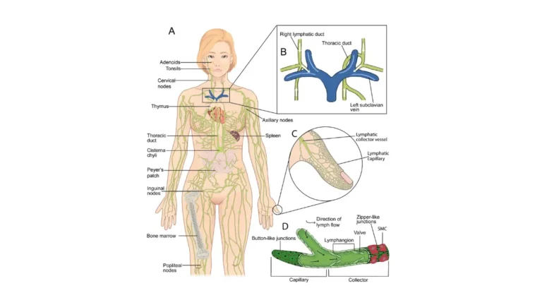
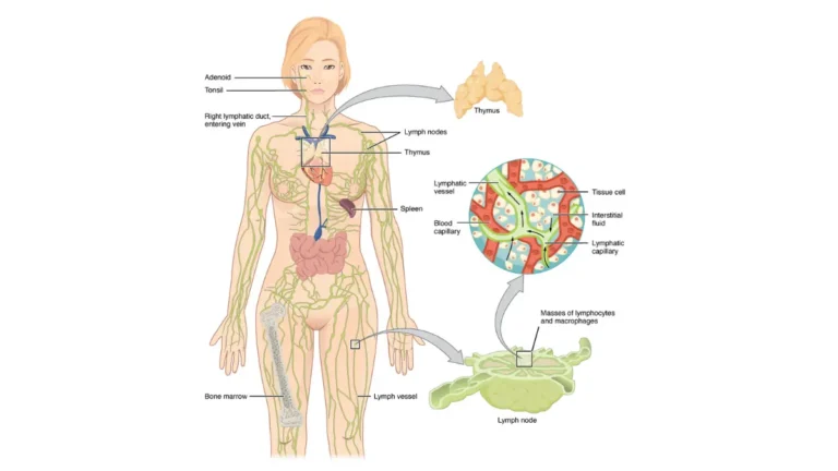
Most breast cancers grow as a discrete mass that enlarges until it becomes palpable. Inflammatory breast cancer forms no such mass. Instead, IBC cells cluster into emboli that obstruct the dermal lymphatic channels — the microscopic drainage vessels running within the breast skin itself — causing fluid accumulation rather than solid tumor growth.
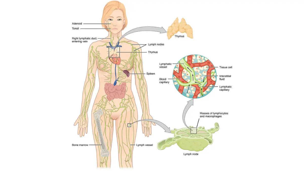
Adapted from Wikimedia Commons Anatomy of the Lymphatic System, licensed under CC BY-SA 4.0.
🔬 How It Works: Tumor emboli block dermal lymphatic vessels, trapping interstitial fluid in the breast skin layers. The result is diffuse skin edema, redness, and thickening — not a lump beneath the skin. Because the cancer lives inside the lymphatic vessels rather than forming a solid mass, mammography frequently returns a “no discrete mass identified” result even when active IBC is present.
This lymphatic blockage mimics a breast infection so convincingly that most IBC patients complete at least one antibiotic course before the correct diagnosis is pursued.
Why IBC is always at least Stage III — the AJCC criterion explained
Under American Joint Committee on Cancer staging criteria, IBC is classified as at minimum Stage IIIB at diagnosis — regardless of tumor size, lymph node count, or imaging findings — because skin involvement by dermal lymphatic invasion is, by definition, an advanced local disease state.
📊 Clinical Data Point: Per the NCCN Clinical Practice Guidelines in Oncology — Breast Cancer (2026), IBC is staged at IIIB minimum based on clinical presentation criteria. Stage IV is assigned when distant metastasis is confirmed. — Source: NCCN, 2026.
This mandatory staging reality determines why IBC treatment begins with systemic therapy rather than surgery — a sequence detailed in our comprehensive guide to breast cancer stages and survival.
IBC symptoms: what to look for when there’s no lump
The seven warning signs of IBC — with no lump present
IBC symptoms typically appear within days to weeks and include:
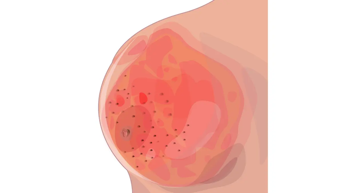
- Breast redness (erythema) covering at least one-third of the breast surface — the skin appears pink, red, or bruised
- Rapid breast swelling — the affected breast enlarges noticeably within days, not weeks
- Peau d’orange skin texture — orange-peel dimpling caused by lymphatic fluid accumulation around hair follicles
- Breast warmth without systemic fever — the skin temperature is elevated, but full-body fever is often absent
- Nipple retraction or inversion — the nipple pulls inward or flattens suddenly
- Breast heaviness or aching — a sensation of fullness or pressure, not a localized sharp pain
- Skin thickening or ridging — the breast skin feels leathery or abnormally firm to the touch
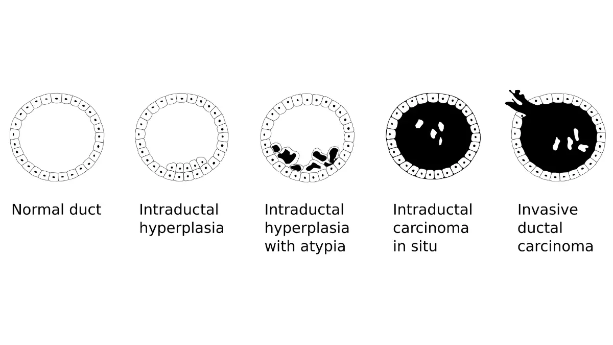
Adapted from Wikimedia Commons Breast cancer progression, licensed under CC BY-SA 4.0.
⚠️ Clinical Warning: IBC symptoms do not resolve on their own and do not respond to antibiotics. If any combination of these signs has persisted for more than one week — or appeared without a preceding infection or injury — contact a board-certified breast surgeon or gynecologic oncologist immediately. Request a skin punch biopsy evaluation by name. Do not wait for a mammogram result before making that call.
What peau d’orange looks and feels like
🔬 How It Works: Peau d’orange — French for “orange-peel skin” — develops when tumor emboli block dermal lymphatic drainage, causing fluid to pool around hair follicle insertion points. The follicles remain tethered to the deeper skin layer while the surrounding tissue swells, creating the characteristic dimpled pattern. This is not an infection sign — it is a direct result of lymphatic obstruction at the cellular level, and it indicates IBC until proven otherwise.
Unlike the smooth redness of mastitis, peau d’orange has a visible, textured quality and may cover part or all of the breast. It tends to develop and worsen within days.
IBC vs. mastitis — how to tell the difference
| Clinical Feature | IBC | Mastitis |
|---|---|---|
| Resolves with antibiotics | No | Yes, within 7–10 days |
| Systemic fever | Often absent | Usually present |
| Peau d’orange present | Common | Rare |
| Lump palpable | No | Sometimes |
| Affects only breastfeeding women | No — any adult | Predominantly |
| Worsens despite treatment | Yes | No |
✅ Patient Action: If breast redness, swelling, or skin texture changes have not resolved after completing a full antibiotic course, do not accept “monitor it” as the next step. Ask your physician by name: “I need a referral to a breast surgeon for a punch skin biopsy to rule out inflammatory breast cancer.” Naming the procedure accelerates the referral.
For a complete overview of how IBC presentation differs from other breast cancer types, see our guide to breast cancer warning signs and symptoms.
How IBC is diagnosed — steps that go beyond a mammogram
Why a normal mammogram does not rule out IBC
Diagnosing inflammatory breast cancer requires clinical evaluation combined with a skin punch biopsy — a normal mammogram result does not rule IBC out. IBC forms no discrete mass, so mammography may report “no discrete mass identified” or note only diffuse skin thickening. That result can be falsely reassuring when IBC symptoms are present.
⚠️ Clinical Warning: If you have IBC symptoms and your mammogram returns a negative or inconclusive result, do not stop there. A clear mammogram in the presence of breast redness, swelling, or peau d’orange is a diagnostic red flag — not clearance. A punch biopsy referral is still required.
The punch skin biopsy — what it is and why it matters
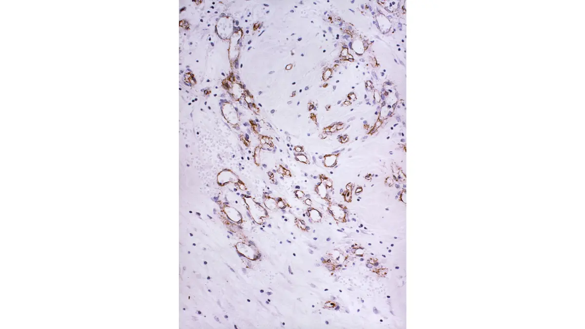
Adapted from Wikimedia Commons Ductal carcinoma histology, licensed under CC BY-SA 3.0.
The IBC diagnostic pathway, in clinical order:
- Clinical assessment — a breast surgeon or gynecologic oncologist evaluates the extent of skin redness, texture changes, and nipple changes across the full breast surface
- Punch skin biopsy — a 3–4mm circular skin sample is removed from the affected skin (not from a breast mass — there is none); pathology examines the sample for dermal lymphatic tumor emboli
- Breast imaging workup — mammography, ultrasound, and MRI assess disease extent and rule out a coexisting mass; they do not make the primary IBC diagnosis
- PET-CT staging — confirms whether distant metastasis is present, determining Stage IIIB versus Stage IV classification
📊 Clinical Data Point: Per NCCN Clinical Practice Guidelines in Oncology — Breast Cancer (2026), punch skin biopsy demonstrating dermal lymphatic tumor involvement is required to confirm IBC. Mammography is a supportive — not confirmatory — diagnostic tool in IBC evaluation. — Source: NCCN, 2026.
✅ Patient Action: Before your specialist appointment, write down this question: “Can we proceed with a punch skin biopsy today, regardless of my mammogram result?” Consult a board-certified breast surgeon or gynecologic oncologist — not a primary care physician — for this evaluation.
Inflammatory breast cancer treatment: the step-by-step protocol
Why chemotherapy comes before surgery in IBC — the neoadjuvant sequence
IBC treatment follows a fixed three-phase sequence mandated by the NCCN Clinical Practice Guidelines in Oncology — Breast Cancer (2026), per the NCCN Breast Cancer Clinical Practice Guidelines
- Neoadjuvant chemotherapy — systemic chemotherapy is administered first, before any surgical intervention, to reduce disease burden in the skin and lymphatic network
- Modified radical mastectomy — surgical removal of the breast and axillary lymph nodes; sentinel lymph node biopsy is contraindicated in IBC because the lymphatic network is diffusely involved; full axillary lymph node dissection is performed instead
- Post-mastectomy radiation therapy — mandatory for all IBC patients regardless of surgical margins; directed at the chest wall and regional lymph node fields
🩺 Physician Note: “The question I hear most often from newly diagnosed IBC patients is: ‘Why can’t we just remove it?’ The answer is that IBC is a lymphatic disease first — the cancer occupies the breast skin’s entire drainage network. Removing the breast before systemic treatment leaves that network intact. Chemotherapy first is not a delay; it is what makes surgery’s goal achievable.” — Dr. Carolyn D. Fairweather, MD, Gynecologic Oncology
HER2-positive IBC — adding targeted therapy to the regimen
When IBC is HER2-positive, dual HER2 blockade is added to the neoadjuvant chemotherapy regimen alongside the anthracycline-taxane backbone. Both trastuzumab and pertuzumab carry FDA approval for use in this setting. Our guide to HER2-positive breast cancer treatment covers the targeted therapy sequence in full detail.
Triple-negative IBC — what changes when there are no targeted options
When IBC is hormone receptor–negative and HER2-negative — triple-negative — treatment relies entirely on chemotherapy, with no hormonal or targeted agents available. Carboplatin is commonly added to the neoadjuvant regimen to increase the probability of achieving pathologic complete response. Our guide to triple-negative breast cancer covers chemotherapy protocols and emerging immunotherapy options for this subtype.
Can IBC go into remission?
Pathologic complete response (pCR) — no residual invasive cancer in the breast tissue or lymph nodes at the time of mastectomy — is a real, achievable clinical endpoint and the strongest documented prognostic indicator after neoadjuvant therapy. Patients who achieve pCR demonstrate consistently improved long-term outcomes compared to those with residual disease at surgery.
✅ Patient Action: Before beginning chemotherapy, ask your medical oncologist: “Is my treatment plan built on the 2026 NCCN IBC-specific protocol, and am I a candidate for clinical trial enrollment?” For currently enrolling IBC studies, search active IBC clinical trials on ClinicalTrials.gov. Consult a board-certified medical oncologist at an NCI-designated comprehensive cancer center for this decision.
For a patient-level explanation of what mastectomy involves and what recovery looks like, see our guide on what your surgeon won’t always tell you about mastectomy.
Inflammatory breast cancer survival rates — what the 2026 data shows
Stage IIIB vs. Stage IV IBC — what the survival data actually means
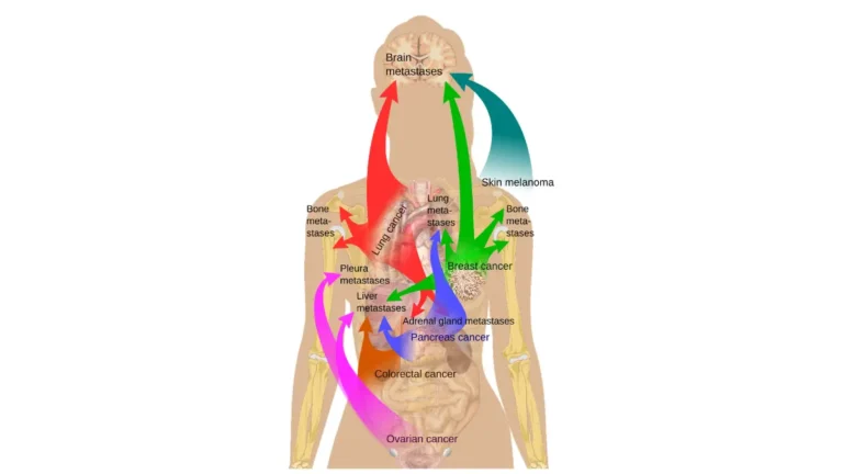
IBC survival rates are tracked by the SEER (Surveillance, Epidemiology, and End Results) program and stratified by stage, because Stage IIIB and Stage IV IBC have meaningfully different outcomes. Stage IIIB — the minimum IBC designation — represents locally advanced disease without confirmed distant spread. Stage IV represents confirmed metastatic disease, most commonly to the liver, lungs, or bone.
For context on how IBC survival compares across all breast cancer subtypes, our guide to breast cancer survival rates by stage provides a full data comparison.
Factors that significantly improve IBC prognosis
Four clinical variables have the strongest documented association with improved IBC outcomes in current oncology literature:
- Achieving pathologic complete response (pCR) after neoadjuvant chemotherapy — the single most powerful modifiable prognostic factor; patients who achieve pCR consistently outperform population-level survival projections for their stage
- HER2-positive receptor status — when treated with dual HER2 blockade and neoadjuvant chemotherapy, HER2-positive IBC patients who achieve pCR have outcomes that approach those of non-IBC breast cancer
- Care at an NCI-designated comprehensive cancer center — associated with access to IBC-specific treatment protocols, clinical trial eligibility, and coordinated multidisciplinary team management
- Absence of distant metastasis at diagnosis — Stage IIIB carries a substantially better prognosis trajectory than Stage IV
📊 Clinical Data Point: Five-year relative survival rates for IBC are tracked by the SEER program and summarized by the American Cancer Society. Current stage-stratified figures are available through the American Cancer Society’s inflammatory breast cancer statistics page. — Source: ACS, citing SEER 2026.
✅ Patient Action: Survival statistics describe population outcomes — not your individual prognosis. To understand what current IBC outcome data means for your specific receptor subtype, treatment response, and disease stage, consult a board-certified medical oncologist and ask: “Given my pathology results, what do the 2026 SEER figures show for patients with my IBC subtype and receptor profile?”
A gynecologic oncologist’s perspective on IBC and the misdiagnosis problem
Why IBC is the most commonly misdiagnosed breast cancer type
In my practice as a gynecologic oncologist, the pattern I encounter most consistently is this: IBC symptoms were present and progressing for weeks before the diagnosis was considered. Most patients had completed one or two antibiotic courses. None of them failed — they applied the framework they had been given, and that framework did not account for a breast cancer with no lump.
The clinical threshold that should trigger immediate biopsy referral is unambiguous. Any breast redness, swelling, or skin textural change that does not resolve within 7–10 days of antibiotic therapy is IBC until a punch biopsy proves otherwise — not a reason to prescribe a second antibiotic course.
🩺 Physician Note: “In every new IBC consultation, the first conversation I have is the same: ‘You were not wrong to think it was an infection. The symptoms are nearly identical. What separated your case from mastitis was that antibiotics didn’t work — and that single fact required a biopsy, not a refill.'” — Dr. Carolyn D. Fairweather, MD, Gynecologic Oncology
Women with a personal or family history of breast or ovarian cancer carry a higher baseline risk for aggressive breast cancer subtypes including IBC. Use our Genetic Risk Assessment Tool to evaluate your hereditary risk profile. If genetic testing has already been performed, our guide to reading your BRCA test results explains what each result means for your ongoing screening plan.
What I tell every patient who asks “should I have waited?”
You did not wait too long. You are here now.
What matters from this point is a confirmed diagnosis, a specialist-led multidisciplinary treatment team, and a treatment plan built on the 2026 NCCN IBC protocol. Pathologic complete response is a documented, achievable clinical endpoint — and the patients who reach it are the ones who move from the correct diagnosis to the correct treatment without further hesitation.
✅ Patient Action: If IBC has been confirmed, contact a board-certified gynecologic oncologist or medical oncologist at an NCI-designated comprehensive cancer center. Ask specifically: “Is my treatment plan built on the 2026 NCCN IBC protocol, and am I eligible for clinical trial enrollment?” This question, asked before the first chemotherapy cycle, is the single most valuable action you can take at this stage of your diagnosis.
Inflammatory breast cancer — frequently asked questions
1. Can you have breast cancer without a lump?
Yes. Inflammatory breast cancer is breast cancer that forms no palpable mass. Instead, IBC cells obstruct the dermal lymphatic vessels, causing skin and lymphatic changes — redness, swelling, orange-peel texture, and nipple changes — rather than a detectable lump. This is why IBC is consistently dismissed as mastitis or infection before the correct diagnosis is reached. No lump being present does not mean no cancer is present.
2. What does inflammatory breast cancer look like?
IBC typically causes one or more of the following: redness or pink discoloration covering at least one-third of the breast, rapid swelling developing over days, orange-peel skin texture (peau d’orange), elevated skin temperature, nipple retraction or flattening, or a heavy aching sensation in the breast. No lump is palpable. Symptoms develop quickly and do not resolve with antibiotics or anti-inflammatory medications.
3. How fast does IBC spread?
IBC progresses significantly faster than most breast cancer types. Visible symptoms can appear and worsen within days to weeks. By the time skin changes are visible, IBC has already invaded the dermal lymphatic network — and by definition, all IBC diagnoses begin at Stage IIIB. Speed of specialist evaluation and treatment initiation directly affects outcomes. Consult a board-certified gynecologic oncologist without delay if IBC symptoms are present.
4. Is inflammatory breast cancer always Stage III at diagnosis?
Yes. Per the NCCN Clinical Practice Guidelines in Oncology — Breast Cancer (2026), IBC is classified as at minimum Stage IIIB at diagnosis, based on clinical presentation criteria — regardless of tumor size or imaging findings. Stage IV is assigned when distant metastasis is confirmed, such as cancer in the liver, lungs, or bone. There is no early-stage IBC. Consult a board-certified medical oncologist for staging confirmation and treatment planning.
5. What causes IBC — is it an infection?
No. Inflammatory breast cancer is not an infection, and antibiotics do not treat it. The term “inflammatory” describes the appearance of the breast skin — caused by cancer cells obstructing dermal lymphatic vessels, not by bacteria or immune activation. The exact biological trigger for IBC development remains an active area of 2026 oncology research, and no single preventable cause has been confirmed.
6. How is IBC diagnosed — is a mammogram enough?
No. A normal mammogram does not rule out IBC. Because IBC forms no discrete mass, mammography may return a negative or inconclusive result even when IBC is active. Diagnosis requires a clinical assessment by a breast surgeon or gynecologic oncologist plus a punch skin biopsy — a small skin sample taken from the affected area to identify dermal lymphatic tumor involvement. Consult a board-certified breast surgeon for this evaluation regardless of mammogram results.
7. What is the survival rate for inflammatory breast cancer?
IBC survival rates are tracked by the SEER program and stratified by stage. Stage IIIB IBC carries a meaningfully better 5-year relative survival rate than Stage IV IBC. The most critical individual prognostic factor is whether pathologic complete response is achieved after neoadjuvant chemotherapy — patients who achieve pCR consistently outperform population-level projections for their stage. Consult a board-certified medical oncologist for survival data specific to your IBC subtype and treatment response.
8. Is IBC the same as mastitis?
No. Both cause breast redness and swelling, but mastitis is a bacterial infection that resolves with antibiotics within 7–10 days. Inflammatory breast cancer does not respond to antibiotics. Mastitis primarily affects breastfeeding women; IBC occurs in any adult. Peau d’orange is a characteristic IBC finding and is rare in mastitis. If breast symptoms have not resolved after a full antibiotic course, contact a breast surgeon immediately — do not wait.
9. Can inflammatory breast cancer be cured or go into remission?
Pathologic complete response — no residual invasive cancer in the breast or lymph nodes at mastectomy — is a real, achievable clinical endpoint with neoadjuvant chemotherapy. Patients who achieve pCR demonstrate consistently improved long-term outcomes compared to those with residual disease at surgery. IBC is not incurable as a category. Consult a board-certified gynecologic oncologist or medical oncologist to discuss your specific remission probability based on your receptor subtype and treatment protocol.
10. Who is at highest risk for IBC?
Inflammatory breast cancer disproportionately affects younger women, Black women, and women who are overweight or obese at diagnosis. It has a higher incidence among women under 40 than other breast cancer types. BRCA mutation carriers face elevated risk for aggressive breast cancer subtypes including IBC. IBC is not limited to breastfeeding or pregnant women — it occurs most commonly in non-pregnant, non-lactating adults.
11. What is the treatment for IBC — and in what order?
IBC treatment follows a mandatory three-phase sequence: (1) neoadjuvant chemotherapy first — an anthracycline-taxane regimen, with trastuzumab and pertuzumab added if HER2-positive; (2) modified radical mastectomy with full axillary lymph node dissection — sentinel lymph node biopsy is contraindicated; (3) post-mastectomy radiation therapy to the chest wall and regional lymph nodes. Adjuvant targeted or endocrine therapy follows based on receptor status. Consult a board-certified medical oncologist before beginning any phase of IBC treatment.
IBC has no lump — but it does have a diagnosis, a treatment path, and a specialist who can help
Inflammatory breast cancer is serious, fast-moving, and frequently misdiagnosed — and it is also treatable, with a clearly defined diagnostic pathway and an evidence-based treatment sequence governed by the 2026 NCCN IBC protocol. Pathologic complete response is a documented, achievable endpoint.
You were right to look this up. The next step is not another search. It is a call to a board-certified breast surgeon or gynecologic oncologist to request a punch skin biopsy evaluation — today.
For the full clinical picture of how IBC staging fits within breast cancer as a whole, return to our comprehensive guide to breast cancer stages and survival. The more accurately you understand your diagnosis, the more effectively you can advocate for the right treatment at the right time.
About this content
How this article was put together: researched from recognised health sources, drafted with the help of AI tools, and edited by hand, with sources linked throughout.

Sameer Patel is the founder and editor of My Medicine Advisor. He is not a doctor or medical professional — before starting this site he worked in banking,…
Medical disclaimer
The content on MyMedicineAdvisor is provided for general informational and educational purposes only and is not a substitute for professional medical advice, diagnosis, or treatment. Health information on this website should not be used to diagnose, treat, cure, or prevent any condition without guidance from a qualified healthcare professional. Always seek the advice of your doctor, physician, or another licensed healthcare provider with any questions you may have regarding a medical condition, symptoms, medications, or treatment decisions.