
On This Page – Quick Medical Summary
What breast cancer symptoms actually mean — and when to act
You noticed something different — a lump, a skin change, a nipple that doesn’t look the way it did — and you turned to Google before calling a doctor. That is the right first step. As a gynecologic oncologist, I see this exact moment every week in clinical practice. The fear behind that search is real, and it deserves a real clinical answer — not a generic list you could find anywhere.
Before your appointment, use our Symptom Checker to record the characteristics of your breast symptoms — it helps your physician respond faster and more precisely. For a full clinical overview of breast cancer — including staging, subtypes, and treatment pathways — see our breast cancer stages and treatment guide.
ℹ️ Medical Disclaimer: The symptom descriptions, diagnostic criteria, imaging references, biopsy procedures, staging information, and treatment details in this article reflect current clinical guidelines and are provided for educational purposes only. Individual diagnostic conclusions, treatment decisions, and procedure choices depend on factors including patient history, comorbidities, imaging results, biopsy findings, and specialist assessment. Consult a board-certified gynecologic oncologist, breast surgeon, or primary care physician before acting on any clinical information in this article. Health insurance coverage for mammography, breast MRI, and biopsy procedures varies by plan; confirm coverage directly with your insurer.
The most common early signs of breast cancer
Breast cancer symptoms in their earliest stages do not always look the way most people expect. The most important clinical fact to understand is this: most early breast cancers are painless.
The early signs of breast cancer, as explained by Dr. Carolyn D. Fairweather, MD, board-certified gynecologic oncologist, include:
- A new palpable mass — firm, with irregular borders, and typically fixed in place rather than mobile
- A lump that feels distinctly different from surrounding breast tissue — not soft or rubbery, but dense and immovable
- Nipple discharge — particularly when it is unilateral, spontaneous, and blood-tinged or clear
- Skin dimpling — a puckering or inward pucker caused by a tumor pulling on the connective tissue beneath the skin
- New nipple retraction — inversion of the nipple that was not present previously
- Unexplained breast asymmetry — one breast changing shape or size without a clear hormonal cause
- A diffuse breast thickening or heaviness in one area with no discrete lump
- Persistent redness or warmth across the breast — especially without signs of infection or fever
📊 Clinical Data Point: The majority of breast cancer diagnoses are first identified as a palpable breast mass. The proportion of cases discovered incidentally on screening mammogram increases significantly with regular screening adherence. Source: National Cancer Institute (2026)
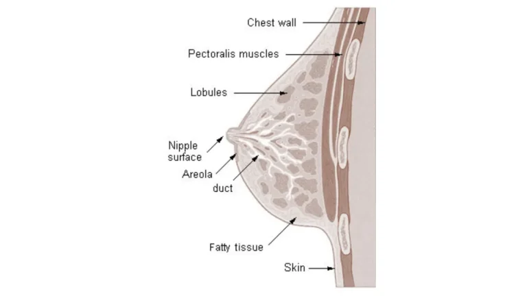
🔬 How It Works: When a tumor grows, it can anchor to the Cooper’s ligaments — fibrous connective bands running from the skin to the chest wall. As the tumor expands, it pulls these ligaments inward, creating the characteristic skin dimpling physicians recognize as a clinical red flag. This explains why dimpling can appear before a lump is large enough to feel.
What a malignant breast lump actually feels like
In my clinical experience, patients describe a malignant lump as an area that feels “different” from the rest of the breast — not necessarily painful, just firm and fixed in a way that was not there before. A benign fibroadenoma, by contrast, feels smooth, rubbery, and moveable.
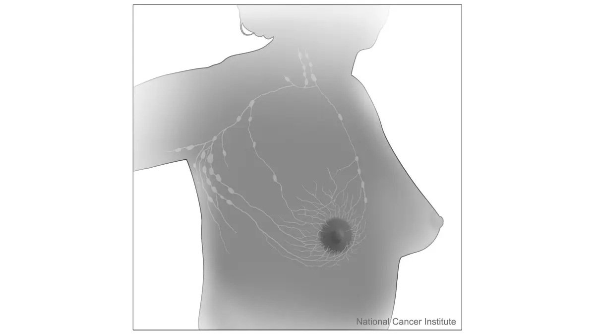
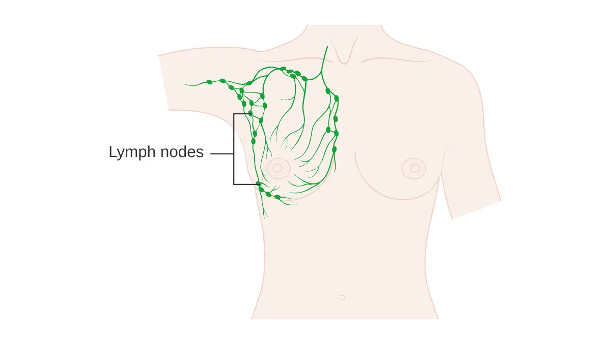
The upper-outer quadrant — the area of the breast closest to the armpit — is the most common location for malignant lesions, because it contains the highest concentration of glandular tissue. This is also where lymph nodes first receive drainage.
Nipple discharge: the clinical threshold that matters
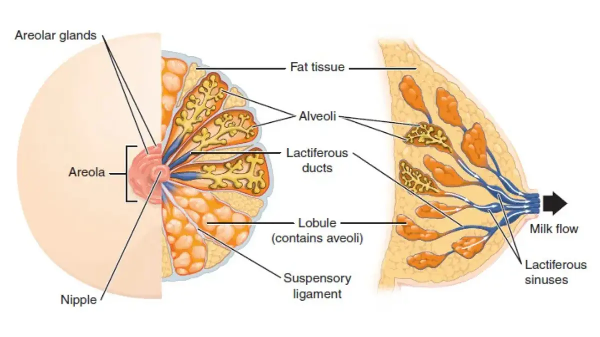
Not all nipple discharge signals cancer. The characteristics that raise concern are: unilateral origin, spontaneous onset, and blood-tinged or clear fluid from a single duct.
Bilateral milky discharge that requires manual expression is far more likely to be hormonal or medication-related. Any discharge that is spontaneous, unilateral, and blood-tinged warrants evaluation within one to two weeks.
✅ Patient Action: Any new breast lump — whether painful or completely painless — should be evaluated by a board-certified gynecologic oncologist or primary care physician within one to two weeks of noticing it. Painlessness does not indicate benignity. Do not wait for the symptom to resolve on its own.
For additional detail on early presentations of breast cancer, see our guide to the first signs of breast cancer symptoms.
For a federal clinical reference, see the NCI’s breast cancer symptoms and diagnosis information.
How breast cancer is diagnosed after symptoms appear
Diagnosing breast cancer after a new symptom appears follows a structured five-step clinical process. Understanding each step reduces the anxiety of not knowing what is happening — or what comes next.
The breast cancer diagnostic pathway, as explained by Dr. Carolyn D. Fairweather, MD:
- Clinical breast exam — your physician palpates all four quadrants, the nipple-areolar complex, and the axillary lymph nodes to map the characteristics of any mass or thickening
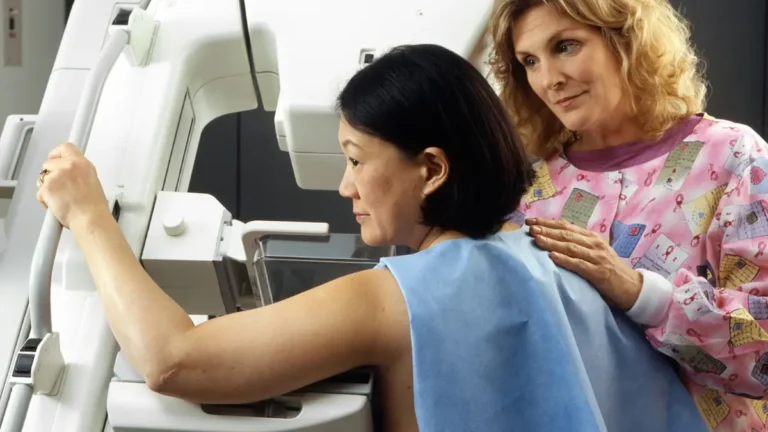
- Diagnostic mammogram — targeted imaging of the area of concern; the radiologist assigns a BI-RADS score from 0 to 6 to communicate malignancy probability
- Breast ultrasound — used alongside or instead of mammogram when dense breast tissue limits X-ray sensitivity, or when a mass needs further characterization
- Breast MRI — ordered for high-risk patients, when mammogram and ultrasound findings are discordant, or when surgical planning requires more precise mapping
- Core needle biopsy — the definitive step; a tissue sample is extracted under ultrasound or stereotactic guidance and sent to pathology to confirm or exclude malignancy
📊 Clinical Data Point: The 5-year relative survival rate for localized breast cancer (confined to the breast) is approximately 99%. For regional disease (spread to nearby lymph nodes), it is approximately 86%. For distant-stage disease, it falls to approximately 30%. Source: American Cancer Society Cancer Facts & Figures (2026).
🔬 How It Works: The BI-RADS classification system — developed by the American College of Radiology — gives your radiologist a standardized way to communicate risk. BI-RADS 1 and 2 indicate normal or benign findings. BI-RADS 4 (subdivided into 4A, 4B, and 4C) carries malignancy probability ranging from approximately 2% to 95%. BI-RADS 5 carries malignancy probability above 95%. A BI-RADS 4 or higher result typically triggers biopsy.
Why a normal mammogram doesn’t always rule out cancer
Mammography sensitivity drops significantly in women with dense breast tissue — from above 85% in fatty breast tissue to as low as 30–50% in extremely dense tissue. This is not a test failure; it is a documented physical limitation of X-ray in denser tissue.
The FDA’s 2023 mammography density notification rule — which took effect in September 2024 — now requires mammography providers to notify patients of their breast density classification. If you have dense breast tissue, ask your radiologist whether supplemental ultrasound or MRI is appropriate for your risk profile.
Understanding your BI-RADS result
| BI-RADS Category | Malignancy Probability | Standard Next Step |
|---|---|---|
| 1–2 | Essentially 0% | Routine annual screening |
| 3 | < 2% | 6-month follow-up imaging |
| 4A | 2–10% | Biopsy recommended |
| 4B | 10–50% | Biopsy recommended |
| 4C | 50–95% | Biopsy strongly indicated |
| 5 | > 95% | Biopsy + surgical planning |
Source: American College of Radiology BI-RADS Atlas (2026 edition)
For a full explanation of your imaging results, see our guide to mammogram BI-RADS results and what each category means.
If biopsy findings confirm cancer, your care team will likely discuss sentinel lymph node evaluation — see our sentinel lymph node biopsy procedure guide for what to expect.
✅ Patient Action: If your mammogram result is BI-RADS 4 or higher, ask your breast radiologist or gynecologic oncologist: “Which BI-RADS subcategory is my result, and what does that specific subcategory mean for my biopsy recommendation?” The subcategory changes the clinical urgency and timeline.
ACS 2026 survival data: the American Cancer Society’s 2026 breast cancer statistics. FDA density rule: the FDA mammography density notification guidance.
Symptoms that differ by breast cancer type and stage
Not every breast cancer looks the same at presentation. Symptom pattern, speed of progression, and clinical urgency vary significantly by subtype — and understanding this is the single most important thing a newly symptomatic or newly diagnosed patient can know.
Inflammatory breast cancer: the subtype most often missed
⚠️ Clinical Warning: Inflammatory breast cancer does not always present as a lump. In approximately 40% of IBC cases, no discrete mass is present. This is why IBC is frequently mistaken for mastitis — and why the average time from first symptom to correct diagnosis is 4 to 6 months. That delay is clinically catastrophic in a subtype with disproportionately poor outcomes.
Inflammatory breast cancer (IBC) — which accounts for approximately 1–5% of US breast cancer diagnoses — presents with a distinct symptom cluster:
- Rapid onset of breast redness (erythema) covering one-third or more of the breast surface
- Warmth and tenderness without fever or systemic infection signs
- Peau d’orange — an orange-peel skin texture caused by dermal lymphatic obstruction
- Breast enlargement or heaviness that develops over days to weeks, not months
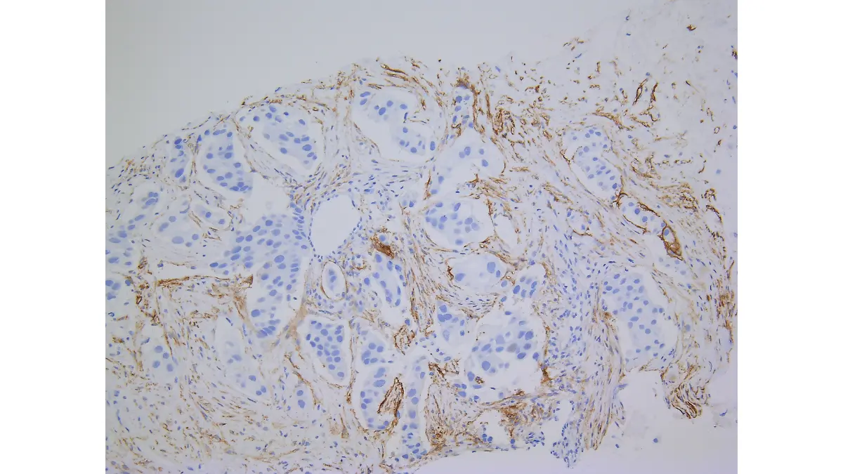
🔬 How It Works: In inflammatory breast cancer, tumor cells invade the dermal lymphatic vessels beneath the skin — blocking lymph drainage and causing fluid to accumulate in the dermis. This trapped fluid creates the characteristic peau d’orange texture. Unlike mastitis (which is caused by bacterial infection), IBC does not respond to antibiotics. If a patient is treated for mastitis and shows no improvement within 7 to 10 days, biopsy — not a second antibiotic course — is the correct next step.
🩺 Physician Note: “The patients who worry me most in my clinical practice are the ones who have been treated for mastitis for two, three, even four months before someone orders a biopsy. If you have been told you have a breast infection, received antibiotics, and the redness and swelling have not meaningfully improved in one to two weeks, that is not mastitis until a biopsy proves it. You need a breast specialist, not another prescription.” — Dr. Carolyn D. Fairweather, MD, Gynecologic Oncology
✅ Patient Action: If you have rapid onset of breast redness, warmth, peau d’orange, and breast enlargement — with or without a palpable lump — see a board-certified breast surgeon or gynecologic oncologist within 24 to 48 hours. Do not wait for a second opinion on antibiotics.
Stage 1 vs. advanced breast cancer: how symptoms change
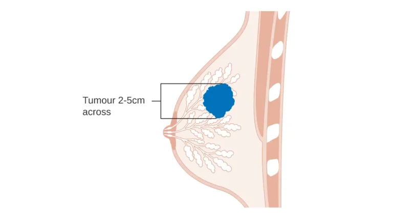
Stage 1A breast cancer (tumor ≤2 cm, no lymph node involvement) is often asymptomatic — discovered only on a screening mammogram, before any palpable change is present. Stage 1B involves micrometastatic clusters in one to three lymph nodes that may not be palpable.
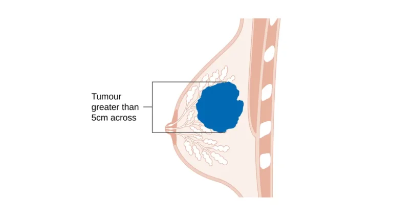
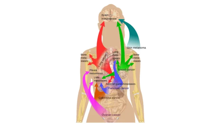
As disease advances to Stage III and IV, additional symptoms emerge: visible skin involvement, arm swelling from axillary lymph node disruption, and — in metastatic disease — persistent bone pain from skeletal involvement.
Triple-negative and HER2-positive: do subtypes feel different?
Triple-negative breast cancer (TNBC) does not feel distinctly different from other subtypes — but it grows faster. It is more likely to present as a rapidly expanding palpable mass, more common in women under 50, and more prevalent in Black women.
Invasive lobular carcinoma (ILC) is the exception: rather than a discrete lump, it creates a diffuse thickening or fullness. This happens because ILC cells infiltrate in a single-file pattern without triggering the fibrotic response that creates a palpable nodule. ILC is more likely to be missed on mammography as a result.
For cure rate context by subtype, see our guide to invasive ductal carcinoma cure rates.
📊 Clinical Data Point: Inflammatory breast cancer represents approximately 1–5% of all US breast cancer diagnoses but carries a disproportionately poor prognosis relative to its incidence. Source: National Cancer Institute (2026).
Who faces the highest risk and what to watch for
Breast cancer risk is not uniformly distributed — and knowing which factors elevate your personal risk changes what symptoms to watch for and how aggressively to screen.
📊 Clinical Data Point: Black women in the United States are diagnosed with breast cancer at later stages at a disproportionately higher rate than white women, contributing to a significantly higher breast cancer mortality rate despite similar or lower incidence. Source: CDC breast cancer surveillance data (2026) via CDC breast cancer data and statistics.
BRCA1 and BRCA2: what the mutation means for your symptoms and screening
Women with a BRCA1 mutation carry a lifetime breast cancer risk of approximately 72–80%, compared to the general population average of approximately 13%. BRCA1-associated cancers are more likely to be triple-negative, to appear at a younger age, and to grow more quickly.
BRCA2 carriers carry a lifetime risk of approximately 45–70% and are more likely to develop hormone receptor-positive tumors. Any woman with a first-degree relative diagnosed with breast cancer — particularly before age 50 — should discuss BRCA testing with a genetic counselor before their next mammogram.
Use our Genetic Risk Assessment Tool to evaluate whether your personal and family history warrants genetic counseling. To understand what a BRCA result means after testing, see our guide to reading BRCA test results.
Age, dense breast tissue, and male breast cancer
Postmenopausal women should treat any new breast lump with elevated urgency — without hormonal fluctuation to explain breast changes, a new finding in a postmenopausal woman carries a higher prior probability of malignancy.
Male breast cancer represents approximately 1% of all US breast cancer cases. The primary presentation in men is a subareolar lump — a firm mass directly behind the nipple — often accompanied by nipple retraction or discharge.
✅ Patient Action: If you have one or more first-degree relatives diagnosed with breast or ovarian cancer — particularly before age 50 — consult a board-certified genetic counselor or gynecologic oncologist about BRCA1/2 testing before your next mammogram. A positive result changes your surveillance protocol immediately.
Breast cancer symptoms that are often overlooked
Some of the most clinically significant breast cancer warning signs are the ones that don’t fit the public’s mental model of what breast cancer looks like. These are the symptoms patients most often dismiss — and the ones I see delayed the longest before evaluation.
Nipple changes beyond discharge: Paget’s disease of the breast
Paget’s disease of the breast presents not as a lump or discharge, but as a persistent rash, scaling, or crusting on and around the nipple. Patients typically treat it with steroid cream for months before someone considers a biopsy.
The clinical threshold is straightforward: nipple scaling or irritation that does not resolve with standard topical treatment within two to four weeks requires evaluation. A nipple punch biopsy can confirm or exclude Paget’s disease in a single outpatient procedure.
Upper arm swelling and back pain in advanced disease
Lymphedema — swelling of the arm or hand caused by axillary lymph node disruption — can be the first noticeable symptom in patients with significant nodal involvement. It is not a sign of early-stage disease, but it is one that caregivers often notice before the patient reports it.
Persistent mid-back or thoracic spine pain in a patient with a known or suspected breast cancer history warrants bone imaging. Skeletal metastasis is the most common site of distant spread in Stage IV breast cancer, and back pain is its most frequent presenting symptom.
Symptoms often missed in older women and in men
In older women, new breast skin thickening or retraction is sometimes attributed to age-related skin changes — but unilateral, asymmetric findings require evaluation regardless of age.
In men, the subareolar location of tumors means that nipple retraction is frequently the first noticed change, not a discrete lump.
✅ Patient Action: Persistent nipple scaling, crusting, or itching that does not resolve with topical treatment within two to four weeks should be evaluated by a board-certified gynecologic oncologist or dermatologist. Ask specifically about Paget’s disease of the breast — a nipple biopsy is the definitive diagnostic step.
When breast symptoms need urgent evaluation
As a gynecologic oncologist, the question I hear most is: “How do I know when this is an emergency?”
Here is the clinical answer.
See a breast specialist within 24–48 hours if you notice:
- Rapid onset of breast redness covering a significant portion of the breast surface — with or without a palpable lump
- Skin thickening, peau d’orange, or warmth in one breast that has developed over days to weeks
- Spontaneous, unilateral, blood-tinged nipple discharge that appears without squeezing
- New nipple retraction that was not present at your last clinical exam
🩺 Physician Note: “The patients I most worry about are the ones who waited three months to come in because their lump was painless. They assumed cancer would hurt. In most early-stage breast cancer, painlessness is the norm — not the exception. A painless lump is not a reassuring lump. It is a lump that needs an evaluation.” — Dr. Carolyn D. Fairweather, MD, Gynecologic Oncology
Go to the emergency room if:
- You have been treated with antibiotics for a presumed breast infection, shown no improvement after 7 to 10 days, and now have systemic signs including fever above 101°F, chills, or worsening skin changes
- You have a confirmed breast cancer history and develop sudden, severe bone pain in the spine or ribs that is unlike your prior experience
When watchful waiting is clinically appropriate
Bilateral cyclical breast pain that worsens before menstruation and resolves after — without any palpable mass — is a recognized pattern called mastalgia. It is most often benign. Watchful waiting is appropriate only when a physician has confirmed the absence of any palpable finding.
A known fibroadenoma that has not changed in size or character at two consecutive clinical exams can also be monitored by agreement with your physician — not on your own assessment.
✅ Patient Action: If you are uncertain whether your symptom requires urgent evaluation, contact a board-certified gynecologic oncologist or breast surgeon and describe the specific characteristics — onset, location, appearance, and any associated changes. Early evaluation does not commit you to treatment. It gives you clinical certainty. If you need same-day guidance, a telehealth consultation with a physician can help you determine the appropriate level of urgency before your in-person appointment.
For guidance on what happens after an evaluation confirms breast cancer, including mastectomy as a treatment option, see our article on what to expect from a mastectomy.
Frequently asked questions about breast cancer symptoms
Q1: What are the first signs of breast cancer?
The first signs of breast cancer are most commonly a firm, painless, irregularly bordered breast lump — often in the upper-outer quadrant — followed by nipple discharge, skin dimpling, or nipple retraction. Not all early breast cancers produce a palpable lump; some are detected first on mammogram. Consult a board-certified gynecologic oncologist if you notice any new or unexplained breast change.
Q2: Can breast cancer cause pain?
Breast cancer is most often painless at early stages — which is why many patients delay evaluation. The exception is inflammatory breast cancer, which causes breast tenderness and warmth as part of its presentation. Cyclic mastalgia — breast pain that tracks with the menstrual cycle — is more commonly benign. Consult a board-certified gynecologic oncologist if pain is new, unilateral, or accompanied by any skin or nipple change.
Q3: What does a breast cancer lump feel like?
A malignant breast cancer lump typically feels firm to hard, has irregular or poorly defined borders, and is fixed in place rather than mobile. It is usually painless. A benign fibroadenoma, by contrast, feels smooth, round, and rubbery. These distinctions are clinically useful but not definitive — any new lump requires evaluation by a board-certified gynecologic oncologist or breast surgeon.
Q4: What are the symptoms of inflammatory breast cancer?
Inflammatory breast cancer does not always present as a lump. Its hallmark signs are rapid-onset breast redness across a significant area, warmth, peau d’orange (orange-peel skin texture), and breast enlargement developing over days to weeks. It accounts for approximately 1–5% of breast cancer diagnoses but carries a disproportionately poor prognosis. See a board-certified breast surgeon or gynecologic oncologist within 24 to 48 hours if these signs are present.
Q5: Can men get breast cancer symptoms?
Male breast cancer accounts for approximately 1% of all US breast cancer cases. The most common presentation is a firm subareolar lump — a mass directly behind the nipple — often accompanied by nipple retraction or discharge. Because breast cancer in men is frequently dismissed or attributed to gynecomastia, diagnosis is often delayed. Consult a board-certified oncologist or primary care physician promptly if any of these signs are present.
Q6: What are breast cancer symptoms in women over 50?
In postmenopausal women, any new breast lump carries higher clinical urgency than in reproductive-age women — hormonal fluctuations no longer explain breast changes. New nipple retraction, skin dimpling, or breast asymmetry after menopause warrants prompt evaluation. A standard annual mammogram is the appropriate first step; dense breast tissue may require supplemental ultrasound. Consult a board-certified gynecologic oncologist regarding any new finding.
Q7: Does breast cancer always show up on a mammogram?
No — breast cancer does not always appear on mammography. In women with extremely dense breast tissue, mammography sensitivity can fall to 30–50%, meaning a significant proportion of cancers may be missed. Supplemental breast ultrasound or MRI is recommended for dense-tissue patients at elevated risk. Under current FDA guidelines, mammography facilities are required to notify patients of their breast density classification. Consult a board-certified gynecologic oncologist or breast radiologist about supplemental imaging if you have dense tissue.
Q8: What is nipple discharge a sign of?
Nipple discharge is most commonly benign — particularly when it is bilateral, milky, and expressed rather than spontaneous. The characteristics that elevate clinical concern are: unilateral origin, spontaneous onset, and blood-tinged or clear fluid from a single duct. Blood-tinged spontaneous discharge from one breast warrants evaluation within one to two weeks. Consult a board-certified gynecologic oncologist to determine whether imaging or ductoscopy is needed.
Q9: What is peau d’orange in breast cancer?
Peau d’orange is a skin texture change that resembles the surface of an orange peel. In breast cancer, it results from tumor cells blocking the dermal lymphatic vessels beneath the skin, causing fluid to accumulate in the dermis and creating the characteristic dimpling. It is a hallmark sign of inflammatory breast cancer and requires urgent evaluation — within 24 to 48 hours — by a board-certified breast surgeon or gynecologic oncologist.
Q10: What are the symptoms of stage 1 breast cancer?
Stage 1A breast cancer involves a tumor measuring 2 centimeters or smaller with no lymph node involvement. It is frequently asymptomatic — discovered on a screening mammogram before any palpable change is present. Stage 1B involves micrometastatic clusters in one to three lymph nodes that may not be felt externally. The 5-year survival rate for localized breast cancer exceeds 99%. Consult a board-certified gynecologic oncologist for stage-specific treatment planning after diagnosis.
Q11: How is breast cancer diagnosed after symptoms appear?
Breast cancer diagnosis follows a structured pathway: clinical breast exam → diagnostic mammogram with BI-RADS scoring → breast ultrasound if dense tissue is present → MRI if findings are discordant or surgical planning is required → core needle biopsy for tissue confirmation. A BI-RADS category 4 or higher result on imaging typically triggers biopsy. Consult a board-certified gynecologic oncologist and breast radiologist to interpret your imaging results and determine whether biopsy is indicated.
Q12: What breast cancer symptoms are most often missed?
The most frequently overlooked breast cancer signs include: persistent nipple scaling or crusting not responding to topical treatment (Paget’s disease of the breast); diffuse breast thickening without a discrete lump (common in invasive lobular carcinoma); new unilateral arm swelling indicating axillary lymph node involvement; and subareolar lump in men. Consult a board-certified gynecologic oncologist or dermatologist if any nipple change fails to respond to standard treatment within two to four weeks.
Q13: Can breast cancer cause back pain?
Yes — but breast cancer causes back pain only in advanced disease, when metastasis reaches the spinal vertebrae or ribs. Back pain is not a symptom of early-stage breast cancer. In Stage IV disease, bone metastasis is the most common site of spread, and thoracic or lumbar spine pain is its most frequent presentation. If you have a known breast cancer history and develop new, unexplained back pain, contact your oncologist immediately — do not attribute it to musculoskeletal causes without imaging.
Q14: What are triple-negative breast cancer symptoms?
Triple-negative breast cancer (TNBC) symptoms overlap with other breast cancer subtypes — typically a rapidly growing palpable mass — but the subtype is characterized by faster growth rate, earlier age of onset, and higher prevalence in Black women. TNBC does not respond to hormone therapy or HER2-targeted drugs; systemic chemotherapy is the primary treatment approach. Consult a board-certified oncologist for molecular subtype profiling and subtype-specific treatment planning as soon as a diagnosis is confirmed.
Q15: How quickly do breast cancer symptoms progress?
Symptom progression in breast cancer depends heavily on subtype: inflammatory breast cancer develops over days to weeks; triple-negative and HER2-positive subtypes may progress over weeks to months; invasive ductal carcinoma (luminal A subtype) may grow over months to years; ductal carcinoma in situ (DCIS) may progress to invasive disease over years without treatment. This variability is exactly why any new or changing breast symptom warrants prompt evaluation rather than a months-long monitoring period.
Q16: What does breast skin dimpling mean?
Skin dimpling in the breast results from a tumor anchoring to the Cooper’s ligaments — connective tissue running between the breast skin and underlying chest wall — and pulling them inward as it expands. It can indicate invasive ductal carcinoma or inflammatory breast cancer. New, unilateral skin dimpling must be distinguished from bilateral age-related skin laxity. Any new unilateral dimpling warrants evaluation within one week. Consult a board-certified gynecologic oncologist or breast surgeon.
Q17: When should I go to the ER for breast symptoms?
Emergency evaluation is warranted for breast cancer symptoms in two scenarios: first, if you have been treated for a presumed breast infection with antibiotics and show no improvement after 7 to 10 days, with worsening redness, skin thickening, or fever — inflammatory breast cancer must be excluded. Second, if you have a confirmed metastatic breast cancer history and develop sudden, severe bone pain suggesting pathological fracture. For non-emergency new symptoms, urgent outpatient evaluation within 24 to 48 hours is preferable to an emergency department visit.
Your next step after reading about breast cancer symptoms
If you have read this far, you now understand something that most online resources on breast cancer symptoms do not give you: the clinical difference between what warrants same-week evaluation, what requires immediate action, and what can be monitored in partnership with your physician.
The single most effective thing you can do right now is act on that information.
If you noticed a breast change before opening this article, schedule a clinical breast exam with your primary care physician or gynecologic oncologist this week. Do not wait for your annual visit. Do not monitor the symptom alone.
As Dr. Fairweather advises her own patients: early evaluation does not mean catastrophe. It means certainty — and clinical certainty is the foundation of every effective treatment plan, at every stage.
For a complete breakdown of what happens after a breast cancer diagnosis — including stages, surgery, and treatment options — see our breast cancer stages and treatment guide.
About this content
How this article was put together: researched from recognised health sources, drafted with the help of AI tools, and edited by hand, with sources linked throughout.

Sameer Patel is the founder and editor of My Medicine Advisor. He is not a doctor or medical professional — before starting this site he worked in banking,…
Medical disclaimer
The content on MyMedicineAdvisor is provided for general informational and educational purposes only and is not a substitute for professional medical advice, diagnosis, or treatment. Health information on this website should not be used to diagnose, treat, cure, or prevent any condition without guidance from a qualified healthcare professional. Always seek the advice of your doctor, physician, or another licensed healthcare provider with any questions you may have regarding a medical condition, symptoms, medications, or treatment decisions.