
On This Page – Quick Medical Summary
Do you actually know when you should get a mammogram?
If you have heard 40 from one doctor and 50 from another, you are not confused — the guidelines themselves have disagreed for years. This article, written by a board-certified gynecologic oncologist, gives you a clear, risk-stratified answer for your specific situation.
If you are here because you noticed a new breast lump, nipple change, skin dimpling, or discharge, check your symptoms before continuing — this guide is written for screening decisions, not active symptom evaluation. For what happens if a screening result comes back abnormal, our complete guide to breast cancer stages and survival explains every stage from initial imaging to treatment planning.
ℹ️ Medical Disclaimer: The breast cancer mammogram scheduling guidelines, risk stratification protocols, genetic testing recommendations, supplemental imaging criteria, insurance coverage descriptions, and clinical data points in this article are provided for educational purposes only and reflect current clinical guidelines as of May 2026. They do not constitute a diagnosis, a personalized treatment recommendation, a prescription directive, a procedure order, or insurance advice.
Screening start age, imaging frequency, supplemental modality selection, and BRCA management plans depend on your personal health history, family history, prior imaging results, insurance plan specifics, and a physician’s direct assessment of your individual case. Consult a board-certified gynecologic oncologist or breast imaging specialist before making any screening, diagnostic, or supplemental imaging decision. If you are experiencing breast symptoms, contact your healthcare provider promptly — do not wait for a scheduled screening appointment.
What age do mammogram guidelines actually recommend?
The three organizations that govern breast cancer mammogram screening in the United States have moved closer together — and all three now agree that screening should begin no later than age 40.
Where they differ is on frequency, and on how to handle the 40–44 window. Understanding each position lets you and your physician identify which protocol fits your situation.
ACS, USPSTF, and ACR recommendations side by side
| Organization | Recommended Start Age | Frequency | Key Notes |
|---|---|---|---|
| ACS (American Cancer Society) | 40 | Annual | Shared decision-making encouraged 40–44; annual recommended 45–54; optional biennial from 55+ |
| USPSTF (U.S. Preventive Services Task Force) | 40 | Every 2 years | Applies to average-risk women; individual patient choice acknowledged for 40–49 |
| ACR / SBI (American College of Radiology) | 40 | Annual | Risk assessment by age 25 recommended to identify women who may need to start earlier |
📊 Clinical Data Point: Current ACS guidelines recommend that average-risk women begin annual mammography no later than age 45, with the option and explicit encouragement to begin at age 40. The USPSTF currently recommends biennial screening beginning at age 40. Source: ACS current mammography guidelines; USPSTF current recommendation — American Cancer Society mammography recommendations.
What “average risk” actually means — and whether that’s you
Average risk is a specific clinical designation. It means: no personal history of breast cancer, no known BRCA1 or BRCA2 mutation, no first-degree relative diagnosed with breast cancer before age 50, no history of chest radiation before age 30, and no high-risk benign breast condition such as atypical ductal hyperplasia or lobular carcinoma in situ.
If none of those apply to you, you are average-risk by current clinical definitions. If even one applies, your protocol changes — and Section 3 tells you exactly how.
Screening mammogram vs diagnostic mammogram: the clinical difference
A screening mammogram is a routine bilateral X-ray performed when you have no symptoms, ordered by your OB-GYN or primary care physician. A diagnostic mammogram is ordered when something requires further evaluation — a lump, nipple discharge, skin change, or an abnormal result from a prior screening — and typically involves additional targeted images.
These are different clinical procedures with different billing codes and different insurance coverage implications. For patients experiencing breast symptoms before their screening age, see our guide to breast cancer warning signs that warrant immediate evaluation.
🩺 Physician Note: In my practice, the single most frequent misunderstanding I encounter is women who are called back after a screening mammogram and believe the callback means cancer. A callback — also called a recall — occurs in approximately 10% of first-time mammograms and the large majority resolve with additional imaging showing no malignancy. A callback is a process step in a quality system. It is not a diagnosis.
✅ Patient Action: Before your appointment, confirm with your imaging center whether your mammogram is coded as screening or diagnostic. Ask specifically: “Is this a screening or diagnostic mammogram, and how does that affect my out-of-pocket cost?” The distinction determines your insurance obligation and what the radiologist does next if something is found.
Risk factors that move your mammogram start date earlier
Your personal risk profile — not a population-level guideline — determines when your breast cancer mammogram should begin and how often it should occur. These are the clinical factors that change the equation.
Family history and BRCA status: the two highest-risk flags
A first-degree relative — mother, sister, or daughter — diagnosed with breast cancer, particularly before age 50, significantly elevates your personal lifetime risk. Two or more affected first-degree relatives, or any relative with a confirmed BRCA1 or BRCA2 gene mutation, may push your estimated lifetime breast cancer risk above the 20% threshold that triggers dual-modality supplemental screening under current NCCN guidelines.
Women with a strong family history who have not yet undergone genetic counseling should ask their physician for a referral. Before that conversation, assess your personal breast cancer risk using our Genetic Risk Assessment Tool to organize your family history into a format your physician can use immediately.
What breast density does to your mammogram accuracy
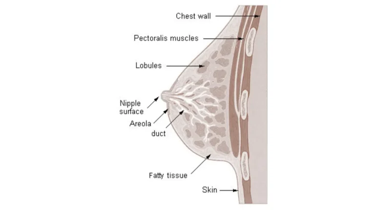
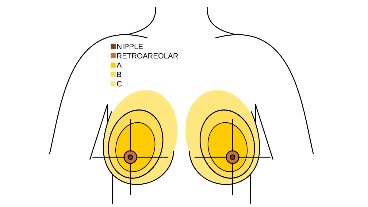
Breast density is determined by the ratio of fibroglandular tissue to fat in your breast, classified by radiologists on the BI-RADS density scale from Category A (almost entirely fatty) to Category D (extremely dense).
Categories C and D both independently elevate cancer risk AND reduce mammogram sensitivity — tumors are harder to detect against dense tissue, and the density itself statistically increases your risk. This dual effect is why Categories C and D are clinical indicators for supplemental screening, not just additional reassurance.
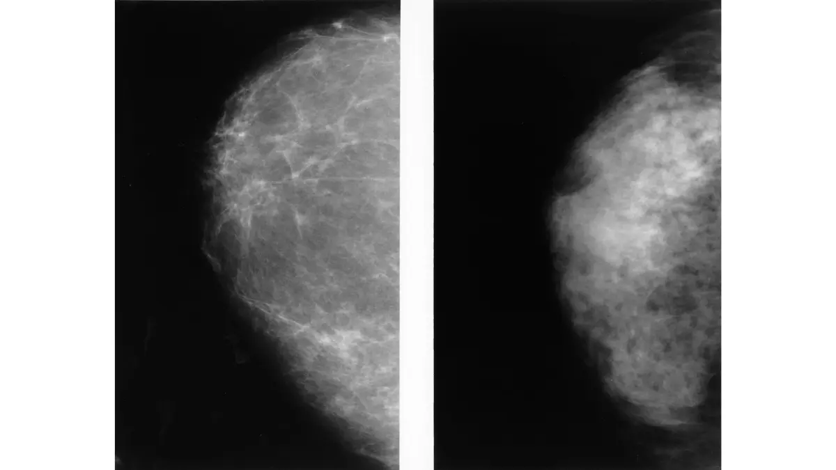
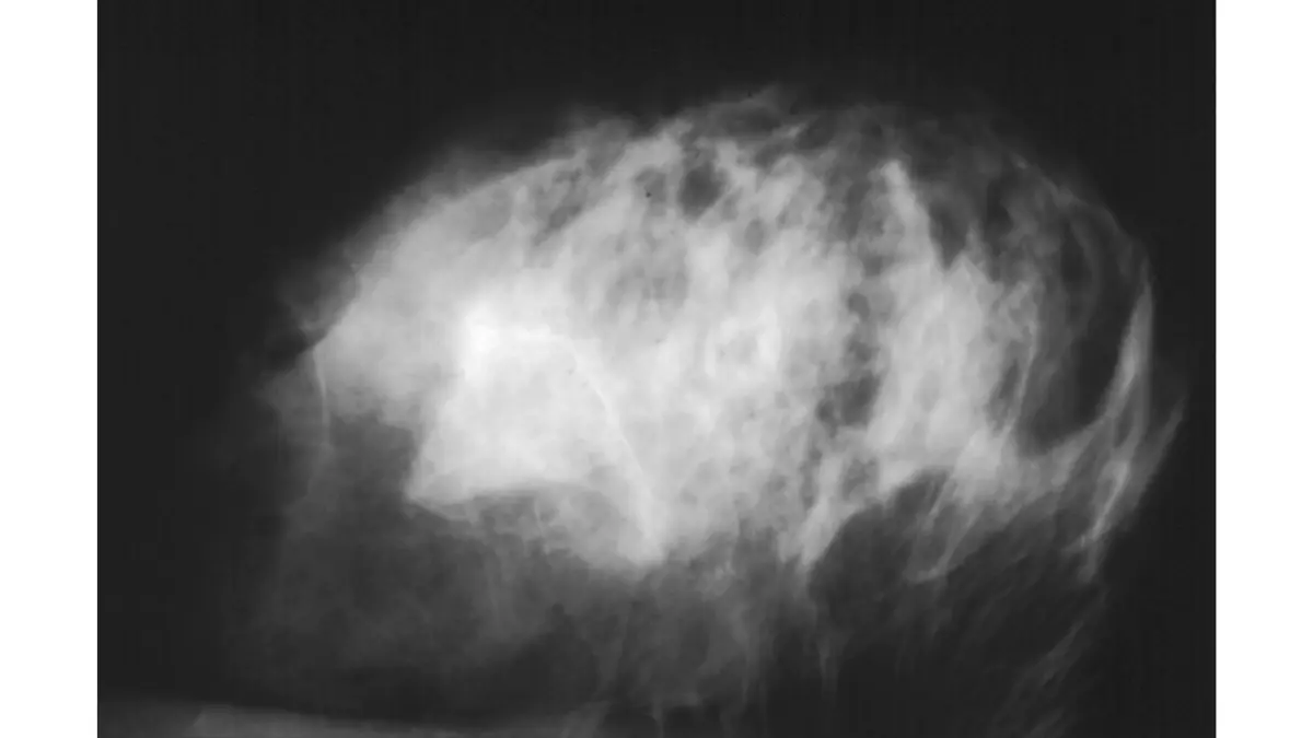
🔬 How It Works: On a standard mammogram, dense breast tissue appears white — and so do tumors. When the two overlap, a tumor can be hidden in plain sight on the image. BI-RADS Category D (extremely dense) tissue reduces mammogram sensitivity to approximately 50–60% in some studies, compared to approximately 85–90% in fatty tissue. This is why supplemental ultrasound or MRI is clinically indicated for women with significant density — it detects abnormalities the mammogram physically cannot see through the tissue.
Other risk factors that may lower your recommended start age
Additional factors that qualify women for earlier or more frequent screening include:
- Prior diagnosis of atypical ductal hyperplasia (ADH) or lobular carcinoma in situ (LCIS) — both are non-cancerous but significantly elevate future risk
- History of chest radiation therapy between ages 10 and 30 (typically for prior lymphoma treatment)
- Ashkenazi Jewish ancestry — associated with higher BRCA1 and BRCA2 gene variant prevalence
- Black race — associated with higher rates of aggressive breast cancer subtypes, including triple-negative breast cancer, diagnosed at younger ages and at more advanced stages
For a complete breakdown of the factors a gynecologic oncologist evaluates when calculating individual risk, see our guide to breast cancer risk factors and how specialists assess them.
📊 Clinical Data Point: The clinical threshold for dual-modality screening — annual mammography combined with annual breast MRI — is a calculated lifetime breast cancer risk of 20% or greater, per current NCCN high-risk screening guidelines. Source: NCI breast cancer screening evidence summary — National Cancer Institute breast cancer screening overview.
✅ Patient Action: Women who identify two or more risk factors in this section should consult a board-certified gynecologic oncologist or breast imaging specialist — not a general practitioner — before their next screening cycle. Ask specifically: “Based on my risk factors, does my calculated lifetime breast cancer risk exceed 20%? If so, what supplemental screening modality do current guidelines recommend for me?”
How to prepare for your mammogram appointment
Knowing exactly what to do before and during your mammogram removes the procedural anxiety that leads many women to delay scheduling — or to cancel on the day.
The five things to do (and not do) before your mammogram
Follow these five steps before every breast cancer mammogram appointment:
- Avoid deodorant, antiperspirant, powder, lotion, or perfume on the day of your exam — residue from these products can appear as calcifications on the mammogram image and trigger an unnecessary callback.
- Time your appointment around your menstrual cycle when possible — scheduling during days 7 through 14 significantly reduces breast tenderness during compression.
- Bring prior mammogram images or records if you are using a new imaging center — radiologists depend on comparison images to detect subtle changes over time.
- Tell the technologist about implants, prior biopsies, or breast surgeries before the exam begins — this changes the imaging protocol and positioning.
- Wear a two-piece outfit — you will remove your top and bra, and a separates outfit makes the process faster and more comfortable.
What happens during the exam, step by step
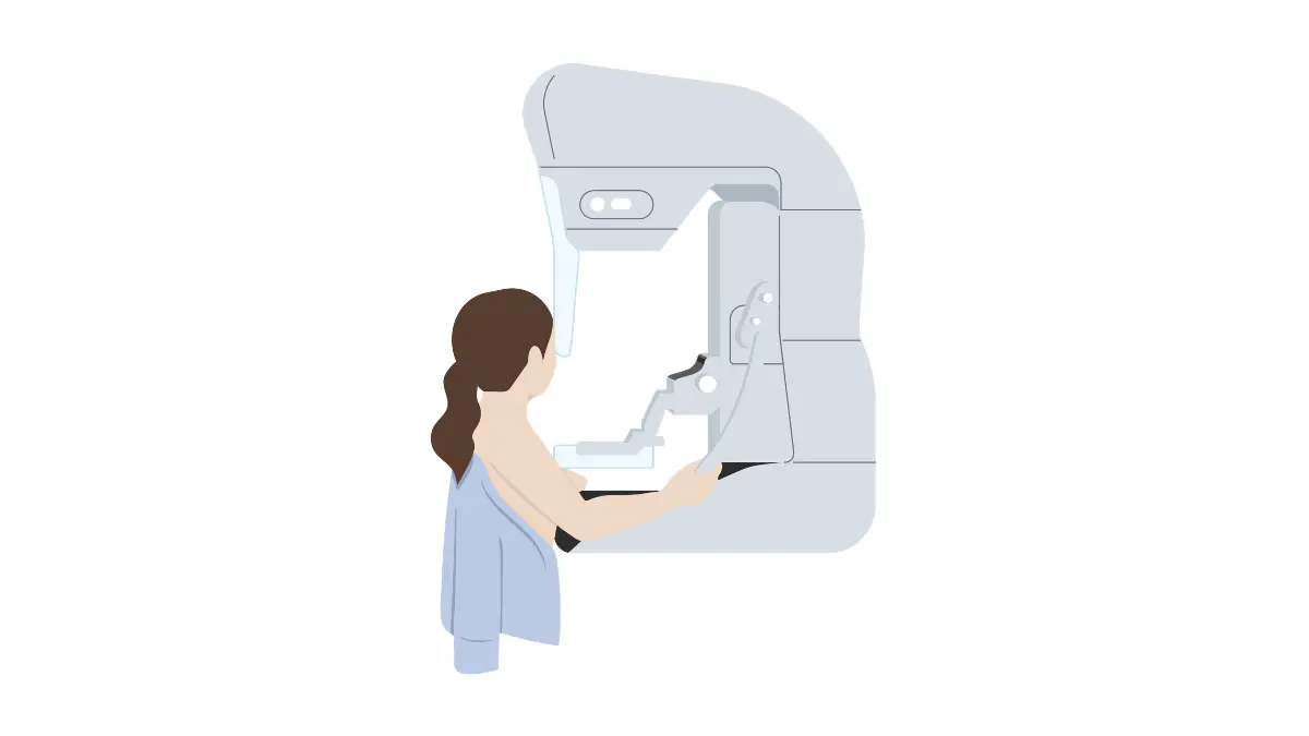
Each breast is positioned on a flat plate and compressed briefly while the X-ray image is captured. The compression lasts approximately 10 to 15 seconds per image. Most women describe it as pressure — momentary and manageable — rather than pain.
A standard screening mammogram takes 15 to 30 minutes from check-in to completion. Most imaging centers now deliver results within 2 to 3 business days, and many provide same-day or next-day notification.
Is a 3D mammogram worth requesting?
Digital breast tomosynthesis — the formal name for a 3D mammogram — captures multiple thin-slice images of breast tissue from multiple angles, rather than a single flat image. In women with dense breast tissue, it detects more cancers at earlier stages and reduces the callback rate compared to standard 2D mammography.
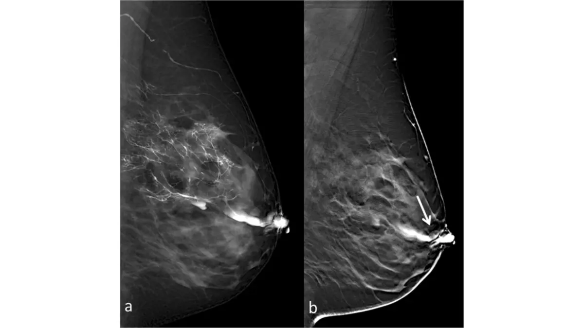
🔬 How It Works: Standard 2D mammography compresses the breast and captures a single image, causing dense tissue layers to overlap — which can obscure small tumors. Digital breast tomosynthesis moves an X-ray arm in a short arc around the breast, capturing a series of low-dose images from slightly different angles. Software reconstructs these into a layered view that allows radiologists to scroll through breast tissue slice by slice, dramatically improving the ability to detect tumors hidden within dense tissue and reducing unnecessary callbacks caused by overlapping normal structures.
High-risk screening: what BRCA, dense breasts, and family history mean for your protocol
If any risk factor from Section 3 applies to you, the standard annual-at-40 recommendation does not fully cover your clinical situation. This section defines the high-risk protocol.
BRCA1 and BRCA2 carriers: your screening protocol is different
Women with a confirmed BRCA1 gene mutation face a lifetime breast cancer risk estimated at 55–72%. Women with a confirmed BRCA2 mutation carry a lifetime risk estimated at 45–69%. Both figures far exceed the average-risk lifetime baseline of approximately 13%.
BRCA1 carriers also have a higher association with triple-negative breast cancer — a subtype that is typically more aggressive and does not respond to hormone or HER2-targeted therapy. This biological distinction is part of why BRCA1 carriers are generally placed at the earlier end of the recommended screening start window.
📊 Clinical Data Point: Current NCCN high-risk screening guidelines recommend that confirmed BRCA1 and BRCA2 carriers begin annual breast cancer mammogram combined with annual breast MRI between ages 25 and 30 — substantially earlier than the age-40 recommendation for average-risk women. Source: NCCN current high-risk guidelines; MedlinePlus BRCA gene information — MedlinePlus BRCA gene testing guide for patients.
If you have already received a BRCA test result and want to understand what it means clinically, see our guide to how to read your BRCA results in plain language.
Dense breasts and supplemental screening options
Women with BI-RADS Category C or D breast density who are not BRCA carriers have two primary supplemental screening options. Breast MRI is preferred for women with dense tissue whose calculated lifetime risk meets or exceeds 20%. Breast ultrasound is the second-line option for women with dense tissue whose lifetime risk falls below the MRI threshold or who cannot undergo MRI.
MRI does not replace the annual mammogram. The two modalities detect different types of abnormalities — mammography is more sensitive to calcifications, while MRI detects lesions that do not calcify — and are used together, not interchangeably.
What to do if your mammogram comes back abnormal
Radiologists report mammogram findings on the BI-RADS classification scale from 0 to 6. BI-RADS 0 means the image was incomplete and additional imaging is needed. BI-RADS 3 means probably benign — a 6-month follow-up imaging is typically recommended. BI-RADS 4 and 5 indicate suspicious or highly suspicious findings where biopsy may be warranted.
A callback is not a diagnosis. For a detailed explanation of every BI-RADS score and what your radiologist is communicating, see our guide to mammogram BI-RADS results from 0 to 6 and what each means. For what a confirmed breast cancer diagnosis means at each stage, see the stages of breast cancer at diagnosis.
✅ Patient Action: Women with a confirmed BRCA1 or BRCA2 mutation, a calculated lifetime breast cancer risk ≥20%, or a personal history of atypical ductal hyperplasia should consult a board-certified gynecologic oncologist or breast imaging specialist — not a general practitioner — to establish a personalized high-risk protocol before their next screening cycle. Ask specifically: “Does my lifetime risk calculation indicate annual breast MRI alongside mammography under current NCCN or ACS guidelines?”
Does insurance cover your mammogram — and what if you’re uninsured?
What the ACA requires health insurers to cover
Under the Affordable Care Act, all non-grandfathered private health insurance plans must cover screening mammograms at no cost-sharing — no deductible, no copay — when the service qualifies as a preventive benefit under current USPSTF grading. The USPSTF’s recommendation for biennial screening beginning at age 40 carries a B grade, which triggers mandatory no-cost coverage under ACA-compliant plans.
The critical distinction: a diagnostic mammogram — ordered after an abnormal screening finding — is billed as a diagnostic procedure, not a preventive service. It may carry standard deductible and copay obligations under your plan. Confirm this distinction with your insurer before your appointment if a prior screening was abnormal.
Medicare Part B and mammogram coverage at 40+
Medicare Part B covers one annual screening mammogram for women age 40 and older at no cost-sharing. There is no cap on age. For current eligibility and coverage details, see Medicare screening mammogram coverage and annual benefit information.
Free and low-cost programs for uninsured women
The CDC’s National Breast and Cervical Cancer Early Detection Program (NBCCEDP) provides free or low-cost breast cancer mammogram screening to uninsured and underinsured women who meet income eligibility criteria. To find your nearest program location and confirm eligibility, see the CDC National Breast and Cervical Cancer Early Detection Program.
A gynecologic oncologist’s take on the mammogram debate
The guideline disagreement — 40 versus 50, annual versus biennial — has genuinely confused patients, and for understandable reasons. Different organizations gave different answers for years, and the debate was covered in the news in ways that made women feel that no clear answer existed.
I want to be direct: the debate is about population-level cost-benefit calculations around callback rates and unnecessary biopsies. It is not a debate about whether mammograms detect breast cancer at earlier stages. They do.
The one question every woman should ask her doctor at her next appointment
Ask your OB-GYN or primary care physician directly: “Based on my personal history and risk factors, which mammogram guideline applies to me — and should I start this year?” Do not leave the appointment with vague reassurance. A specific protocol — start age, frequency, modality — is what you need. Vague reassurance is not a screening plan.
Why delaying your first mammogram is the decision I see patients regret most
Early-stage breast cancer — diagnosed at Stage I or Stage II — carries dramatically different outcomes than Stage III or IV. The clinical difference between finding a one-centimeter tumor in year one and a three-centimeter tumor two years later is not a statistical abstraction. For a clear picture of what survival rates look like at each stage, see breast cancer survival rates by stage explained.
🩺 Physician Note: Every patient I have treated who found an early-stage tumor through a routine breast cancer mammogram — including women who almost cancelled that appointment — has told me the same thing afterward. The decision to be in that chair was the decision that mattered. Guidelines inform the population. Your physician’s recommendation, based on your specific history, guides your individual decision.
✅ Patient Action: If you have read this guide and remain uncertain whether your personal risk factors require a modified screening protocol, that uncertainty is itself a reason to act. Consult a board-certified gynecologic oncologist or breast imaging specialist before your next birthday — not at your next annual physical. Ask: “Do I meet the clinical threshold for supplemental screening? What protocol do current guidelines recommend specifically for my risk profile?”
Breast cancer mammogram: frequently asked questions
1. At what age should women start getting mammograms?
Current ACS guidelines recommend that average-risk women begin annual breast cancer mammogram screening no later than age 45, with explicit encouragement to start at 40. The USPSTF recommends biennial screening beginning at 40. The ACR recommends annual mammography from 40 for all women. If any risk factor applies — family history, dense breasts, BRCA status — your start age may be earlier. Consult a board-certified gynecologic oncologist to determine your personal start age.
2. How often should you get a mammogram after 40?
Frequency depends on your risk profile. Average-risk women screening under ACS guidelines do so annually from 45 (or 40 by choice); the USPSTF recommends every two years. Women with dense breasts or elevated lifetime risk screen annually, typically with supplemental imaging. Women with a calculated lifetime risk ≥20% or a confirmed BRCA mutation screen annually with both breast cancer mammogram and breast MRI. Consult a board-certified gynecologic oncologist for your personalized frequency.
3. Do I need a mammogram every year or every two years?
It depends on your risk level and which guideline your physician follows. The ACS recommends annual breast cancer mammogram screening from 45; the USPSTF recommends every two years from 40. Higher-risk women — those with dense breasts, significant family history, or a BRCA mutation — typically screen annually regardless of which guideline their physician references. The right frequency for you depends on your individual risk calculation. Consult a board-certified gynecologic oncologist for a specific protocol.
4. What is the difference between a screening and diagnostic mammogram?
A screening mammogram is a routine bilateral X-ray performed when no symptoms are present — ordered by your primary care physician or OB-GYN and covered at no cost under most ACA-compliant plans. A diagnostic mammogram is ordered when a symptom, prior abnormal result, or clinical concern requires further evaluation. Diagnostic mammograms involve additional targeted imaging views and are billed differently. For help identifying symptoms that warrant a diagnostic referral, see our guide to the first signs of breast cancer that doctors take seriously.
5. Should I get a mammogram at 40 if I have no family history?
Yes. The ACS recommends that average-risk women — including those with no family history — can begin annual breast cancer mammogram screening at age 40 and should begin by 45. The ACR recommends annual mammography from 40 for all women, family history or not. No family history does not mean no risk: approximately 75% of women diagnosed with breast cancer have no first-degree relative with the disease. Consult a board-certified gynecologic oncologist if you are uncertain which guideline applies to you.
6. What happens if I have dense breasts?
Dense breast tissue — classified as BI-RADS Category C or D — both reduces standard mammogram sensitivity and independently elevates cancer risk. If your mammogram report notes heterogeneous or extremely dense tissue, ask your radiologist whether supplemental screening is indicated for your risk level. Breast ultrasound is second-line; breast MRI is preferred for women with dense tissue plus a calculated lifetime risk ≥20%. Consult a board-certified breast imaging specialist about your personal supplemental breast cancer mammogram protocol.
7. Can I wait until 50 to get my first mammogram?
Waiting until 50 reflects an older interpretation of USPSTF guidelines that has since been updated. Current USPSTF guidance recommends biennial breast cancer mammogram screening beginning at age 40, not 50. Cancers can develop and progress substantially in the decade between 40 and 50. All major clinical organizations now agree that 50 is too late as a universal starting point for average-risk women. Consult a board-certified gynecologic oncologist to confirm your specific start age.
8. What is a 3D mammogram and is it better than a regular mammogram?
Digital breast tomosynthesis (3D mammogram) captures multiple thin-slice images from different angles, producing a layered view that improves cancer detection and reduces unnecessary callbacks compared to standard 2D mammography. Whether the benefit is significant for you depends on breast density — women with dense tissue gain the most from 3D. Women with fatty breast tissue gain less. Ask your imaging center whether 3D breast cancer mammogram is available, and whether your insurance covers it.
9. What does BRCA positive mean for mammogram screening?
A confirmed BRCA1 or BRCA2 mutation places you in the highest-risk screening category. Current NCCN high-risk guidelines recommend that BRCA carriers begin annual breast cancer mammogram combined with annual breast MRI between ages 25 and 30 — decades earlier than average-risk protocols. BRCA1 carriers face a higher association with triple-negative breast cancer, reinforcing the earlier start age. Consult a board-certified gynecologic oncologist immediately after a positive BRCA result to establish your personalized high-risk screening protocol.
10. Does health insurance cover annual mammograms?
Under the ACA, all qualifying health plans must cover screening mammograms at no cost-sharing when they qualify as a preventive service. Medicare Part B covers one annual breast cancer mammogram for women 40 and older at no cost. Critical distinction: a diagnostic mammogram — ordered after an abnormal screening result — may carry standard deductible and copay obligations. Verify with your insurer before your appointment whether your planned mammogram is coded as screening or diagnostic, as the billing code determines your out-of-pocket cost.
11. How do I prepare for a mammogram appointment?
Preparing for a breast cancer mammogram involves five key steps: avoid deodorant, antiperspirant, powder, or lotion on the day of the exam; schedule during cycle days 7 through 14 to minimize breast tenderness; bring prior mammogram images if switching imaging centers; tell the technologist about any implants or prior breast surgeries; and wear a two-piece outfit for easy undressing. No physician consultation is needed for standard preparation — following these five steps ensures your appointment produces a clean, readable image.
12. Are mammograms painful?
Most women describe a mammogram as momentary pressure — approximately 10 to 15 seconds of compression per image — rather than pain. Women with fibrocystic breast tissue or natural breast tenderness may experience more discomfort. Scheduling your breast cancer mammogram during days 7 through 14 of your menstrual cycle significantly reduces tenderness. If you have concerns about discomfort, tell the technologist before the exam begins — compression can be adjusted within limits that still produce a diagnostically adequate image.
13. What happens if my mammogram results are abnormal?
An abnormal breast cancer mammogram is reported on the BI-RADS scale from 0 to 6. BI-RADS 0 means additional imaging is needed to complete the assessment. BI-RADS 3 means probably benign — a 6-month follow-up is typically recommended. BI-RADS 4 and 5 indicate suspicious or highly suspicious findings where biopsy may be warranted. A callback is not a diagnosis — the majority resolve with additional imaging. For a full explanation of every score, see mammogram BI-RADS results from 0 to 6 decoded.
14. Can women under 40 get mammograms?
Women under 40 can receive a breast cancer mammogram, though routine screening is not recommended for average-risk women in this age group. High-risk women — those with a confirmed BRCA mutation, a calculated lifetime risk ≥20%, or a history of chest radiation before age 30 — may begin annual mammography combined with annual breast MRI as early as ages 25 to 30, per current NCCN guidelines. Consult a board-certified gynecologic oncologist to determine whether early screening is clinically indicated for your risk profile.
15. What are the risks of getting too many mammograms?
The two primary risks associated with frequent breast cancer mammogram screening are false-positive callbacks — which can lead to unnecessary additional imaging or biopsy — and cumulative low-dose radiation exposure, estimated at approximately 0.4 millisieverts per exam (comparable to several weeks of natural background radiation). Current guidelines weigh these risks against the proven benefit of early cancer detection. For average-risk women, annual mammography is considered clinically safe. Consult a board-certified gynecologic oncologist if you have specific concerns about cumulative exposure.
16. Is MRI better than a mammogram for breast cancer screening?
Breast MRI is more sensitive than mammography — particularly in dense tissue — but produces more false-positive findings and is not recommended as a standalone screening tool for average-risk women. For high-risk women — confirmed BRCA carriers or those with a calculated lifetime risk ≥20% — current NCCN guidelines recommend annual breast MRI combined with annual breast cancer mammogram, not as a replacement. Both modalities detect different abnormalities and work as a clinical pair. Consult a board-certified gynecologic oncologist or breast imaging specialist to determine if MRI is indicated for you.
17. What is a baseline mammogram and do I need one?
A baseline mammogram is simply your first mammogram — it establishes your personal imaging baseline, which radiologists compare against all future exams to detect changes over time. It is not a separate type of test or imaging protocol. Every woman’s first mammogram is her baseline by definition. Current ACS guidelines recommend establishing your baseline breast cancer mammogram no later than age 45 — and no later than 40 if any risk factors apply to your individual history.
Your next step is one phone call
You now know when your breast cancer mammogram should start, how often it should occur, and what changes if you are high-risk. The only question remaining is whether you have made the appointment.
Call your OB-GYN or book directly with a local imaging center this week. Insurance covers it at no cost for most women. Preparation takes five minutes. The appointment takes thirty.
If any risk factor in this guide applies to you — a family history, dense breast tissue, an unresolved BRCA question — schedule an appointment with a board-certified gynecologic oncologist before your next birthday, not at your next annual physical. Ask for your personalized screening protocol by name.
For what comes after a screening result — including what each stage of a breast cancer diagnosis means clinically and what treatment decisions follow — our complete guide to what to expect after a breast cancer diagnosis, stage by stage covers every step from initial imaging to treatment planning.
About this content
How this article was put together: researched from recognised health sources, drafted with the help of AI tools, and edited by hand, with sources linked throughout.

Sameer Patel is the founder and editor of My Medicine Advisor. He is not a doctor or medical professional — before starting this site he worked in banking,…
Medical disclaimer
The content on MyMedicineAdvisor is provided for general informational and educational purposes only and is not a substitute for professional medical advice, diagnosis, or treatment. Health information on this website should not be used to diagnose, treat, cure, or prevent any condition without guidance from a qualified healthcare professional. Always seek the advice of your doctor, physician, or another licensed healthcare provider with any questions you may have regarding a medical condition, symptoms, medications, or treatment decisions.