On This Page – Quick Medical Summary
What your breast cancer type means — and why it matters
The most disorienting moment after a breast cancer biopsy is reading a type name on a pathology report — invasive ductal carcinoma, triple-negative, HER2-positive — without knowing what it means for your treatment or prognosis.
As a gynecologic oncologist, I hear this exact question after nearly every new biopsy result: What does my type mean for me? Your breast cancer type is the first chapter of your diagnosis — not the final word. Understanding it is the essential foundation before interpreting breast cancer stages and survival data, which together define your treatment roadmap.
ℹ️ Medical Disclaimer: The breast cancer type descriptions, diagnostic criteria, treatment options, medication names, and survival statistics in this article reflect current 2026 clinical guidelines and are provided for educational purposes only. Individual diagnostic conclusions, treatment decisions, medication choices, surgical procedures, and insurance coverage determinations depend on patient history, tumor characteristics, comorbidities, test results, and specialist assessment. Consult a board-certified breast oncologist or gynecologic oncologist before acting on any clinical information in this article.
The main breast cancer types and how they’re classified
Breast cancer is classified into more than a dozen distinct types based on where in the breast the cancer originates and whether cancer cells have grown beyond the original tissue layer — a distinction that directly determines which treatments your oncologist will recommend and what your prognosis looks like.
The two most foundational questions your pathology report answers: Is the cancer invasive or non-invasive? Did it originate in the milk ducts or the milk-producing lobules?
For a complete clinical listing of recognized breast cancer subtypes in the United States, see the NCI’s breast cancer classification and treatment resource.
Invasive vs non-invasive: what the terms mean for your diagnosis
Non-invasive breast cancer — including ductal carcinoma in situ (DCIS) and LCIS — means abnormal cells remain confined within the duct or lobule where they started and have not penetrated the surrounding breast tissue. Invasive breast cancer means cells have crossed that boundary, with the potential to reach lymph nodes or distant organs.
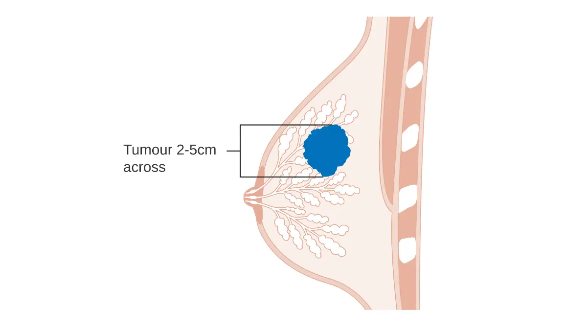
🩺 Physician Note: In my clinical practice, the most common misunderstanding I encounter at a patient’s first appointment is confusing grade with type. A report reading “invasive ductal carcinoma, Grade 2” prompts the question: “Does Grade 2 mean Stage 2?” It does not. Grade describes how abnormal the cancer cells appear under a microscope. Type describes where the cancer originated. Stage describes how far it has spread. All three are distinct variables — and all three matter for treatment planning.
Where breast cancer starts: ductal vs lobular origin
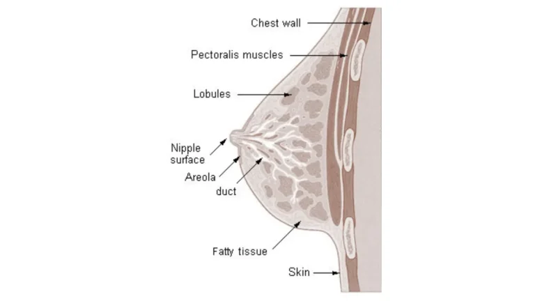
Ductal cancers begin in the milk ducts that carry milk from the lobules toward the nipple. Lobular cancers begin in the milk-producing lobules themselves. Most breast cancers are ductal in origin, with invasive lobular carcinoma (ILC) accounting for the second largest share of invasive diagnoses.
| Type | Where It Starts | Invasive or Non-Invasive | Relative Frequency |
|---|---|---|---|
| Invasive Ductal Carcinoma (IDC) | Milk ducts | Invasive | Most common — majority of all US diagnoses |
| Invasive Lobular Carcinoma (ILC) | Milk lobules | Invasive | Second most common invasive type |
| Ductal Carcinoma In Situ (DCIS) | Milk ducts | Non-invasive (Stage 0) | Most common non-invasive type |
| Lobular Carcinoma In Situ (LCIS) | Milk lobules | Non-invasive (risk marker) | Less common; not classified as cancer |
| Inflammatory Breast Cancer (IBC) | Ductal | Invasive | Approximately 1–5% of US diagnoses |
| Paget Disease of the Nipple | Nipple skin / ducts | Usually associated with DCIS or IDC | Rare |
| Angiosarcoma | Blood/lymph vessel cells | Invasive | Less than 1% of breast malignancies |
Source: 2026 NCI breast cancer data
📊 Clinical Data Point: Invasive ductal carcinoma is the most commonly diagnosed breast cancer type in the United States, accounting for the large majority of all US breast cancer diagnoses. — Source: NCI, 2026
For information on what IDC outcomes look like when caught early, see invasive ductal carcinoma cure rates and treatment data.
Rare and special-type breast cancers
Phyllodes tumors are rare fibroepithelial tumors — most are benign, but a small proportion are malignant and require surgical excision. Angiosarcoma arises from blood or lymph vessel lining cells and may develop as a late complication of prior breast radiation therapy. Paget disease of the nipple causes scaling, redness, and discharge at the nipple and is almost always associated with an underlying breast malignancy.
DCIS and LCIS: what non-invasive breast cancer means for you
A DCIS or LCIS diagnosis is one of the most commonly misunderstood findings in breast oncology. Patients frequently arrive at my clinic either under-alarmed — “It’s not even real cancer, right?” — or over-alarmed: “If it has ‘carcinoma’ in the name, shouldn’t I have a mastectomy?”
Both reactions are understandable. Both are usually inaccurate.
What DCIS actually is — and what it is not
Ductal carcinoma in situ is classified as Stage 0 breast cancer. Abnormal cells are present inside a milk duct but have not grown through the duct wall into surrounding breast tissue. DCIS is not invasive — but it is real, it requires treatment in most cases, and certain high-grade forms carry meaningful risk of progressing to invasive cancer without intervention.
Treatment for DCIS typically involves lumpectomy with or without radiation, and hormone therapy for estrogen receptor-positive DCIS. The specific approach depends on tumor grade, lesion size, and hormone receptor status — three variables your surgical oncologist will weigh before recommending a plan.
✅ Patient Action: Before deciding between lumpectomy, mastectomy, or active surveillance for a DCIS diagnosis, ask your breast surgical oncologist these three specific questions: What is the grade of my DCIS? What is the size of the lesion on imaging? Is my DCIS estrogen receptor-positive? The answers determine whether radiation and hormone therapy should be added to surgery.
For guidance on reading your pathology results once they arrive, understanding your breast cancer pathology report walks through each finding step by step.
LCIS: a cancer risk marker, not a cancer diagnosis
Lobular carcinoma in situ (LCIS) describes abnormal cell changes inside the milk-producing lobules that have not invaded surrounding tissue. Despite the word “carcinoma” in its name, LCIS is not classified as breast cancer — it is a pathologic risk marker that tells your oncologist your lifetime breast cancer risk is significantly elevated.
LCIS typically requires no surgery. It does require enhanced surveillance: more frequent mammograms, possible breast MRI, and a discussion of risk-reduction strategies including medication (tamoxifen or raloxifene in eligible patients) or, in select high-risk cases, preventive surgery. Women with LCIS who also carry a BRCA1 or BRCA2 mutation face compounded lifetime risk that warrants individualized genetic counseling.
If you have received BRCA test results you need help interpreting, see how to read your BRCA genetic test results. You can also use our Genetic Risk Assessment Tool to evaluate your broader hereditary breast cancer risk profile.
For current screening recommendations for women living with LCIS, see the American Cancer Society’s breast cancer screening guidelines.
📊 Clinical Data Point: Women with LCIS face an elevated lifetime risk of developing invasive breast cancer compared to the general population — the magnitude varies by additional risk factors including BRCA status, family history, and breast density. — Source: ACS 2026 breast cancer data
How your pathology report describes non-invasive cancer
Your pathology report will use specific language to classify your finding: “in situ” means confined, “non-invasive” confirms no tissue invasion has occurred, and nuclear grade (low, intermediate, or high) describes how abnormal the cells look. Understanding these terms before your oncology appointment helps you ask more targeted questions and make a more informed treatment decision.
Ongoing familiarity with early breast cancer symptoms remains important for all women with LCIS, who are often entirely asymptomatic and rely on screening rather than symptom recognition.
⚠️ Clinical Warning: Women with LCIS who have a first-degree relative with breast cancer — or who have not had BRCA genetic testing — should discuss genetic counseling with their oncologist before deciding on a surveillance-only approach. An at-home hereditary breast cancer genetic test can provide a starting point, but results require clinical interpretation before they change a management plan. Consult a board-certified breast surgical oncologist or geneticist before making any treatment or surveillance decision based on LCIS alone.
Invasive breast cancer types: ductal, lobular, and inflammatory
Most patients who receive a breast cancer diagnosis are diagnosed with an invasive breast cancer type — meaning cancer cells have grown beyond the original duct or lobule into surrounding tissue. The three most clinically significant invasive types are IDC, ILC, and IBC. They share the “invasive” classification but differ profoundly in how they grow, how they’re detected, and in the case of IBC, how urgently they require evaluation.
Invasive ductal carcinoma: the most common breast cancer diagnosis
Invasive ductal carcinoma begins in the milk ducts and has grown through the duct wall into surrounding breast tissue. It typically forms a palpable mass and is usually visible on mammogram. IDC is the most common breast cancer type, and its treatment is determined not by type alone but by the combination of tumor grade, stage, and receptor status — particularly ER, PR, and HER2 expression.
For many IDC patients, understanding what sentinel lymph node biopsy reveals is an important next step — this procedure determines whether the cancer has reached the nearby lymph nodes and directly influences staging and treatment decisions.
Invasive lobular carcinoma and why it’s often harder to detect
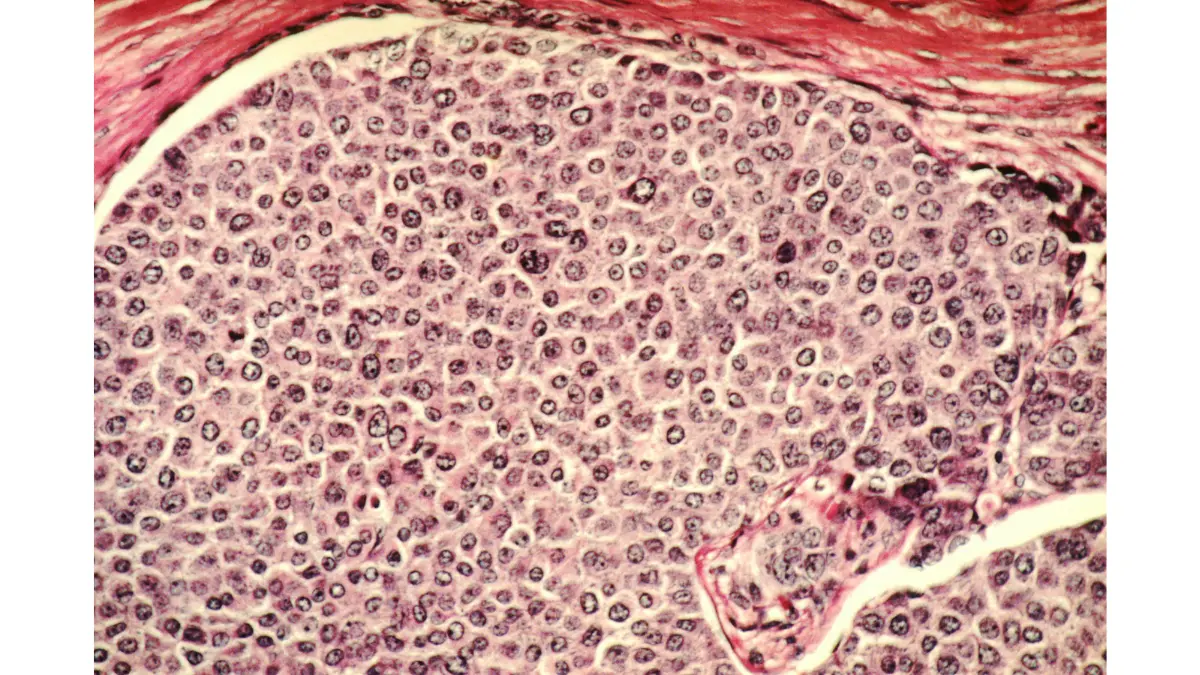
Invasive lobular carcinoma begins in the milk-producing lobules and grows in a distinctive diffuse, single-file sheet pattern — rather than forming a discrete lump. This growth pattern makes ILC harder to feel on physical exam and, in many cases, harder to see on a standard mammogram than IDC.
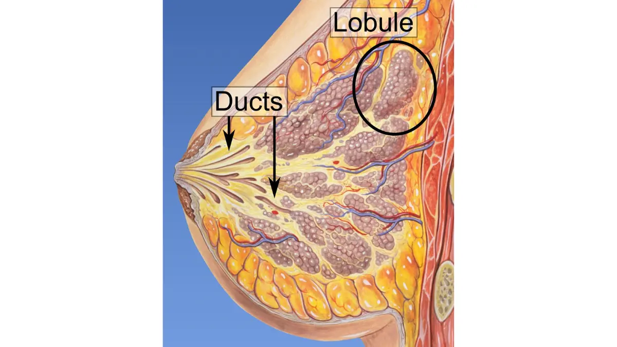
ILC is frequently estrogen receptor-positive, which means hormone therapy is typically part of the treatment plan. Because of its atypical growth pattern, some ILC patients are referred for breast MRI to more accurately assess tumor size and any additional disease sites before surgery.
🔬 How It Works: ILC cancer cells lose a protein called E-cadherin, which normally keeps cells anchored together. Without this anchor, ILC cells detach from each other and infiltrate surrounding tissue in single-file strands — creating the diffuse, sheet-like growth pattern that standard imaging can miss. This is a biological feature of the cancer itself, not a failure of your mammogram.
Inflammatory breast cancer: symptoms, urgency, and what to do now
Inflammatory breast cancer (IBC) is a rare but rapidly progressing form of invasive breast cancer diagnosed primarily by clinical skin signs — not by a palpable lump. IBC accounts for approximately 1–5% of US breast cancer diagnoses per 2026 NCI data, but it carries a lower 5-year survival rate than most other invasive breast cancers.
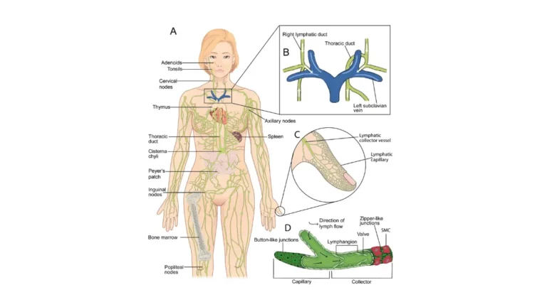
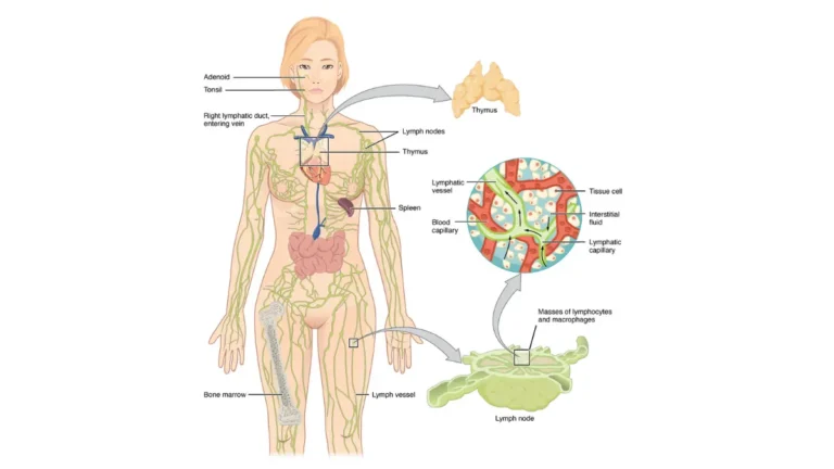
The defining clinical signs of IBC are skin redness covering more than one-third of the breast, warmth, rapid swelling, and a peau d’orange (orange-peel) texture caused by lymphatic obstruction. These signs develop over days to weeks — not months. A standard mammogram frequently appears normal in IBC. The diagnosis is confirmed by skin punch biopsy, not by imaging alone.
⚠️ Clinical Warning: IBC is frequently misdiagnosed as mastitis or a breast infection in primary care because the skin signs are similar. If you have been prescribed antibiotics for a suspected breast infection and the redness and swelling have not improved within 7–10 days, request an urgent referral to a breast oncologist — not a general surgeon. IBC can progress to lymph node involvement and distant metastasis within weeks without appropriate staging and treatment.
If you are experiencing new breast skin changes, use our Symptom Checker as a first step — but any sudden redness or rapid swelling warrants a same-day call to a physician, not a wait for tool results.
✅ Patient Action: If you notice breast skin redness, warmth, rapid swelling, or peau d’orange texture that developed over days to weeks — with or without a palpable mass — contact a breast oncologist immediately and request an urgent skin punch biopsy and full staging workup. Do not wait for a scheduled mammogram appointment. Inflammatory breast cancer requires urgent evaluation.
HER2+, triple-negative, and hormone receptor breast cancer
Molecular receptor status is the second layer of breast cancer classification — and it is the layer that most directly determines which drugs and treatment classes your oncologist will recommend. Every breast cancer, regardless of type, is tested for three biological targets: estrogen receptors (ER), progesterone receptors (PR), and HER2 protein overexpression. The combination of positive and negative results defines your molecular subtype.
ER-positive and PR-positive breast cancer: hormone therapy targets
Estrogen receptor-positive (ER+) breast cancer — the most common molecular subtype — means cancer cells carry receptors that bind estrogen, using it as a growth signal. This receptor profile makes the tumor responsive to hormone-blocking therapies including tamoxifen and aromatase inhibitors (letrozole, anastrozole, exemestane).
For early-stage ER-positive, HER2-negative breast cancer, the Oncotype DX genomic assay — a 21-gene expression test performed on the tumor tissue — can predict the benefit of adding chemotherapy to hormone therapy. A low Oncotype DX recurrence score indicates chemotherapy can often be safely omitted, sparing patients its side effects without compromising survival outcomes.
HER2-positive breast cancer: what overexpression means for treatment
HER2-positive breast cancer overexpresses the HER2 protein, which drives rapid cell growth. HER2+ tumors tend to grow more aggressively than ER+ tumors, but they also respond specifically to HER2-targeted drugs. The combination of trastuzumab (Herceptin) and pertuzumab (Perjeta) paired with chemotherapy has transformed HER2+ outcomes — many patients now achieve a complete pathologic response (no detectable cancer remaining at surgery) with neoadjuvant treatment.
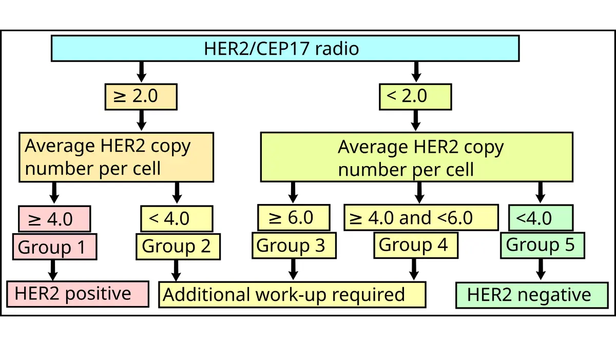
📊 Clinical Data Point: Trastuzumab (Herceptin) for HER2-positive breast cancer and pembrolizumab (Keytruda) for eligible triple-negative breast cancer are among the FDA-approved treatments whose approval status and current labeling can be confirmed through the FDA’s breast cancer drug resources page. — Source: FDA, 2026
Triple-negative breast cancer: why it requires a different approach
Triple-negative breast cancer (TNBC) tests negative for estrogen receptors, progesterone receptors, and HER2 overexpression, making it ineligible for hormone therapy and HER2-targeted drugs and requiring chemotherapy — and, in eligible patients, pembrolizumab (Keytruda) — as its primary systemic treatment strategy.
TNBC tends to be more aggressive than hormone receptor-positive subtypes and has a higher risk of early recurrence and distant metastasis. However, TNBC also tends to respond more dramatically to chemotherapy than other subtypes — a property called chemosensitivity — and recent advances in immunotherapy have meaningfully improved outcomes for eligible patients.
For a detailed breakdown of what TNBC chemotherapy regimens involve and what to expect from treatment, see how chemotherapy works for breast cancer and its side effects.
🔬 How It Works: TNBC lacks the three most common biological “locks” that targeted therapies use as entry points. This is why there is no hormonal key (tamoxifen) or HER2 key (trastuzumab) that fits a TNBC tumor. Chemotherapy and immunotherapy work through different mechanisms — attacking cell division directly and activating the immune system — which is why they remain the treatment backbone for TNBC.
How your molecular subtype and breast cancer type combine in treatment planning
| Molecular Subtype | Receptor Profile | First-Line Treatment Class | FDA-Approved Drug Example |
|---|---|---|---|
| ER+/PR+, HER2− | ER+, PR+, HER2− | Hormone therapy ± Oncotype DX-guided chemotherapy | Tamoxifen; Letrozole |
| HER2+, ER− | HER2+, ER− | HER2-targeted therapy + chemotherapy | Trastuzumab + Pertuzumab |
| HER2+, ER+ | HER2+, ER+ | HER2-targeted therapy + hormone therapy | Trastuzumab + Aromatase inhibitor |
| Triple-Negative | ER−, PR−, HER2− | Chemotherapy ± immunotherapy | Pembrolizumab (if PD-L1+) |
Source: FDA drug approvals 2026; NCCN Breast Cancer Clinical Practice Guidelines 2026
✅ Patient Action: Treatment selection for triple-negative or HER2-positive breast cancer requires multidisciplinary tumor board input. Before starting any systemic therapy, ask your oncologist specifically: Has my case been presented to a breast cancer tumor board? What is my PD-L1 status if I have TNBC? The answers determine whether immunotherapy belongs in your treatment plan.
Breast cancer survival rates by type — what the 2026 data shows
Survival statistics are the numbers patients most want — and most often misread. Among all breast cancer types, hormone receptor-positive (ER+/PR+) cancers detected at stage I consistently show the highest 5-year relative survival rates, often exceeding 95% per current NCI data. Non-invasive DCIS at stage 0 carries an even more favorable outlook. Inflammatory breast cancer and triple-negative breast cancer at advanced stages carry lower survival estimates.
For a full breakdown of how survival estimates are structured and what they mean for your specific diagnosis, see breast cancer survival rates by stage and subtype.
5-year relative survival rates for the most common breast cancer types
| Breast Cancer Type/Subtype | Approximate 5-Year Relative Survival | Stage Note |
|---|---|---|
| DCIS (Stage 0) | Highly favorable — near 100% | Non-invasive; localized |
| ER+/PR+ (Stage I–II) | Often >95% | Localized to breast |
| HER2+ (with targeted therapy) | Substantially improved with trastuzumab | Stage-dependent |
| Triple-Negative | Lower than ER+ overall | Varies significantly by stage |
| Inflammatory Breast Cancer | Lower than non-IBC invasive types | Frequently stage III at diagnosis |
Source: 2026 NCI/SEER data
📊 Clinical Data Point: 5-year relative survival rates for breast cancer are derived from the NCI SEER database and represent population-level outcomes — not individual predictions. Current 2026 data should be verified against the most recent NCI SEER annual release at publish. — Source: NCI SEER Program, 2026
How stage and molecular subtype interact to affect prognosis
Stage and receptor status together produce a more accurate prognosis signal than type alone. An ER-positive IDC caught at stage I carries a profoundly different prognosis than an ER-negative IDC caught at stage III, even though both are “IDC.” This is why oncologists evaluate type, grade, stage, and receptor status as a combined clinical picture — not individual variables in isolation.
What survival statistics cannot tell you about your outcome
Population-level 5-year relative survival rates describe how a large group of patients diagnosed with a specific cancer type fared over five years. They cannot tell you how you will fare — because your outcome depends on variables that aggregate statistics cannot capture: your tumor’s specific grade, its lymph node involvement, your age, your treatment facility’s experience, and how your tumor responds to the first line of treatment.
✅ Patient Action: Survival statistics are starting points for a conversation — not a prognosis handed down. Before your next oncology appointment, ask your oncologist: What specific factors in my tumor profile modify the population survival estimate that applies to my diagnosis? What does the combination of my type, grade, stage, and receptor status mean for my individual risk picture?
What a breast cancer specialist wants you to know about your type
In my clinical practice, the patients who navigate their diagnosis with the most clarity are those who ask their oncologist four specific questions alongside their type result: What is my tumor grade? What is my receptor status? What is my stage? And what does the combination of these four variables mean for my treatment plan?
Breast cancer type is the first answer on that list — not the only one.
The most important question to ask after you learn your type
The most useful question you can ask your oncologist at your first appointment is not “Is my type bad?” It is: “What does the combination of my type, grade, stage, and receptor status tell you about how aggressive this cancer is, and what treatment protocol it points to?” That question — not type alone — produces the clinical picture your oncologist is actually working from.
Why “rare type” doesn’t always mean worse outcome
A diagnosis of phyllodes tumor, mucinous carcinoma, or tubular carcinoma often triggers immediate fear in patients who assume “rare” equals “more dangerous.” It does not. Several rare breast cancer types — including mucinous (colloid) carcinoma and tubular carcinoma — carry more favorable prognoses than typical IDC in comparable staging contexts. The biology of the individual tumor matters far more than how common the type is.
Patients diagnosed with rare types, IBC, or molecularly complex subtypes who want a specialist second opinion before treatment begins may benefit from a telehealth oncology consultation — accessing a board-certified breast cancer specialist without requiring travel or long referral waits.
For patients who are offered surgical treatment, what your surgeon may not tell you about mastectomy provides detailed preparation information before any decision is made.
Patients with rare or aggressive breast cancer types may also find relevant open studies listed through active breast cancer clinical trials on ClinicalTrials.gov.
✅ Patient Action: Every newly diagnosed patient should ask their oncologist to explain the combination of type, grade, stage, and receptor status together — not type alone — as the complete clinical picture that determines your treatment approach and individual prognosis estimate.
Breast cancer types — answers from our medical advisory board
Q1: What is the most common type of breast cancer?
Invasive ductal carcinoma (IDC) is the most frequently diagnosed breast cancer type in the United States, accounting for the large majority of all diagnoses per 2026 NCI data. IDC begins in the milk ducts and has grown into surrounding breast tissue. Receptor status and tumor grade — not type alone — drive the specific treatment protocol. Consult a board-certified breast oncologist about your complete diagnostic profile.
Q2: What is ductal carcinoma in situ (DCIS)?
DCIS is a Stage 0 breast cancer in which abnormal cells are confined within a milk duct and have not grown into surrounding tissue. It is classified as non-invasive and is highly treatable. Treatment typically involves lumpectomy with or without radiation and, in ER-positive cases, hormone therapy. Consult a board-certified breast surgical oncologist before deciding on a treatment approach for your specific DCIS grade and lesion size.
Q3: Is LCIS considered cancer?
Lobular carcinoma in situ (LCIS) is not cancer — it is a pathologic risk marker showing abnormal cell changes in the lobules that do not invade surrounding tissue. LCIS indicates elevated lifetime breast cancer risk and requires enhanced surveillance rather than surgery in most cases. Discuss your monitoring plan, screening frequency, and risk-reduction options with a board-certified breast specialist.
Q4: What is invasive lobular carcinoma?
Invasive lobular carcinoma (ILC) is the second most common invasive breast cancer type. ILC cells grow in a diffuse, single-file sheet pattern rather than a discrete lump, making it harder to detect on mammogram and by physical exam. ILC is frequently estrogen receptor-positive. Consult a board-certified breast oncologist about whether dedicated breast MRI is appropriate for your ILC imaging workup.
Q5: What is inflammatory breast cancer?
Inflammatory breast cancer (IBC) is diagnosed by clinical skin signs — redness, warmth, swelling, and peau d’orange texture — rather than by a palpable lump. Mammograms frequently appear normal. IBC accounts for approximately 1–5% of US breast cancer diagnoses and is among the most aggressive breast cancer types. If you notice sudden breast skin changes, contact a breast oncologist immediately for urgent evaluation and biopsy.
Q6: What is triple-negative breast cancer?
Triple-negative breast cancer (TNBC) tests negative for estrogen receptors, progesterone receptors, and HER2 overexpression, making it ineligible for hormone therapy and HER2-targeted drugs. Chemotherapy — and pembrolizumab in PD-L1-positive eligible patients — serves as the primary systemic treatment. TNBC tends to be more aggressive than hormone receptor-positive types. Consult a board-certified oncologist to determine whether immunotherapy is appropriate for your specific TNBC profile.
Q7: Is HER2-positive breast cancer aggressive?
HER2-positive breast cancer grows more rapidly than hormone receptor-positive types but responds specifically to targeted therapy. Trastuzumab combined with pertuzumab and chemotherapy has significantly improved survival outcomes, with many patients achieving complete pathologic response after neoadjuvant treatment. Ask your oncologist whether your HER2+ tumor qualifies for pre-surgical targeted therapy before any surgical decision is made.
Q8: What type of breast cancer has the best survival rate?
Among all breast cancer types, hormone receptor-positive (ER+/PR+) cancers detected at stage I consistently show the highest 5-year relative survival rates — often exceeding 95% per 2026 NCI data. Non-invasive DCIS at stage 0 carries an even more favorable outlook. Prognosis depends on grade, lymph node involvement, and molecular subtype. Ask your oncologist how your individual tumor factors modify the population estimate for your diagnosis.
Q9: What is the difference between invasive and non-invasive breast cancer?
Non-invasive breast cancer — DCIS and LCIS — means abnormal cells remain confined within the duct or lobule and have not penetrated surrounding tissue. Invasive breast cancer means cells have crossed that boundary, with potential to reach lymph nodes or distant organs. Ask your oncologist to walk through each finding in your pathology report and its specific treatment implication before your next appointment.
Q10: What is Paget disease of the nipple?
Paget disease of the nipple affects the skin of the nipple and areola, causing scaling, redness, and sometimes discharge. It is almost always associated with an underlying breast cancer — DCIS in the majority of cases, invasive cancer in others. Consult a board-certified breast surgical oncologist if you notice persistent, unexplained changes to nipple skin or texture that do not resolve within a few weeks.
Q11: How is breast cancer type determined?
Your breast cancer type is determined through core needle biopsy — a tissue sample analyzed by a pathologist. The pathology report identifies cell origin, invasiveness, tumor grade, and receptor status (ER, PR, HER2). Genomic assays such as Oncotype DX may follow for early-stage ER-positive cases. Consult a board-certified breast oncologist to interpret your complete pathology findings and understand their treatment implications.
Q12: Can breast cancer type change over time or after treatment?
Breast cancer type itself does not change, but receptor status can shift — particularly after chemotherapy or hormonal therapy. A tumor initially classified as ER-positive may lose estrogen receptor expression at recurrence, changing the next treatment approach. Oncologists often repeat receptor testing at recurrence for this reason. Consult a board-certified oncologist to confirm current receptor status before proceeding with any treatment for a breast cancer recurrence.
Q13: What type of breast cancer is most likely to metastasize?
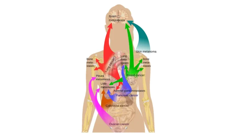
Triple-negative and inflammatory breast cancers carry the highest risk of distant metastasis. TNBC commonly spreads to the lungs, liver, and brain. IBC frequently presents with lymph node involvement and a higher rate of distant spread at diagnosis. Hormone receptor-positive cancers also metastasize — typically to bone, liver, and lungs — but generally at a slower rate. Ask your oncologist about your individual metastatic risk profile based on type, grade, and stage.
Q14: What is LCIS and why isn’t it classified as cancer?
LCIS describes abnormal cells within breast lobules that have not invaded surrounding tissue — making it a risk marker, not a true cancer diagnosis. Despite the word “carcinoma” in its name, LCIS does not require surgery in most cases. It elevates lifetime breast cancer risk and requires enhanced surveillance. Consult a board-certified breast specialist to establish the right monitoring schedule and risk-reduction strategy for your individual profile.
Q15: How does breast cancer type affect treatment decisions?
Breast cancer type determines the biological target for treatment: ER-positive tumors respond to tamoxifen or aromatase inhibitors, HER2-positive tumors respond to trastuzumab-based regimens, and triple-negative tumors require chemotherapy with possible immunotherapy. Type works alongside grade, stage, and lymph node status — not in isolation. Consult your oncologist about the full combination of factors that together define your optimal treatment plan.
Q16: What is angiosarcoma of the breast?
Angiosarcoma is a rare breast malignancy arising from cells lining blood or lymph vessels, accounting for less than 1% of all breast cancers. It may develop as a primary tumor or — more commonly — as a late complication years after breast radiation therapy. Treatment involves wide surgical excision. Consult a board-certified surgical oncologist with experience in rare breast tumors or sarcomas for this diagnosis.
Q17: What questions should I ask my oncologist about my breast cancer type?
After a breast cancer type diagnosis, ask your oncologist: What is my tumor grade alongside my type? What are my ER, PR, and HER2 receptor results? Is Oncotype DX genomic testing appropriate for my case? Has my case been reviewed by a multidisciplinary tumor board? Am I eligible for any active clinical trials? A board-certified breast oncologist should address each of these before any treatment decision is finalized.
Your breast cancer type is the starting point — not the final word
Receiving a breast cancer type diagnosis is one of the most disorienting experiences a person can face. What feels like a verdict is, clinically, a starting point — the first piece of a complete diagnostic picture that your oncologist will build alongside grade, stage, and receptor status.
Every patient deserves to understand that picture fully before any treatment decision is made. Ask the questions. Request the pathology walkthrough. Push for the tumor board review. And consult a board-certified breast oncologist or gynecologic oncologist for the individualized guidance that no article — however detailed — can replace.
This article was written by Dr. Carolyn D. Fairweather, MD (Gynecologic Oncology) and reviewed by Dr. Nathaniel J. Hargrove, MD (Oncology) and Dr. Alicia M. Thornton, MD (Internal Medicine and Preventive Health), reflecting 2026 clinical guidelines.
About this content
How this article was put together: researched from recognised health sources, drafted with the help of AI tools, and edited by hand, with sources linked throughout.

Sameer Patel is the founder and editor of My Medicine Advisor. He is not a doctor or medical professional — before starting this site he worked in banking,…
Medical disclaimer
The content on MyMedicineAdvisor is provided for general informational and educational purposes only and is not a substitute for professional medical advice, diagnosis, or treatment. Health information on this website should not be used to diagnose, treat, cure, or prevent any condition without guidance from a qualified healthcare professional. Always seek the advice of your doctor, physician, or another licensed healthcare provider with any questions you may have regarding a medical condition, symptoms, medications, or treatment decisions.